Thyroid Disease

Published on 06/06/2015 by admin

Filed under Pediatrics

Last modified 22/04/2025

Print this page

rate 1 star rate 2 star rate 3 star rate 4 star rate 5 star
Your rating: none, Average: 0 (0 votes)

This article have been viewed 3432 times

68 Thyroid Disease

The follicular cells of the thyroid gland produce triiodothyronine (T3) and thyroxine (T4), thyroid hormones that have important roles in growth and development, as well as influential effects on metabolism, the central nervous system (CNS), and the cardiovascular system. This chapter reviews thyroid physiology, as well as the most common childhood thyroid disorders.

Thyroid Hormone Production

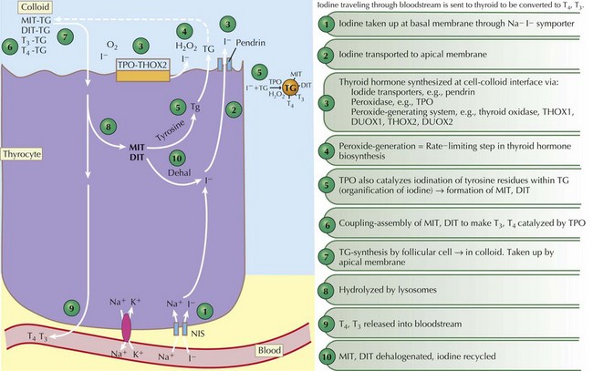
Thyroid hormones are produced by the coupling of iodine molecules to the amino acid tyrosine (Figure 68-1). The principal secreted thyroid hormone is T4. The normal thyroid secretes only small amounts of T3, and most circulating T3 (≈70%-90%) is derived from peripheral deiodination of T4. Thyroid hormones circulate bound to carrier proteins, including thyroid-binding globulin (TBG), transthyretin (thyroxine-binding prealbumin), and albumin. Less than 1% of circulating T4 and T3 are unbound or “free.”

Hypothalamic thyrotropin-releasing hormone (TRH) stimulates thyrotrope cells in the anterior pituitary to release thyroid-stimulating hormone (TSH), which stimulates production and release of thyroid hormone. When T4 levels are inadequate, a negative feedback loop activates the hypothalamic–pituitary axis and results in increased secretion of TSH, which then acts on the thyroid gland to stimulate increased hormone synthesis. When circulating levels of T4 and T3 are high, as in Graves’ disease or overtreatment with exogenous T4, this negative feedback loop acts to reduce or suppress TSH secretion. Circulating levels of T4 are also influenced by peripheral conversion to either T3 or reverse T3 (rT3), an inactive form of thyroid hormone.

Circulating T4 and T3 enter cells by diffusion and carrier-mediated transport processes; inside the cell, T4 is then converted to T3. T3 is the most active thyroid hormone because it binds thyroid hormone receptors with approximately 10 times the affinity of T4. The T3–receptor complex is transported to the nucleus and regulates transcription of a variety of genes, ultimately leading to the synthesis of proteins that manifest thyroid hormone action in peripheral tissues. T4 is necessary for normal growth and development and is absolutely critical for brain development in utero as well as during the first 2 years of life.

Congenital Hypothyroidism

Etiology and Pathogenesis

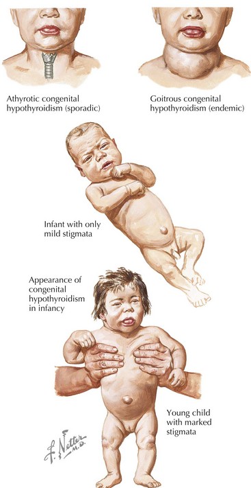
Congenital hypothyroidism (CH) is one of the most common causes of preventable mental retardation (Figure 68-2). Fortunately, early identification and rapid treatment lead to normal neurocognitive development. Although approximately 10% of cases of CH are transient and caused by factors such as iodine exposure, prematurity, or maternal transfer of antithyroid antibodies, in most cases, hypothyroidism is permanent. Worldwide, iodine deficiency is the most common cause of CH. However, in areas of the world where iodine deficiency is uncommon, CH most commonly results from thyroid dysgenesis (≈75% of cases); thyroid dyshormonogenesis (≈10%), TSH deficiency (5%), and genetic defects in the TSH receptor are much less common. The incidence of CH is approximately one in 3000 to 4000 births. In most cases, CH is sporadic, but mutations in genes encoding transcription factors that are required for normal development of the thyroid gland are present in about 10% to 15% of cases. Circulating levels of TSH are elevated and thyroid hormone levels are low in those with CH.

A clinical picture that is similar to CH can occur in children who have genetic defects in the thyroid hormone receptor or in the MCT8 protein required to transport thyroid hormone into cells. Unlike CH, however, these patients have elevated serum levels of both TSH and thyroid hormones.

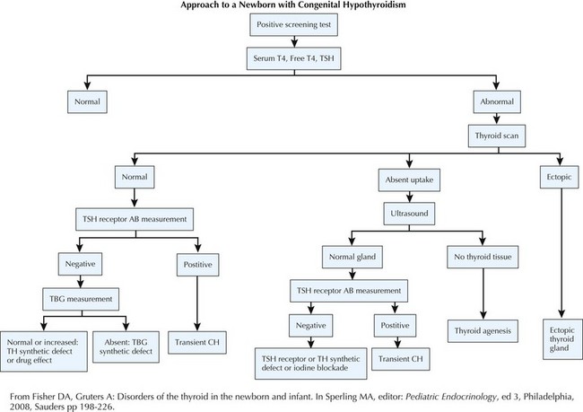
Evaluation (Figure 68-3)

NBS for CH is based on collection of a blood sample from the newborn between 48 hours and 4 days of life. Earlier measurement may lead to erroneous results because of the normal physiologic surge in TSH that occurs soon after birth. NBS programs use blood spots collected on filter paper and use one of two strategies to identify infants with CH: a primary TSH with backup T4 or a primary T4 with a backup TSH method. An abnormal result should be further evaluated immediately using serum-based assays for TSH and T4 or free T4. Premature or ill infants may have false-negative or false-positive results and should be retested by 7 days of age. Any infant with a TSH level above 40 mU/L with low T4 is considered to have primary hypothyroidism. In addition to T4 and TSH measurements, a thyroglobulin (TG) level may be helpful because elevation suggests dyshormonogenesis.

image

Figure 68-3 Evaluation of congenital hypothyroidism.

AB, antibody; CH, congenital hypothyroidism; TBG, thyroid-binding globulin; TH, thyroid hormone; TSH, thyroid-stimulating hormone.

When there is history of maternal autoimmune thyroid disease, measurement of TSH–receptor binding antibodies in the infant or the mother may identify transient CH. Other diagnostic tests include thyroid ultrasonography and technetium or iodine (I-123) scans to identify functional thyroid tissue, as well as the perchlorate washout test to detect iodine organification defects that might indicate Pendred’s syndrome. Treatment with levothyroxine (L-T4) should not be delayed to perform imaging.

Treatment

To avoid neurocognitive deficit, newborns with CH must be treated promptly with L-T4 and monitored closely by a pediatric endocrinologist. L-T4 is instituted at a dosage of 10 to 17 µg/kg/d initially, with a goal to normalize the serum T4 level within 2 weeks (fT4 >2 ng/dL) and serum TSH by 1 month of age. There are no suitable liquid preparations of L-T4, so tablets must be used. Tablets should be crushed and mixed with a few milliliters of formula, breast milk, or water. Soy-based formula, fiber, or iron may reduce absorption of L-T4 and should be given separately.

Serum levels of T4, T4 index or free T4, and TSH should be measured every 1 to 2 months during the first 6 months of life, every 3 to 4 months until 3 years of age, and then every 6 to 12 months until growth is complete. The half-life of T4 in the circulation is 1 week, so levels of TSH and T4 should be repeated 4 to 5 weeks after dose changes to ensure appropriate steady-state levels. An appropriately treated child will have serum T4 levels that are at or above the upper limit of normal with a serum TSH level of 1 to 2 mU/mL. The serum TSH level is not a reliable indicator of euthyroidism in children with pituitary or hypothalamic disorders, and in these patients, the T4 level should be maintained at the upper limit of the assay’s normal range. Infants with suspected transient hypothyroidism should continue L-T4 therapy until at least age 2 years, when thyroid-dependent CNS myelinization is complete.

Acquired Hypothyroidism

Etiology and Pathogenesis

Hypothyroidism is defined as a deficiency in thyroid hormone and in most cases is associated with an elevated TSH level. Hypothyroidism is more common in females than males and has an increased incidence in adolescents. The most common cause of hypothyroidism is chronic lymphocytic (Hashimoto’s) thyroiditis, which results in autoimmune destruction of the thyroid gland. Goiter, caused by lymphocytic infiltration of thyroid tissue, and circulating antithyroid antibodies (antitissue peroxidase and anti-TG) are common but are not universally present. Hashimoto’s thyroiditis is common in children who have the type 2 autoimmune polyglandular syndrome, which includes Addison’s disease of the adrenal gland, pernicious anemia, celiac disease, type 1 diabetes mellitus, and juvenile rheumatoid arthritis. Children with chromosomal defects, such as trisomy 21, Turner’s syndrome, 22q11 deletion syndrome, and Klinefelter’s syndrome, have an increased incidence of autoimmune diseases, including autoimmune thyroid disease.

Other causes of hypothyroidism include chronic iodine deficiency, excessive iodine exposure, hypothalamic–pituitary dysfunction, acute infection, and medications. Excessive iodine exposure may lead to acute blockage of thyroid hormone release or thyroid hormone synthesis, known as the Wolff-Chaikoff effect. Children with CNS disease are at risk of central hypothyroidism. Medications such as amiodarone, antiepileptics, nitroprusside, and lithium can all affect thyroid hormone production or metabolism; dopamine can reduce TSH secretion.

Clinical Presentation (Figure 68-4)

Symptoms of hypothyroidism include dry skin, constipation, cold intolerance, and a decreased energy level. Severe thyroxine deficiency is associated with linear growth failure and delayed bone age, coarse hair, myxedema, galactorrhea, delayed or rarely precocious puberty, bradycardia, depressed reflexes, pallor, and hyperlipidemia. Carotenemia is more common in children than adults with hypothyroidism. Children with weight gain are often referred for evaluation of hypothyroidism. Although weight gain can occur in children with hypothyroidism, height is reduced, which is in contrast to exogenous obesity, in which height is often increased. On examination, the thyroid gland may be enlarged, asymmetric, and/or bosselated. In severe hypothyroidism, the gland may be atrophic. Reflexes are often slowed, the heart rate may be reduced, and the pulse pressure can be decreased. There is a family history of thyroid disease in 30% to 40% of patients.

Occasionally, patients with Hashimoto’s thyroiditis present with elevations in T4 levels and suppressed TSH, mimicking Graves’ disease, but without the eye symptoms. This is termed Hashitoxicosis, and it may occur because of excessive release of thyroid hormone from thyroid destruction. Patients with Hashimoto’s thyroiditis can have TG antibodies or blocking TSH-receptor antibodies, both of which are known to destroy thyroid cells. TG antibodies are seen in Hashimoto’s thyroiditis, and blocking TSH-receptor antibodies are seen in patients with atrophic thyroiditis. In addition, patients typically have thyroid peroxidase (TPO) antibodies, which are considered markers of inflammation. TPO antibodies also cause thyroid inflammation and thyroid disease to persist, interfering with the healing process. Patients with Hashitoxicosis can also have stimulating TSH-receptor antibodies, although their levels may not reach the high levels that cause hyperthyroidism in patients with Graves’ disease.

In some ways, these patients can be described as having both Hashimoto’s thyroiditis and Graves’ disease because the antibodies associated with both diseases are often present. Thyroid uptake scans can differentiate between Hashitoxicosis with decreased uptake and true Graves’ disease with increased uptake.

Acquired Hyperthyroidism

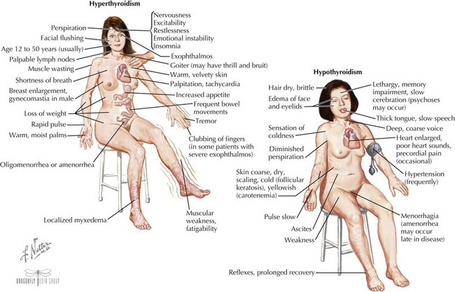
Clinical Presentation (see Figure 68-4)

Children with hyperthyroidism often have hyperactivity with periods of fatigue and poor sleeping habits. Emotional lability, poor concentration, and a marked decrease in school performance are common. Children often come to medical attention because of cardiovascular complaints such as persistent tachycardia, palpitations, or syncopal episodes. Heat intolerance, weight loss, tremors, and diaphoresis are seen regularly. Although many children with hyperthyroidism indeed lose weight, paradoxical weight gain is common. Hyperthyroidism will also result in an increased appetite, and in some patients, an increased appetite and increased food intake may lead to weight gain despite the increase in metabolic rate that usually accompanies hyperthyroidism.

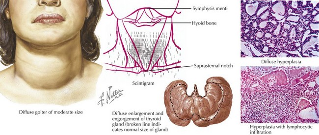
The eyes may reveal a “lid lag” or prominent eyes, with or without proptosis (exophthalmos) in which the eyes are pushed forward (may be asymmetric). In Graves’ disease, the more severely hyperthyroid patients have the largest goiters (Figure 68-5). Mild polyuria may be seen. Frequent bowel movements, rather than diarrhea, are occasionally present.

Heart rate and blood pressure may be elevated with a widened pulse pressure. The degree of tachycardia parallels the severity of the hyperthyroidism. There may be normal to accelerated linear growth with concomitant weight loss. The skin is warm to the touch. The thyroid is smooth and diffusely enlarged, and there may be a palpable thrill or audible bruit caused by increased blood flow. Tremors and generalized restlessness are common. A change in mentation associated with hypertension and cardiovascular instability could indicate thyroid storm, which requires urgent hospitalization.

Subacute, or De Quervain’s, thyroiditis is thought to be viral in origin and lasts weeks to months. In the first few months, hyperthyroidism may be seen because of leakage of thyroid hormone in a damaged gland. In time, hypothyroidism commonly develops.

Treatment

Treatment of children with hyperthyroidism should be made in collaboration with a pediatric endocrinologist. Families should be informed of three treatment modalities: medication, radioactive iodine (I-131) ablation, and surgery. Most endocrinologists believe that initial treatment with antithyroid medication is indicated, especially because approximately 20% of children may go into a remission within 2 years of starting pharmacologic therapy.

The two antithyroid medications that are available in the United States are propylthiouracil (PTU) and methimazole. Both of these agents block synthesis of thyroid hormone, but only PTU can also prevent conversion of T4 to T3. The dosing of PTU is commonly two to three times per day compared with once or twice daily with methimazole. Methimazole has emerged as the preferred treatment based on recent reports that PTU usage, particularly in children, is associated with the occasional development of severe liver failure that often requires transplantation. Side effects from both medications occur in 5% to 14% of children and include skin rash, arthralgia, arthritis, lupus-like reaction, thrombocytopenia, and agranulocytosis. A β-blocker is used in patients with more severe cardiovascular signs.

Radioactive iodine ablation of the thyroid is an effective therapy for destroying the thyroid, although it does not ameliorate proptosis of the eyes. The destruction of the thyroid occurs 2 to 4 months after therapy with I-131. Surgery is used occasionally to treat children with Graves’ disease, although complications include hypothyroidism, hypoparathyroidism, and damage to the laryngeal nerves.

Monitoring thyroid function frequently is particularly important in children with hyperthyroidism because of the fluctuating nature of the disease. Monthly laboratory assessments are common. An occasional complete blood count and chemistry panel, including hepatic function tests, may be useful to monitor for medication side effects.