23 Thrombosis
Venous thromboembolism (VTE)
Venous thromboembolism
Aetiology
Sluggishness of blood flow may be related to bed rest, surgery or reduced cardiac output, for example in heart failure. Factors increasing the risk of hypercoagulability include surgery, pregnancy, oestrogen administration, malignancy, myocardial infarction and several acquired or inherited disorders of coagulation (for further detail of genetic factors, see Rosendaal and Reitsma, 2009).
Other risk factors
There are several other patient-related risk factors for VTE. Age over 60 years is an important factor. Critical care admission, dehydration, and one or more significant medical comorbidities such as heart disease, metabolic, endocrine or respiratory pathologies, acute infectious diseases and inflammatory conditions are all important risk factors for VTE. A full list can be found in the relevant National Institute for Health and Clinical Excellence (2010) guideline.
Investigations
Treatment
Prophylaxis
Prevention of initial episodes of VTE in those at risk is clearly of great importance. It was estimated that around 25,000 people in the UK die from preventable hospital-acquired VTE annually, including patients admitted to hospital for medical care as well as surgery. There is also widespread evidence of inconsistent use of prophylactic measures for VTE in hospital patients, including mechanical as well as pharmacological means of VTE prophylaxis. Some of the medicines described below contribute to those pharmacological measures. Guidelines on this are available (National Institute for Health and Clinical Excellence, 2010).
Heparins
The major adverse effect of all heparins is haemorrhage, which is commoner in patients with severe heart or liver disease, renal disease, general debility and in women aged over 60 years. The risk of haemorrhage is increased in those with prolonged clotting times and in those given heparin by intermittent intravenous bolus rather than by continuous intravenous administration. UFH is monitored by derivatives of the activated partial thromboplastin time (APTT), for example the kaolin–cephalin clotting time (KCCT); in those patients with a KCCT three times greater than control, there is an eightfold increase in the risk of haemorrhage. The therapeutic range for the KCCT during UFH therapy, therefore, appears to be between 1.5 and 2.5 times the control values. Rapid reversal of the effect of heparin can be achieved using protamine sulphate, but this is rarely necessary because of the short duration of action of heparin. LMWHs may produce fewer haemorrhagic complications, and monitoring of effect is not routinely required. At doses normally used for treatment, they do not significantly affect coagulation tests and routine monitoring is not necessary (British Committee for Standards in Haematology, 2006a).
Heparins, particularly UFH, may also cause thrombocytopenia (low platelet count). This may occur in two forms. The first occurs 3–5 days after treatment and does not normally result in complications. The second type of thrombocytopenia occurs after about 6 days of treatment and often results in much more profound decreases in platelet count and an increased risk of thromboembolism. LMWHs are thought to be less likely to cause thrombocytopenia but this complication has been reported, including in individuals who had previously developed thrombocytopenia after UFH. For these reasons, patients should have a platelet count on the day of starting UFH and the alternate-day platelet counts should be performed from days 4 to 14 thereafter. For patients on LMWH, the platelet counts should be performed at 2–4 day intervals from day 4 to 14 (British Committee for Standards in Haematology, 2006b). If the platelet count falls by 50% and/or the patient develops new thrombosis or skin allergy during this period, heparin-induced thrombocytopenia (HIT) should be considered, and if strongly suspected or confirmed, heparin should be stopped and an alternative agent such as a heparinoid or hirudin commenced.
It has been shown that there is a non-linear relationship between the dose of UFH infused and the KCCT. This means that disproportionate adjustments in dose are required depending on the KCCT if under- or over-dosing is to be avoided (Box 23.1). Since the half-life of UFH is 1 h, it would take 5 h (five half-lives of the drug) to reach a steady state. A loading dose is, therefore, administered to reduce the time to achieve adequate anticoagulation. UFH in full dose can also be given by repeated subcutaneous injection, and in these circumstances the calcium salt appears to be less painful than the sodium salt. Opinions differ as to whether the subcutaneous or intravenous route is preferable. The subcutaneous route may take longer to reach effective plasma heparin concentrations but avoids the need for infusion devices.
Box 23.1 Guidelines to control unfractionated heparin (UFH) treatment
Source: Modified from Fennerty et al. (1986) and reproduced in British Committee for Standards in Haematology (1998).
| >7.0 | Discontinue for 30 min to 1 h and reduce by >500 iu/h |
| >5.0 | Reduce by 500 iu/h |
| 4.1–5.0 | Reduce by 300 iu/h |
| 3.1–4.0 | Reduce by 100 iu/h |
| 2.6–3.0 | Reduce by 50 iu/h |
| 1.5–2.5 | No change |
| 1.2–1.4 | Increase by 200 iu/h |
| <1.2 | Increase by 400 iu/h |
Heparin is normally used in the immediate stages of venous thrombosis and PE until the effects of warfarin become apparent. In the past, it has been continued for 7–10 days, but recent evidence indicates that around 5 days of therapy may be sufficient in many instances. This shorter treatment may also reduce the risk of the rare but potentially very serious complications of severe HIT, which normally occurs after the sixth day. LMWH should be administered for at least 5 days or until the INR has been in the therapeutic range for two successive days, whichever is the longer. They have largely replaced UFH, since they can be given subcutaneously (without a loading dose), and without routine monitoring. A full blood count should be ordered after 5 days on LMWH and throughout the duration of LMWH treatment to monitor for heparin-related thrombocytopenia. Patients with previous exposure to heparin within the past 100 days should also have a platelet count performed before the second dose of heparin is administered (Winter et al., 2005).
Hirudins
Lepirudin, a recombinant hirudin, is licensed for anticoagulation in patients with type II (immune) HIT who require parenteral antithrombotic treatment. The dose of lepirudin is adjusted according to the APTT, and it is given intravenously by infusion. Haemorrhage is greater in those with poor renal function. Severe anaphylaxis occurs rarely in association with lepirudin treatment and is more common in previously exposed patients (British Committee for Standards in Haematology, 2006b). Bivaluridin is an analogue of hirudin, but acts as a direct thrombin inhibitor. It is licensed for anticoagulation in patients undergoing percutaneous coronary intervention (PCI). It has to be administered parenterally and the activated clotting time (ACT) is used to assess its activity. Haemorrhage is also an important adverse effect of this agent.
Oral anticoagulants
Warfarin
The effect of warfarin is monitored using the one-stage prothrombin time, for example the international normalised ratio (INR). This test is sensitive chiefly to factors VII, II and X (and to a lesser extent factor V, which is not a vitamin K-dependent clotting factor). However, factor VII, to which the INR is sensitive, is the most important factor in the extrinsic pathway of clotting. The optimum therapeutic range for the INR differs for different clinical indications since the lowest INR consistent with therapeutic efficacy is the best in reducing the risk of haemorrhage. Examples of therapeutic ranges recommended for certain indications are given in Table 23.1 (British Committee for Standards in Haematology, 1998, 2006c).
Table 23.1 Recommended target INRs for different conditions and grade of recommendation
| Indication | Target INR | Grade of recommendation |
|---|---|---|
| Pulmonary embolus | 2.5 | A |
| Proximal deep vein thrombosis | 2.5 | A |
| Calf vein thrombus | 2.5 | A |
| Recurrence of venous thromboembolism when no longer on warfarin therapy | 2.5 | A |
| Recurrence of venous thromboembolism while on warfarin therapy | 3.5 | C |
| Symptomatic inherited thrombophilia | 2.5 | A |
| Antiphospholipid syndrome | 3.5 | A |
| Non-rheumatic atrial fibrillation | 2.5 | A |
| Atrial fibrillation due to rheumatic heart disease, congenital heart disease, thyrotoxicosis | 2.5 | C |
| Cardioversion | 2.5 | B |
| Mural thrombus | 2.5 | B |
| Cardiomyopathy | 2.5 | C |
| Mechanical prosthetic heart valve (caged ball or caged disk), aortic or mitrala | 3.5 | B |
| Mechanical prosthetic heart valve (tilting disc or bileaflet), mitrala | 3.0 | B |
| Mechanical prosthetic heart valve (tilting disc), aortica | 3.0 | B |
| Mechanical prosthetic heart valve (bileaflet), aortica | 2.0 | B |
| Bioprosthetic valve | 2.0 (if anticoagulated) | A |
| Ischaemic stroke without atrial fibrillation | Not indicated | C |
| Retinal vessel occlusion | Not indicated | C |
| Peripheral arterial thrombosis and grafts | Not indicated | A |
| Arterial grafts | 2.5 (if anticoagulated) | |
| Coronary artery thrombosis | 2.5 (if anticoagulated) | |
| Coronary artery graft | Not indicated | A |
| Coronary angioplasty and stents | Not indicated | A |
INR, international normalised ratio; A, at least one randomised controlled trial (RCT); B, well-conducted clinical trials but no RCT; C, expert opinion but no studies.
a If the valve type is not known, a target INR of 3.0 is recommended for valves in the aortic position and 3.5 in the mitral position.
Source: British Committee for Standards in Haematology (1998, 2006c).
Warfarin is metabolised by the liver via the cytochrome P450 system. Only very small amounts of the drug appear unchanged in the urine. The average clearance is 4.5 L/day and the half-life ranges from 20 to 60 h (mean 40 h). It, thus, takes approximately 1 week (around five half-lives) for the steady state to be reached after warfarin has been administered. The enantiomers of warfarin are metabolised stereo-specifically, (R)-warfarin being mainly reduced at the acetyl side chain into secondary warfarin alcohols while (S)-warfarin is predominantly metabolised at the coumarin ring to hydroxywarfarin. The clearance of warfarin may be reduced in liver disease as well as during the administration of a variety of drugs known to inhibit either the (S) or (R), or both, enantiomers. These are shown in Table 23.2 which is not exhaustive. The number of possible interactions and the potential severity of their outcome mean that it is essential not to prescribe any medicine concomitantly with warfarin until a thorough check on all possible interactions has been undertaken. The British National Formulary contains comprehensive tables listing possible interactions between warfarin and other medicines.
Table 23.2 Some clinically important drug interactions with warfarin
| Interacting drug | Effect of interaction on anticoagulant effect | Probable mechanism(s) |
|---|---|---|
| Colestyramine | Reduced anticoagulant effect | Impaired absorption and increased elimination of warfarin. N.B. Long-term treatment may cause impaired vitamin K absorption and enhance anticoagulant effect |
| Colestipol | ||
| Barbiturates | Reduced anticoagulant effect | Induction of warfarin metabolism |
| Carbamazepine | ||
| Griseofulvin | ||
| Phenytoin (see also below) | ||
| Primidone | ||
| Rifampicin | ||
| Rifabutin | ||
| St John’s wort | ||
| Amiodarone | Increased anticoagulant effect | Inhibition of warfarin metabolism |
| Azapropazone | ||
| Chloramphenicol | ||
| Cimetidine | ||
| Ciprofloxacin | ||
| Clarithromycin | ||
| Dextropopoxyphene | ||
| Erythromycin | ||
| Fluconazole | ||
| Fluvastatin | ||
| Itraconazole | ||
| Ketoconazole | ||
| Mefenamic acid | ||
| Metronidazole | ||
| Miconazole | ||
| Nalidixic acid | ||
| Norfloxacin | ||
| Ofloxacin | ||
| Phenylbutazone | ||
| Sulfinpyrazone | ||
| Sulphonamides (e.g. in co-trimoxazole) | ||
| Voriconazole | ||
| Zafirlukast | ||
| Anabolic steroids | Increased anticoagulant effect | Pharmacodynamic potentiation of anticoagulant effect |
| Bezafibrate | ||
| Danazol | ||
| Gemfibrozil | ||
| Levothyroxine | ||
| Phenytoin (see also above) | ||
| Salicylates/aspirin (high dose) | ||
| Stanozolol | ||
| Tamoxifen | ||
| Testosterone | ||
| Cranberry juice | Increased anticoagulant effect | Mechanism unknown |
| NSAIDs (including aspirin at all doses) | Increased risk of bleeding | Additive effects on coagulation and haemostasis |
| Clopidogrel | ||
| Oral contraceptives, oestrogens and progestogens | Reduced anticoagulant effect | Pharmacodynamic antagonism of anticoagulant effect |
Vitamin K (e.g. in some enteral feeds)
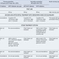
The major adverse effect of warfarin is haemorrhage, which often occurs at a predisposing abnormality such as an ulcer and a tumour. The risk of bleeding is increased by excessive anticoagulation, although this may not need to be present for severe haemorrhage to occur. Close monitoring of the degree of anticoagulation of warfarin is, therefore, important, and guidelines for reversal of excessive anticoagulation are shown in Table 23.3.
Table 23.3 Recommendations for management of bleeding and excessive anticoagulation in patients receiving warfarin
| Cause | Recommendation |
|---|---|
| 3.0<INR<6.0 (target INR 2.5) | 1. Reduce warfarin dose or stop |
| 4.0<INR<6.0 (target INR 3.5) | 2. Restart warfarin when INR <5.0 |
| 6.0<INR<8.0, no bleeding or minor bleeding | 1. Stop warfarin |
| 2. Restart when INR <5.0 | |
| INR>8.0, no bleeding or minor bleeding | 1. Stop warfarin |
| 2. Restart warfarin when INR <5.0 | |
| 3. If other risk factors for bleeding give 0.5–2.5 mg of vitamin K (oral) | |
| Major bleeding | 1. Stop warfarin |
| 2. Give prothrombin complex concentrate 50 units/kg or FFP 15 mL/kg | |
| 3. Give 5 mg of vitamin (oral or i.v.) |
INR, international normalised ratio; FFP, fresh frozen plasma.
Source: British Committee for Standards in Haematology (1998).
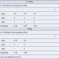
Skin reactions to warfarin may also occur but are rare. The most serious skin reaction is warfarin-induced skin necrosis, which may occur over areas of adipose tissue such as the breasts, buttocks or thighs, especially in women, and which is related to relative deficiency of protein C or S. This is important because these deficiencies result in an increased risk of thrombosis, and therefore warfarin may more often be used in such subjects. Preventing excessive anticoagulation in the initial stages of induction of therapy may reduce the severity of the reaction. A dosing schedule which helps to achieve this is shown in Table 23.4.
Table 23.4 Suggested warfarin induction schedule
| Day | INR | Warfarin dose (mg) |
|---|---|---|
| First | <1.4 | 10 |
| Second | <1.8 | 10 |
| 1.8 | 1 | |
| >1.8 | 0.5 | |
| Third | <2.0 | 10 |
| 2.0–2.1 | 5 | |
| 2.2–2.3 | 4.5 | |
| 2.4–2.5 | 4 | |
| 2.6–2.7 | 3.5 | |
| 2.8–2.9 | 3 | |
| 3.0–3.1 | 2.5 | |
| 3.2–3.3 | 2 | |
| 3.4 | 1.5 | |
| 3.5 | 1 | |
| 3.6–4.0 | 0.5 | |
| >4.0 | 0 (predicted maintenance dose) | |
| Fourth | <1.4 | >8 |
| 1.4 | 8 | |
| 1.5 | 7.5 | |
| 1.6–1.7 | 7 | |
| 1.8 | 6.5 | |
| 1.9 | 6 | |
| 2.0–2.1 | 5.5 | |
| 2.2–2.3 | 5 | |
| 2.4–2.6 | 4.5 | |
| 2.7–3.0 | 4 | |
| 3.1–3.5 | 3.5 | |
| 3.6–4.0 | 3 | |
| 4.1–4.5 | Miss out next day’s dose then give 2 mg | |
| >4.5 | Miss out 2 days’ doses then give 1 mg |
INR, international normalised ratio.
Source: Modified from Fennerty et al. (1984).
Although other coumarin anticoagulants are available, in the vast majority of cases these have not been shown to have any clear benefits over warfarin. They may be used occasionally where a patient does not tolerate warfarin. The necessary duration of anticoagulation in venous thrombosis and pulmonary embolus is still uncertain. On the basis of the available evidence, therapy may be required for approximately 6 months after the first deep vein thrombosis or pulmonary embolus. It may be possible to reduce the duration of therapy in patients who have had a postoperative episode since it is likely that the risk factor has been reversed (unless immobility continues). In patients with a second episode, therapy may be required for even longer and in patients with more than two episodes, life-long treatment may be necessary to reduce the risk of recurrence (British Committee for Standards in Haematology, 1998).
Dabigatran
Dabigatran is an orally active inhibitor of both free and clot-bound thrombin (Wittkowsky, 2010). It has a rapid onset of action and does not require laboratory monitoring. Dabigatran etexilate is a pro-drug which is hydrolysed to active dabigatran in the liver. Since 80% of activated dabigatran is excreted unchanged through the kidneys, it should be avoided in patients with severe renal impairment (creatinine clearance < 30 mL/min) and the dose should be reduced in moderate renal impairment (creatinine clearance 30–50 mL/min). Dabigatran is a substrate for the transport protein p-glycoprotein (p-GP), which facilitates renal elimination of certain drugs. Amiodarone, an inhibitor of p-GP, reduces the clearance of dabigatran and so doses should be reduced in patients who are on concurrent treatment with amiodarone. In patients who are on strong p-GP inhibitors such as verapamil and clarithomycin, dabigatran should be used with caution and it should not be used together with quinidine. Drugs such as rifampicin and St John’s Wort, which are potent p-GP inducers, may potentially reduce its efficacy. Dabigatran can be used for prophylaxis of VTE in adults after total hip replacement or total knee replacement surgery (National Institute for Health and Clinical Excellence, 2008). Haemorrhage is the major adverse effect.
Rivaroxaban
Rivaroxaban is an orally active inhibitor of both the ‘free’ and prothombinase complex-bound forms of activated factor X (Xa) (Wittkowsky, 2010). Two thirds of the dose is metabolised, principally by CYP450 enzymes and the remaining third is excreted unchanged in the urine. Like dabigatran, rivaroxaban also appears to be a p-GP substrate and it should be used with caution when prescribed concomitantly with p-GP inhibitors and potent p-GP inducers. It should also be used with caution in patients with creatinine clearance less than 30 mL/min (severe renal impairment) and is contraindicated in those with creatinine clearance less than 15 mL/min. Several CYP3A4 inhibitors and inducers have been shown to affect its metabolism. Some CYP3A4 inhibitors significantly increase the AUC of rivaroxaban, particularly ketoconazole and other azole-antimycotics such as itraconazole, voriconazole and posaconazole and also HIV protease inhibitors such as ritonavir. Therefore, the use of rivaroxaban is not recommended in patients receiving concomitant systemic treatment with these agents. The CYP3A4 inducer rifampicin (and possibly other inducers of this cytochrome) reduces the AUC for rivaroxaban. It is recommended as an option for prophylaxis of VTE in adults after hip or knee replacement surgery (National Institute for Health and Clinical Excellence, 2009). It also does not require laboratory monitoring. Haemorrhage is the major adverse effect.
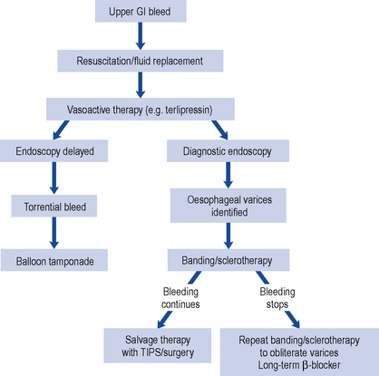
Fibrinolytic drugs
Alteplase
Tissue plasminogen activator (rt-PA) or alteplase was developed using recombinant DNA technology. Although this agent is much more expensive than streptokinase, it can be used in those situations where streptokinase may be less effective because of development of antibodies, for example within 1 year of previous streptokinase use or where allergy to streptokinase has previously occurred. Because it produces a lesser degree of systemic anticoagulation (it is more active against plasminogen associated with the clot), immediate use of heparin subsequently is necessary to prevent recurrence of thrombosis. Alteplase is also used for acute ischaemic stroke, where its prompt use may improve outcome in carefully selected individuals (National Institute for Health and Clinical Excellence, 2007). At the time of writing, it is the only thrombolytic licensed for this indication (see arterial thromboembolism).
Reteplase and tenectaplase
Reteplase, and more recently tenecteplase, are also fibrin-specific agents and so heparin is required to prevent rebound thrombosis. They are indicated for the treatment of acute myocardial infarction. In this clinical situation, reteplase is administered as an intravenous bolus, followed by a second bolus 30 min later (double bolus), and tenecteplase is given as a single intravenous bolus. They, therefore, have the advantage of convenience of administration compared with alteplase, and are the preferred option in pre-hospital settings, particularly when administered by paramedics (National Institute for Health and Clinical Excellence, 2002a).
Patient care
The patient on oral anticoagulants should be given full information on what to do in case of problems and what circumstances and drugs to avoid. An anticoagulant card with previous INR values and doses should also be provided. The patient should be told of the colour code for the different strengths of warfarin tablet and advised to carry their treatment card at all times. The likely duration of anticoagulant therapy should be made clear to the patient to avoid unnecessary and potentially dangerous prolongations of treatment. Patients who have received a fibrinolytic agent should also carry a card identifying the drug given and the date of administration.
Arterial thromboembolism
Treatment and prevention
Clopidogrel
Clopidogrel is also licensed for combination use with low-dose aspirin in the management of acute coronary syndrome without ST-segment elevation, when it is given for up to 12 months after the initial event (National Institute for Health and Clinical Excellence, 2004, 2005). Most benefit is obtained in the first 3 months and there is no evidence of benefit of clopidogrel after 12 months in this indication. In combination with low-dose aspirin, clopidogrel is also licensed for acute myocardial infarction with ST-segment elevation. It is recommended for at least 4 weeks in this indication, but the optimum treatment duration has not been established. Finally clopdogrel is sometimes used (with aspirin) in stenting procedures and this sometimes results in long-term use.
Prasugrel
Prasugrel inhibits platelet activation and aggregation. Its active metabolite binds to the P2Y12 class of ADP receptors on platelets. It is recommended for use in combination with aspirin as an option for the prevention of atherothrombotic events in patients with acute coronary syndromes undergoing PCI, only when immediate primary PCI is necessary for STEMI, or stent thrombosis occurred during treatment with clopidogrel, or the patient has diabetes mellitus (National Institute for Health and Clinical Excellence, 2009).
Patient care
Answer
The patient should be asked about any new medications which might have been introduced recently, including over-the-counter and herbal preparations. Some proprietary medicines may contain vitamin K which could cause resistance by pharmacodynamic mechanisms. Other medicines, including the herbal medicine St John’s wort, might induce warfarin metabolism and result in resistance as a result of a pharmacokinetic interaction (see Table 23.2). One other cause of apparent resistance to warfarin is poor adherence and this should, therefore, be considered. Supervised administration of the dose and/or measurement of plasma warfarin concentrations may be of value if the latter is suspected.
British Committee for Standards in Haematology. Guidelines on oral anticoagulation (warfarin): third edition. Br. J. Haematol.. 1998;132:277-285.
British Committee for Standards in Haematology. Guidelines on the use and monitoring of heparin. Br. J. Haematol.. 2006;133:19-34.
British Committee for Standards in Haematology. The management of heparin induced thrombocytopenia. Br. J. Haematol.. 2006;133:259-269.
British Committee for Standards in Haematology. Guidelines on oral anticoagulation (warfarin): third edition, 2005 update. Br. J. Haematol.. 2006;132:277-285.
Fennerty A., Dolben J., Thomas P., et al. Flexible induction dose regimen for warfarin and prediction of maintenance dose. Br. Med. J.. 1984;288:1268-1270. Available at: http://www.ncbi.nlm.nih.gov/pmc/articles/PMC1441080/?tool=pubmed
Fennerty A.G., Renowden S., Scolding N., et al. Guidelines for the control of heparin treatment. Br. Med. J.. 1986;292:579-580. Available at: http://www.ncbi.nlm.nih.gov/pmc/articles/PMC1339561/?tool=pubmed
National Institute for Health and Clinical Excellence. The Clinical Effectiveness and Cost Effectiveness of Early Thrombolysis for Treatment of Myocardial Infarction, Technology appraisal 52.. London: NICE. 2002. . Available at: http://www.nice.org.uk/guidance/index.jsp?action=byID&r=true&o=11480
National Institute for Health and Clinical Excellence. Acute Coronary Syndromes – Glycoprotein IIb/IIIa Inhibitors (Review), Technology appraisal 47. London: NICE. 2002. . Available at: http://www.nice.org.uk/guidance/index.jsp?action=byID&r=true&o=11470
National Institute for Health and Clinical Excellence. Clopidogrel in the Treatment of Non-ST-Segment-Elevation Acute Coronary Syndrome, Technology appraisal 80. London: NICE. 2004. . Available at: http://www.nice.org.uk/guidance/index.jsp?action=byID&r=true&o=11536
National Institute for Health and Clinical Excellence. Clopidogrel and Dipyridamole for the Prevention of Atherosclerotic Events, Technology appraisal 90. London: NICE. 2005. . Available at: http://www.nice.org.uk/guidance/index.jsp?action=byID&r=true&o=11558
National Institute for Health and Clinical Excellence. Alteplase for the Treatment of Acute Ischaemic Stroke, Technology Appraisal TA122. London: NICE. 2007. . Available at: http://guidance.nice.org.uk/TA122
National Institute for Health and Clinical Excellence. Dabigatran Etexilate for the Prevention of Venous Thromboembolism After Hip or Knee Replacement Surgery in Adults, Technology Appraisal TA157. London: NICE. 2008. . Available at: http://guidance.nice.org.uk/TA157
National Institute for Health and Clinical Excellence. Prasugrel for the Treatment of Acute Coronary Syndromes with Percutaneous Coronary Intervention, Technology Appraisal TA182.. London: NICE. 2009. . Available at: http://guidance.nice.org.uk/TA182
National Institute for Health and Clinical Excellence. Reducing the Risk of Venous Thromboembolism (Deep Vein Thrombosis and Pulmonary Embolism) in Patients Admitted to Hospital, Clinical guideline 92. London: NICE. 2010. . Available at: http://guidance.nice.org.uk/CG92
Rosendaal F.R., Reitsma P.H. Genetics of venous thrombosis. J. Thromb. Haemostasis. 2009;7(Suppl. 1):301-304.
Winter M., Keeling D., Sharpen F., et al. Procedures for the outpatient management of patients with deep vein thrombosis. Clin. Lab. Haematol.. 2005;27:61-66.
Wittkowsky A.K. New oral anticoagulants: a practical guide for clinicians. J. Thromb. Thrombolysis. 2010;29:182-191.