15 Three-Dimensional Ultrasound
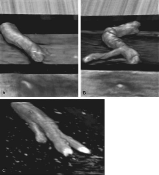
There are several reports of the use of three- or four-dimensional imaging to image nerves and guide regional blocks.1–3 The complexity of the surrounding echoes in musculoskeletal tissue can make rendering clear three-dimensional images challenging. Injected anechoic fluid can improve the interface for three-dimensional imaging of the nerve surface. Rendered volumes are often shown with sepia coloring to improve contrast resolution.
One potential advantage of three-dimensional imaging is to avoid partial line-ups of the block needle that can occur with two-dimensional in-plane technique. Because line-up is not necessary, performance time and accuracy of the procedure would benefit. One study found that the use of higher-dimensional imaging improved needle tip identification.4 However, another study found that multiplanar reformatted displays improved needle conspicuity compared with volume-rendered displays.5 Serious considerations for this developing technology balance obtaining more useful information with unnecessary distraction. Interventional procedure times tend to be longer with this technology.6
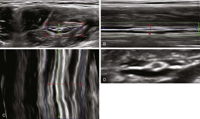
Manual acquisition of images by sliding the transducer at a constant velocity is difficult. Subsequent rendering of three-dimensional images is then done offline. Another problem is that some probes used for three-dimensional imaging using automated sweeps of acquisition are large and bulky. The biggest benefit of three-dimensional imaging may be the detection of injections that would be out-of-plane with two-dimensional imaging (whether it is within a vessel or along a nerve). One of the newer three-dimensional imaging display formats is “niche” format in which all three orthogonal imaging planes are shown in the single image. This format has been reported to improve imaging of the proximal sciatic nerve.7
Clinical Pearls
• Three-dimensional ultrasound has the same artifacts as two-dimensional imaging. Additional artifacts can arise from acquisition and rendering of three-dimensional images. As a rule, three-dimensional imaging is more subject to shadowing artifacts than two-dimensional imaging.8
• Three-dimensional ultrasound may be useful for imaging the local anesthetic distribution or catheter when it tracks out of the plane with two-dimensional imaging.
1 Foxall GL, Hardman JG, Bedforth NM. Three-dimensional, multiplanar, ultrasound-guided, radial nerve block. Reg Anesth Pain Med. 2007;32:516–521.
2 Clendenen SR, Robards CB, Clendenen NJ, et al. Real-time 3-dimensional ultrasound-assisted infraclavicular brachial plexus catheter placement: implications of a new technology. Anesthesiol Res Pract. 2010. 2010. Epub 2010, Jun 1
3 Belavy D, Ruitenberg MJ, Brijball RB. Feasibility study of real-time three-/four-dimensional ultrasound for epidural catheter insertion. Br J Anaesth. 2011;107(3):438–445. Epub 2011, Jun 9
4 Won HJ, Han JK, Do KH, et al. Value of four-dimensional ultrasonography in ultrasonographically guided biopsy of hepatic masses. J Ultrasound Med. 2003;22:215–220.
5 Rose SC, Nelson TR, Deutsch R. Display of 3-dimensional ultrasonographic images for interventional procedures: volume-rendered versus multiplanar display. J Ultrasound Med. 2004;23:1465–1473.
6 Tonni G, Centini G, Rosignoli L, et al. 4D vs 2D ultrasound-guided amniocentesis. J Clin Ultrasound. 2009;37:431–435.
7 Karmakar M, Li X, Li J, et al. Three-dimensional/four-dimensional volumetric ultrasound imaging of the sciatic nerve. Reg Anesth Pain Med. 2012;37(1):60–66.
8 Cohen L, Mangers K, Grobman WA, et al. Three-dimensional fast acquisition with sonographically based volume computer-aided analysis for imaging of the fetal heart at 18 to 22 weeks’ gestation. J Ultrasound Med. 2010;29:751–757.