Chapter 5 Thoracic Outlet Syndrome
Overview
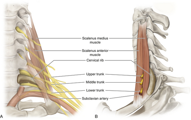
• Every vertebra has a costal element. In the thorax, the costal elements break free and form the ribs. If the C7 vertebra also frees that element, a cervical rib is formed; its size is variable. Bands run from the tip of the bone to the cervical rib and may compress or irritate the lower trunk.
• A variety of surgical approaches have been described for those few patients who suffer from thoracic outlet syndrome and require surgery. We favor an anterior dissection, because we believe that the irritation and compression of plexus elements is at a proximal level and from a variety of causes, and that the affected elements of the plexus should be safely decompressed under direct vision.
Surgery
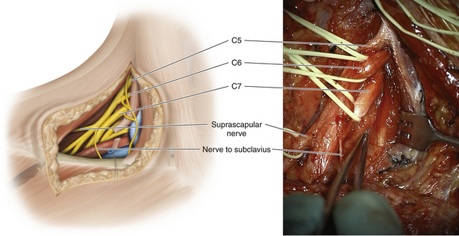
• The skin incision is made slightly above the clavicle and parallel to it (Figure 5-1). Depending on preexisting incisions and the patient’s body habitus, the medial end of the incision may be extended upward or the lateral end of the incision may be extended downward over the deltopectoral groove.
• The clavicular head of the sternocleidomastoid is divided, leaving a cuff on the clavicle for subsequent reattachment. There is no need to divide the sternal head. This maneuver is preceded by passage of a finger behind the lower lateral border of the clavicular head to ensure that all venous structures are separated from the deep surface of that muscle.
• If the external jugular vein impedes progress, it should be divided.
• The phrenic nerve is dissected free of the underlying scalenus anterior and is guarded throughout the operation.
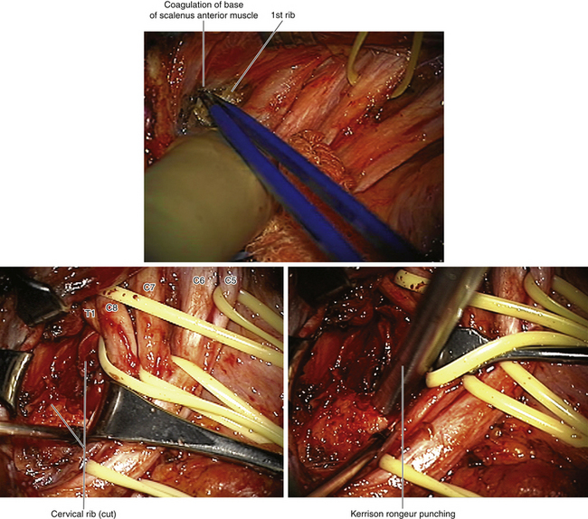
• The scalenus anterior is divided close to its tendon. The divided muscle springs apart, and all bleeding points within the muscle belly are secured (Figure 5-2). It is not reattached at the end of the operation.
• The upper border of the first rib has a very characteristic feel. The surgeon’s fingertip sweeps backward and medially on this border, thus pushing the pleura and suprapleural membrane away.
• The C8 and T1 nerve roots embrace the neck of the first rib, and the stellate ganglion is palpated in front of the neck of the first rib.
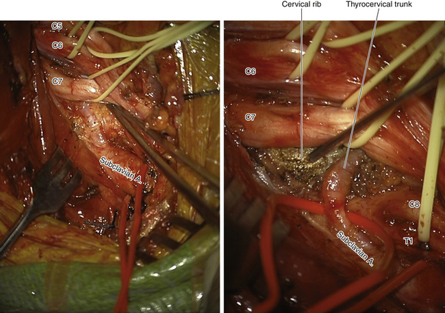
• The subclavian artery must be mobilized forward, so that the lower trunk and the T1 nerve can be clearly seen. Branches of the thyrocervical trunk may have to be divided to allow the artery to be gently retracted forward and downward with a vein retractor (Figure 5-3). The vertebral artery is, of course, respected as it runs from the artery up to the transverse process of the C6 vertebra.
• On occasion, it is necessary to mobilize the proximal medial cord to allow full display of the lower trunk. In that circumstance, the lateral 2 inches of the clavicular head of pectoralis major are detached from the clavicle and the medial cord is defined, adjacent to the artery.
• If excellent vision of the medial neural elements is impaired, a surgical sponge should be passed around the clavicle. The assistant can then pull the clavicle forward and downward and thus improve the view.
• We frequently use the operating microscope for the proximal dissection of the C7, C8, and T1 nerves. Magnification is not required, but better illumination of the field for both the surgeon and the assistant enhances precision.
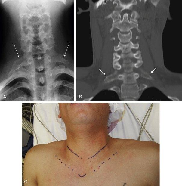
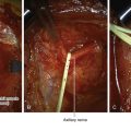
Figure 5-1 A and B, Cervical rib (arrows)C, Skin incisions for bilateral thoracic outlet syndrome approach.
Decompression of Neural Elements
• The C7, C8, and T1 spinal nerves must be clearly seen from their foramina lying on the appropriate transverse processes. The middle trunk and, particularly, the lower trunk are carefully dissected.
• The compression and irritation may be caused by a variety of structures, and during the process of dissecting out the elements as described, the bands arising from the C6 or C7 transverse process are divided. Sharp, tendinous components of the scalenus medius’ medial border are also divided.
• Every vertebra has a costal element. In the thorax these elements are free and constitute the ribs. If the costal element of the C7 vertebra is free, a cervical rib is formed. The size of the costal rib can vary from an enlargement of the transverse process to an extravagant bony rib. It is emphasized that it is usually the bands arising from the tip of the bony structure that cause the neural compression or irritation (Figure 5-4).
• When the cervical rib is resected, the bone is nibbled or punched away under direct vision, with concomitant guarding of the adjacent neural structures (Figure 5-5). Do not strive to remove the rib in one piece, because it is much safer to remove it piecemeal.
• At the conclusion of the procedure, C7, C8, and T1 and the middle and lower trunks should be completely free of any compressive bands and should pass laterally and downward to the apex of the axilla.

