[level-membership-for-cardiothoracic-surgery-category]
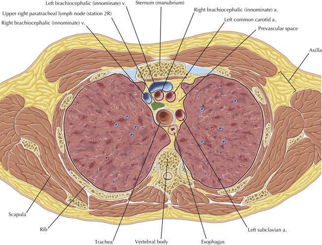
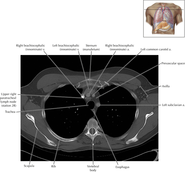
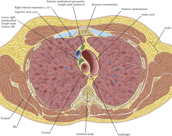
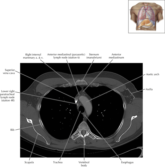
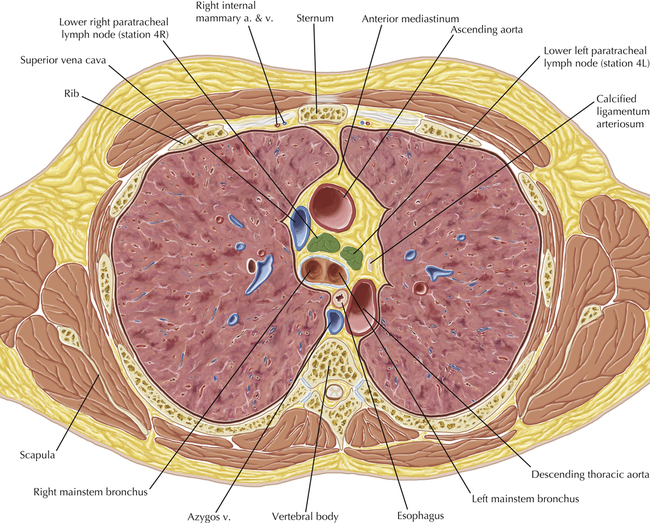
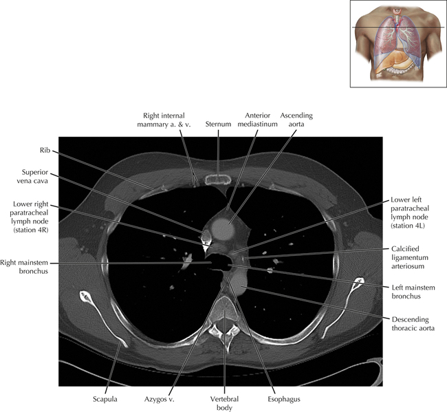
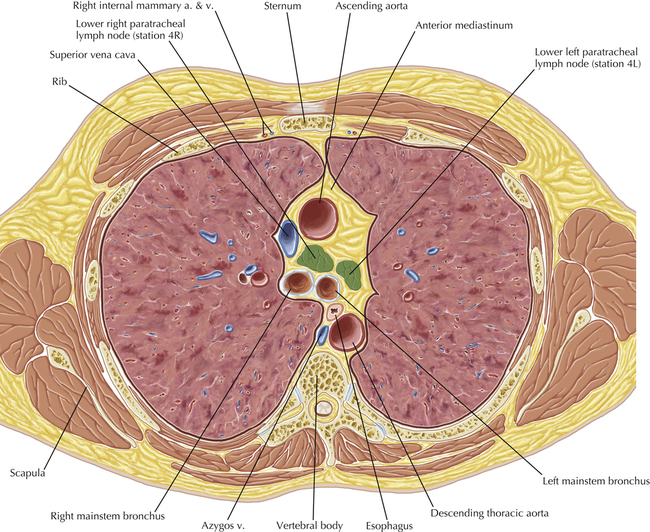
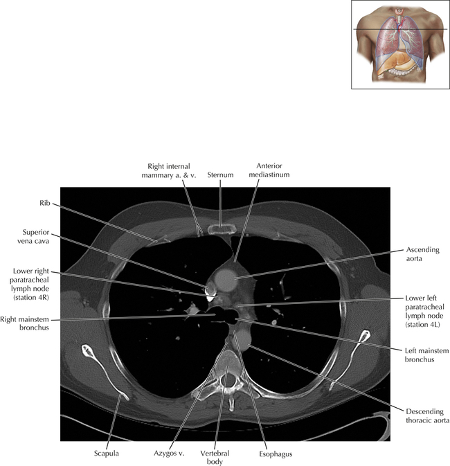
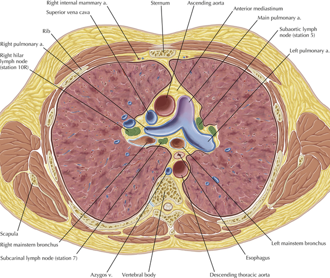
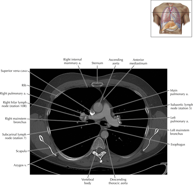
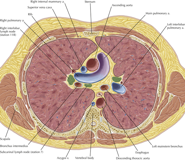
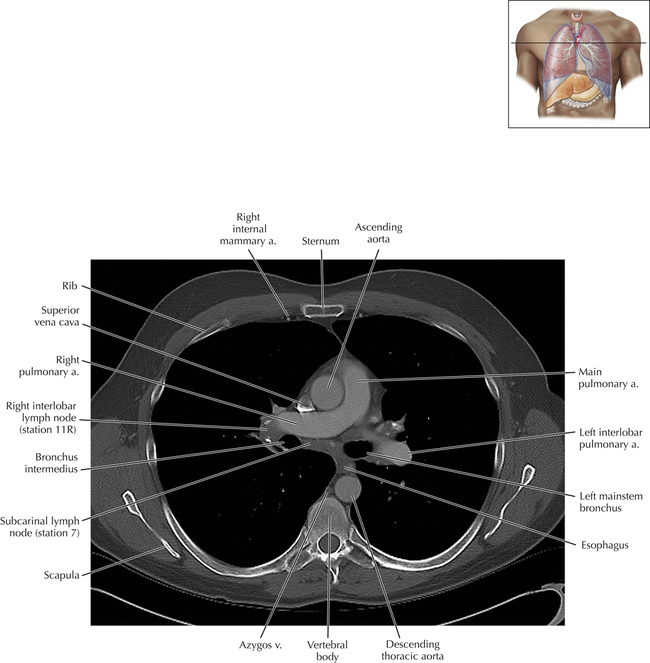
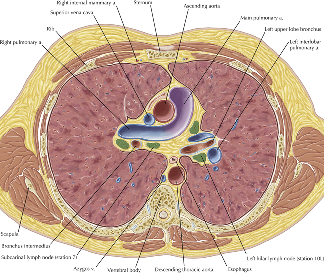
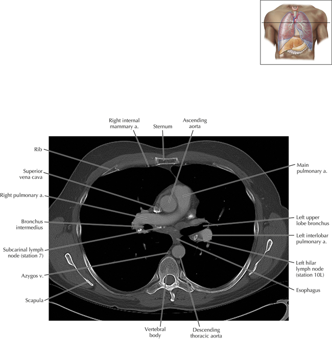
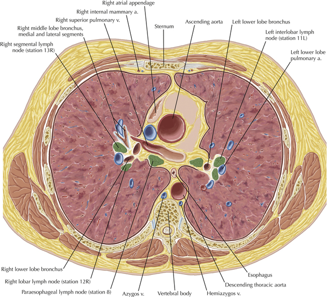
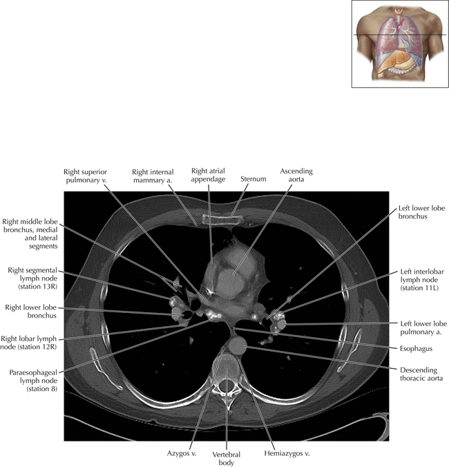
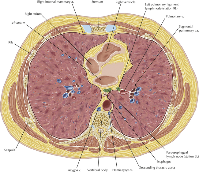
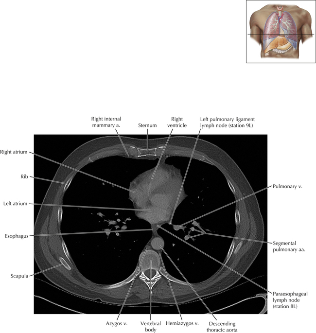
Thoracic Lymph Nodes
[/level-membership-for-cardiothoracic-surgery-category][not-level-membership-for-cardiothoracic-surgery-category]
Published on 13/02/2015 by admin
Filed under Cardiothoracic Surgery
Last modified 22/04/2025
Print this page[level-membership-for-cardiothoracic-surgery-category]
[/level-membership-for-cardiothoracic-surgery-category][not-level-membership-for-cardiothoracic-surgery-category]
