The thalassaemias

Published on 03/04/2015 by admin

Filed under Hematology, Oncology and Palliative Medicine

Last modified 22/04/2025

Print this page

rate 1 star rate 2 star rate 3 star rate 4 star rate 5 star
Your rating: none, Average: 0 (0 votes)

This article have been viewed 1348 times

16

The thalassaemias

The thalassaemias are a heterogeneous group of inherited disorders of haemoglobin synthesis. They are characterised by a reduction in the rate of synthesis of either alpha or beta chains and are classified accordingly (i.e. α-thalassaemia, β-thalassaemia). The basic haematological abnormality in the thalassaemias is a hypochromic microcytic anaemia of variable severity. Unbalanced synthesis of α- and β-globin chains can damage red cells in two ways. Firstly, failure of α and β chains to combine leads to diminished haemoglobinisation of red cells to levels incompatible with survival. Even those hypochromic cells released into the circulation transport oxygen poorly. The second mechanism for red cell damage is the aggregation of unmatched globin chains – the inclusion bodies lead to accelerated apoptosis of erythroid precursors in the bone marrow (ineffective erythropoiesis) and destruction of more mature red cells in the spleen (haemolysis). In general, the clinical severity of any case of thalassaemia is proportionate to the degree of imbalance of α- and β-globin chain synthesis.

Thalassaemias are among the most common inherited disorders. Gene carriers have some protection from falciparum malaria. Cases occur sporadically in most populations but the highest thalassaemia gene frequency is in a broad geographical region extending from the Mediterranean through the Middle East and India to South-East Asia.

Classification

The classification illustrated in Table 16.1 is based on the mode of inheritance of thalassaemia.

Table 16.1

Classification of thalassaemia

Type of thalassaemia Heterozygote Homozygote
α-Thalassaemia1    
α0 (– –/) Thal. minor Hydrops fetalis
α+ (–α/) Thal. minor Thal. minor
β-Thalassaemia    
β0 Thal. minor Thal. major
β+ Thal. minor Thal. major or intermedia

1Compound heterozygosity (– –/–α) leads to HbH disease.

As the α-globin chain gene is duplicated on each chromosome there may be total loss of α-globin chain production (termed α0 or – –/haplotype) or partial loss of α-chain production resulting from loss of only one gene (termed α+ or –α/haplotype).

The most important clinical syndromes are haemoglobin (Hb)-Barts hydrops syndrome (– –/– –), which is incompatible with life, and Hb H disease (–α/– –). At the molecular level the α-thalassaemias result from loss of α-gene function due to gene deletion or non-deletional mutations; different types of mutations may be co-inherited.

β-Thalassaemias are autosomal recessive disorders characterised by reduced (β+) or absent (β0) production of β chains. The heterozygous (‘trait’ or ‘minor’) form of the disease is usually symptomless while homozygosity is associated with the clinical disease β-thalassaemia ‘major’. Homozygous mild (β+) thalassaemia may, however, lead to a less severe clinical syndrome termed ‘thalassaemia intermedia’. The β-thalassaemias are very heterogeneous at the molecular level – the large majority of mutations are single base substitutions (point mutations) and insertions or deletions of one to two bases.

Although molecular analysis may be needed, diagnosis of the major syndromes is normally possible from consideration of the clinical features and simple laboratory tests. The latter must include a blood count and blood film, and haemoglobin electrophoresis with quantification of the different types of haemoglobin (i.e. HbA, HbA2, HbF).

Other structural Hb variants may coexist with thalassaemias giving rise to a wide range of clinical disorders. Only the more common thalassaemia syndromes are discussed here.

Clinical syndromes

α-Thalassaemias

β-Thalassaemias

β-Thalassaemia major

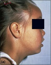
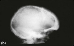
The characteristic severe anaemia (Hb less than 70 g/L) is caused by α-chain excess leading to ineffective erythropoiesis and haemolysis. Anaemia first becomes apparent at 3–6 months when production of HbF declines. The child fails to thrive and develops hepatosplenomegaly. Compensatory expansion of the marrow space causes the typical facies with skull bossing and maxillary enlargement (Fig 16.1a). The ‘hair-on-end’ radiological appearance of the skull (Fig 16.1b) is due to expansion of bone marrow into cortical bone. If left untreated further complications can include repeated infections, bone fractures and leg ulcers. Red cell membrane abnormalities contribute to hypercoagulability.

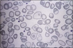
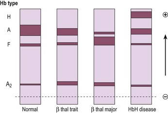
Laboratory testing should precede blood transfusion. There is a severe hypochromic microcytic anaemia with a characteristic blood film (Fig 16.2) and Hb electrophoresis demonstrates absence or near absence of HbA with small amounts of HbA2 and the remainder HbF (Fig 16.3).

With intense supportive therapy, increasing numbers of patients in the developed world survive into adulthood. Blood transfusion remains the mainstay of management. Raising the haemoglobin concentration both reduces tissue hypoxia and suppresses endogenous haematopoiesis which is largely ineffective. There is improved growth and development and reduced hepatosplenomegaly. Transfusion is generally given to maintain a haemoglobin level of at least 90–100 g/L. Splenectomy can reduce the transfusion frequency. With such regular transfusion iron chelation is necessary to minimise iron overload. Without chelation, accumulation of iron damages the liver, endocrine organs and heart with death in the second or third decades. The most commonly used regimen is subcutaneous desferrioxamine given for 5–7 days per week. Compliance may be problematic (especially in teenagers) but where good there is a considerably improved life expectancy. Oral iron chelators (e.g. deferiprone, deferasirox) are emerging as an acceptable alternative. Endocrine disturbances related to iron overload will require appropriate therapy.

Allogeneic stem cell transplantation is a serious option. In ‘best risk’ patients the probability of survival exceeds 90%. Experimental therapies include drugs designed to stimulate fetal haemoglobin production (e.g. erythropoietin, hydroxycarbamide) and gene therapy (see p. 103).

Thalassaemia intermedia

Thalassaemia intermedia is a clinical syndrome which may result from a variety of genetic abnormalities (Table 16.2). The clinical features are less severe than in β-thalassaemia major as the α/β-globin chain imbalance is less pronounced. Patients usually present later than is the case for β-thalassaemia major (often at 2–4 years), and have relatively high haemoglobin levels (80–100 g/L), moderate bone changes and normal growth. Transfusion may be required but requirements are less than in β-thalassaemia major.

Prenatal diagnosis

This depends on early identification of couples at risk and sensitive counselling. Adequate amounts of fetal DNA can be obtained around the 10th week of gestation by chorionic villus sampling. Current technologies allow reliable identification of single point mutations from very small DNA samples. Techniques are being developed to analyse fetal DNA obtained from maternal plasma or peripheral blood.