10 The skull
Skull
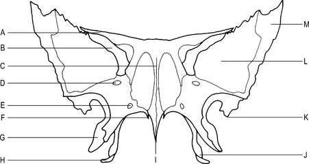
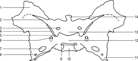
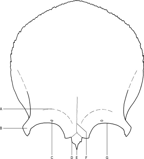
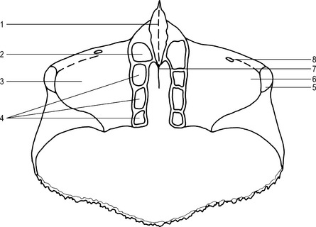
The skull can be divided into 2 sections: the cranium and the face (Figs 10.1, 10.2 and 10.3).
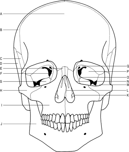
Fig. 10.1 Position of the bones of the skull (anterior aspect).
C–Greater wing of sphenoid bone
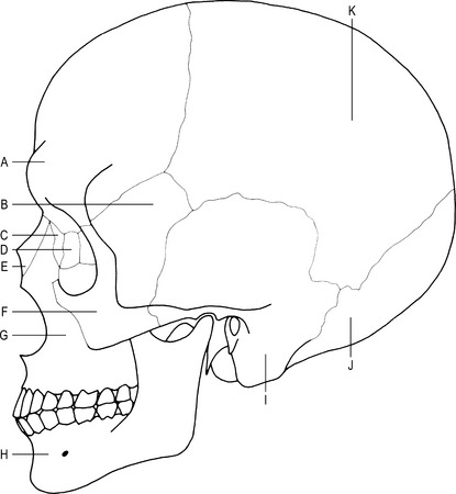
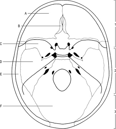
Fig. 10.3 Position of the bones of the skull (cranial cavity).
The floor of the cranial cavity is divided into three distinct sections:
1 – Anterior cranial fossa containing the frontal lobes of the brain.
3 – Posterior cranial fossa containing cerebellum, pons and medulla oblongata.


Orbital cavity (Fig. 10.4)
Bones forming the orbital cavity
Nasal cavity (Fig. 10.7)
Forms the upper part of the respiratory tract and is irregular in shape.
Floor
Forms the division between the oral and nasal cavities. Formed by:
Lateral wall
Irregular as it forms the 3 nasal conchae. Formed by:
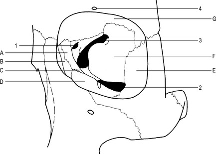
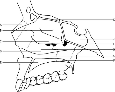
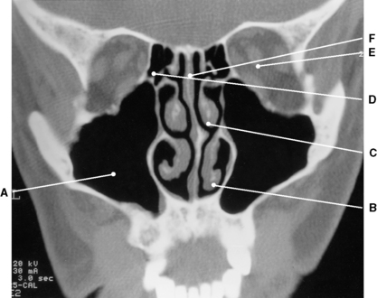
Paranasal sinuses (Figs 10.8 and 10.9)
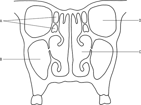
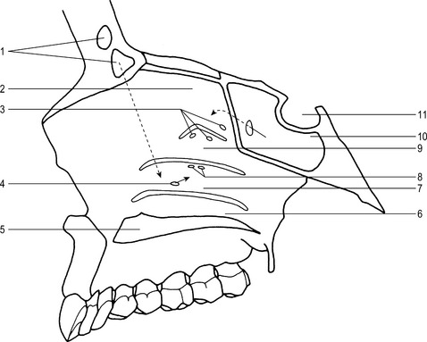
Fig. 10.9 Paranasal sinuses and associated structures (sagittal section).
3 –Opening for the ethmoidal sinuses
4 –Opening for the maxillary sinuses
The sinuses are lined with mucous membrane, which is continuous with that of the nasal cavity.
Sphenoidal sinuses (Fig. 10.9)
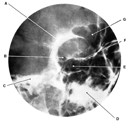
Radiographic appearances of the paranasal sinuses (Figs 10.10 to 10.18
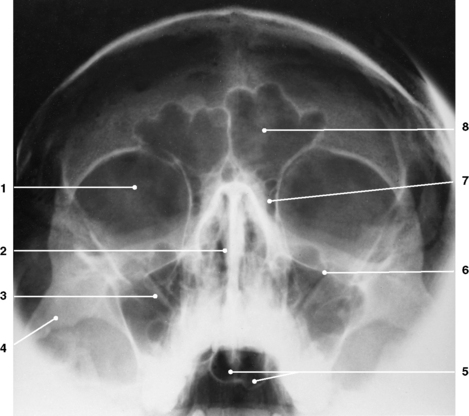
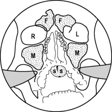
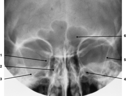
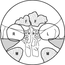
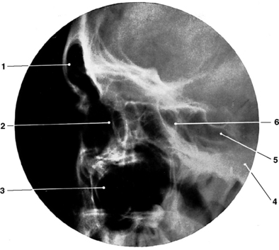
Fig. 10.14 Nasal sinuses: lateral projection.
2 – Anterior margin of ethmoidal sinuses
6 – Posterior margin of ethmoidal sinuses.
(From Bryan 1996.)
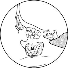
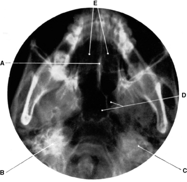
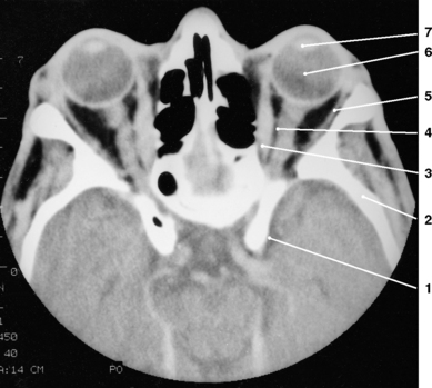
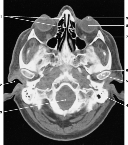
Fig. 10.16 Nasal sinuses: submentovertical projection.
E – Posterior ethmoidal sinuses.
(From Bryan 1996.)
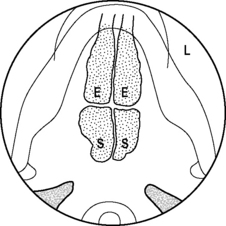
Fig. 10.17 Radiographic projection of the nasal sinuses: submentovertical.
L – Left (Normal radiographic label).
(From Bryan 1996.)
Features of the skull (Figs 10.19 to 10.23)
Radiographic appearances of the skull (Figs 10.24 to 10.30)
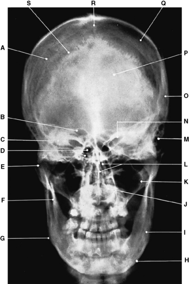
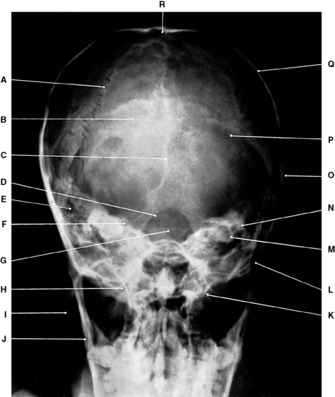
Fig. 10.24 Cranial bones: occipitofrontal projection.
C– Petrous part of temporal bone
L– Ethmoid bone in nasal cavity
O– Squamous part of temporal bone
(From Bryan 1996.)
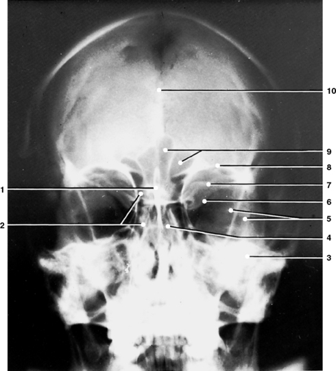
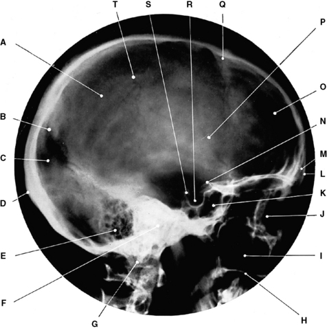
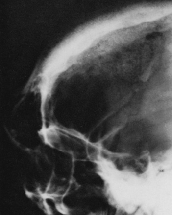
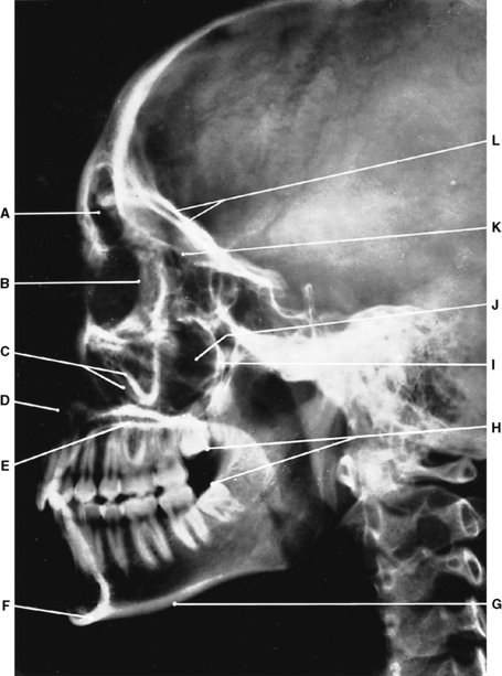
Fig. 10.26 Cranial bones: lateral projection.
D– External occipital protuberance
F– Petrous part of temporal bones
P– Groove for middle meningeal artery
(From Bryan 1996.)
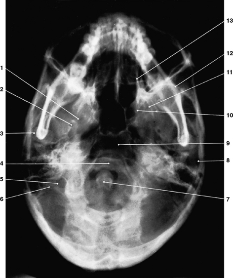
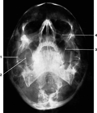
Fig. 10.27 Cranial bones: submentovertical projection.
6 – Transverse process of atlas
12 – Anterior wall of middle cranial fossa
13 – Lateral wall of nasal cavity.
(From Bryan 1996.)
Individual bones of the skull
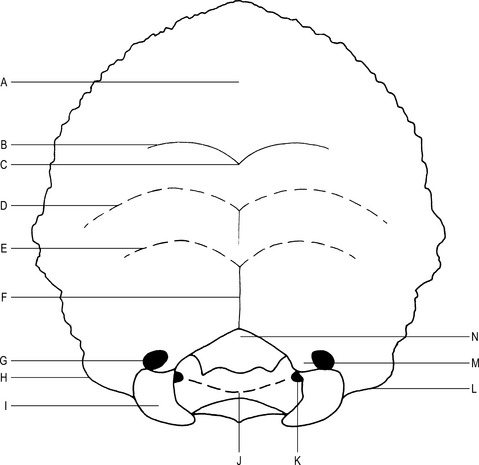
Occipital bone (Fig. 10.31)
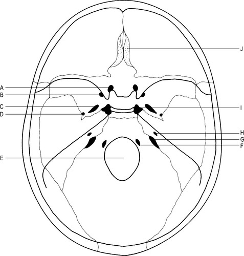
Fig. 10.19 Foramina in the base of the skull (internal aspect).
A – Optic canal, for the optic nerve and the ophthalmic artery.
B – Foramen rotundum, for the maxillary division of the trigeminal nerve.
C – Foramen ovale, for the mandibular division of the trigeminal nerve.
D – Foramen spinosum, for the middle meningeal artery.
H – Internal acoustic (auditory) meatus, for the facial and auditory nerves.
J – Cribriform plate, for the olfactory nerve filaments.
Carotid canal – lies on the external aspect, for the internal carotid artery.
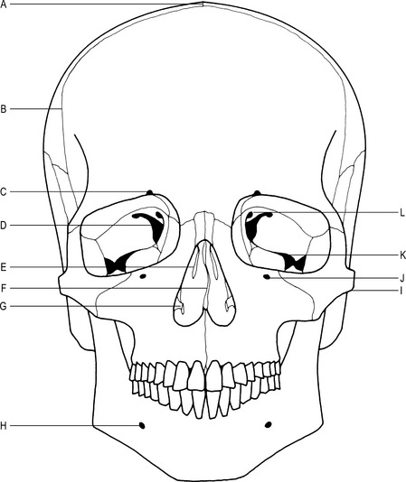
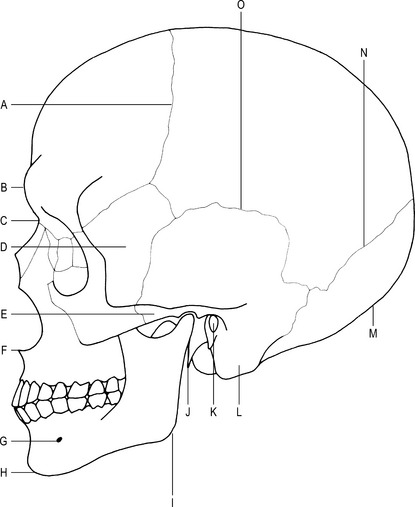
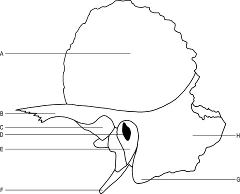
Fig. 10.21 Features of the skull (lateral aspect).
K–External acoustic (auditory) meatus
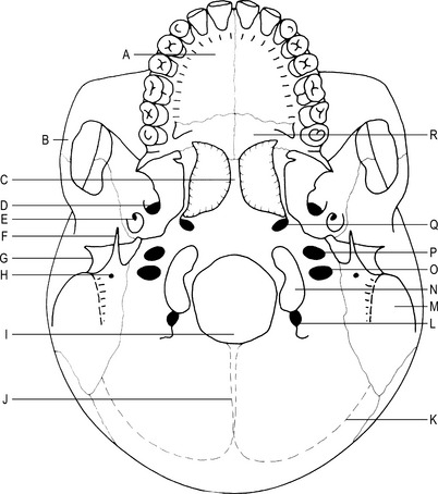
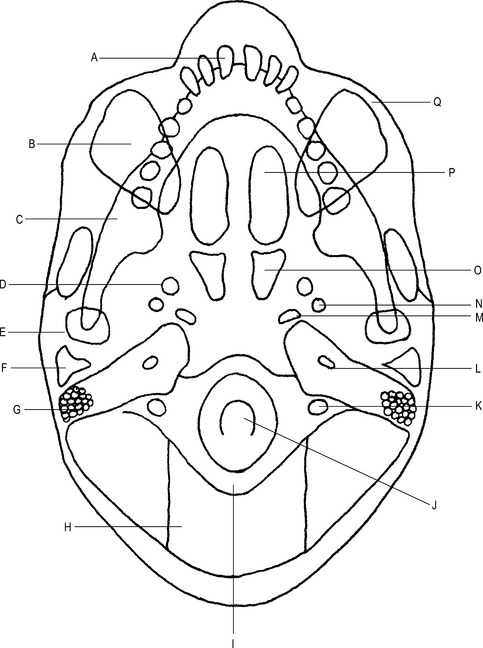
Fig. 10.22 Features of the skull (inferior aspect).
A–Palatine process of the maxilla
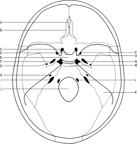
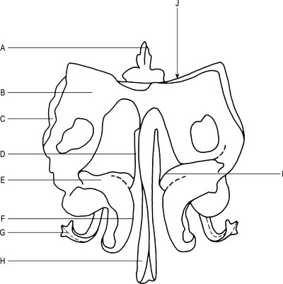
Fig. 10.23 Features of the skull (cranial cavity).
A– Crista galli of the ethmoid bone
B– Cribriform plate of the ethmoid bone
D– Tuberculum sellae of the sphenoid bone
E– Sella turcica (hypophyseal fossa, pituitary fossa) of the sphenoid bone
H– Internal acoustic (auditory) meatus
K– Petrous portion of the temporal bone
Articulations
The occipital condyles with the 1st cervical vertebra to form the atlanto-occipital joint.
The squamous part with the temporal bone and the parietal bones to form the lambdoid suture.
Main parts
The bone can be divided into 3 areas:
Squamous part
This lies posterior to the foramen magnum.
Internal surface –


External occipital crest –
midline structure from the foramen magnum to the external occipital protuberance.
Lateral (condylar) parts
These lie on either side of the foramen magnum.
Occipital condyles –
lie on the inferior surface, on either side of the foramen magnum, and articulate with the atlas.
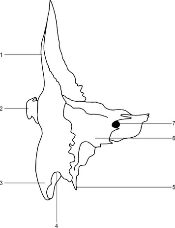
Sphenoid bone (Figs 10.32 and 10.33)
Position
Forms part of the base of the skull, lying between the frontal, temporal and occipital bones.
Articulations
The greater wings with the temporal bone to form the sphenosquamosal suture.
With the vomer, ethmoid, occipital, frontal, zygomatic and palatine bones.
Main parts
The bone consists of a body, 2 pairs of wings and 2 pterygoid processes.
Body
Tuberculum sellae –
posterior to the optic canals, forming the anterior boundary of the sella turcica.
Sella turcica –
posterior to the tuberculum sellae; contains the hypophysis cerebri (pituitary gland).


Greater wings
Two processes from the sides of the body.
Sphenosquamosal suture –
junction between the greater wings and the squamous part of the temporal bone.
Cerebral surface –
forms part of the middle cranial fossa; presents with 3 foramina:


Ossification
Articulations
Mandibular fossa with the head of mandible to form the temporomandibular joint.
With the greater wings of sphenoid to form the sphenosquamosal suture.
Main parts
The bone is formed by 3 parts:
Petromastoid part
This is formed by a mastoid and a petrous part.
Petrous part –
lies between the sphenoid and occipital bones in the base of the skull:
internal acoustic (auditory) meatus –
lies on the posterior surface of the petrous part and carries the facial and auditory nerves.
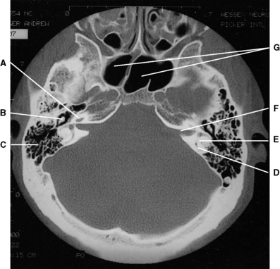
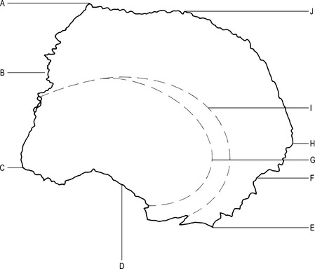
Radiographic appearances of the temporal bone (Figs 10.36, 10.37 and 10.38)
Parietal bones (Fig. 10.39)
Type
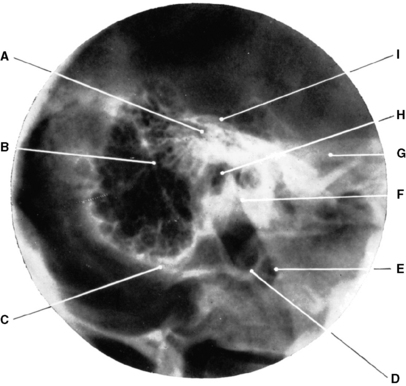
Fig. 10.36 Temporal bone: lateral oblique projection.
E– Posterior border of ramus of mandible
(From Bryan 1996.)
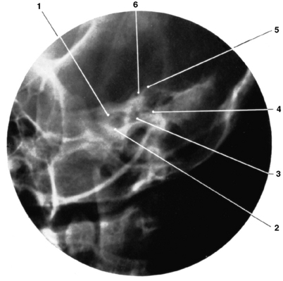
Fig. 10.37 Temporal bone: modified Stenver’s projection.
4 – Lateral semicircular canal
6 – Superior semicircular canal.
(From Bryan 1996.)
Articulations
With the sagittal border of the opposite parietal bone to form the sagittal suture.
With the frontal bone to form part of the coronal suture.
With the occipital bone to form part of the lambdoid suture.
Frontal bone (Figs 10.40 and 10.41)
Ethmoid bone (Fig. 10.42)
Maxillae (Fig. 10.43)
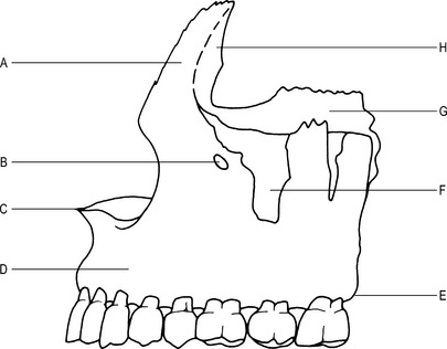
Main parts
Each bone has a body and 4 processes.
Anterior nasal spine –
a pointed process, at the junction of the 2 bodies. Infratemporal surface – convex; lies laterally.
Maxillary tuberosity –
lies on the posterior aspect of the roots of the 3rd molar tooth (upper eight).
Inferior orbital fissure –
anterior border is formed by the posterior border of the orbital surface.
Maxillary hiatus –
opening on the posterior part of the nasal surface; leads to the maxillary sinus.
Mandible (Figs 10.44 and 10.45)
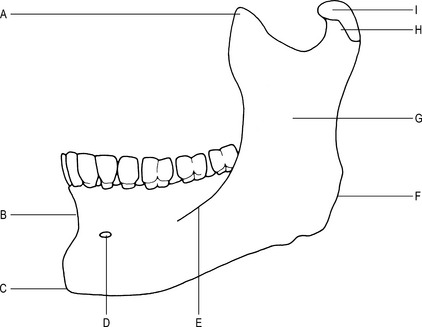
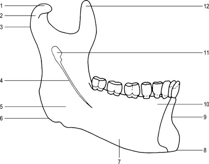
Main parts
The bone is formed by a body and 2 rami.
Body


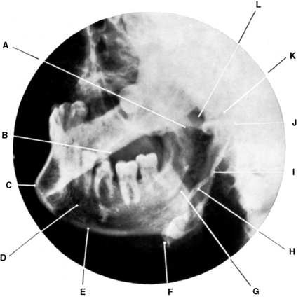
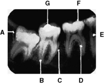
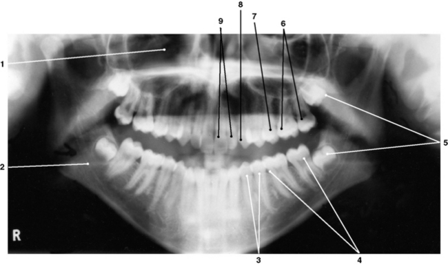
Radiographic appearances of the mandible (Fig. 10.46)
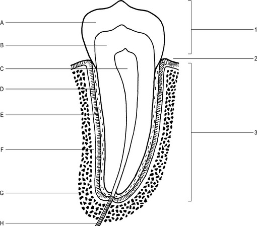
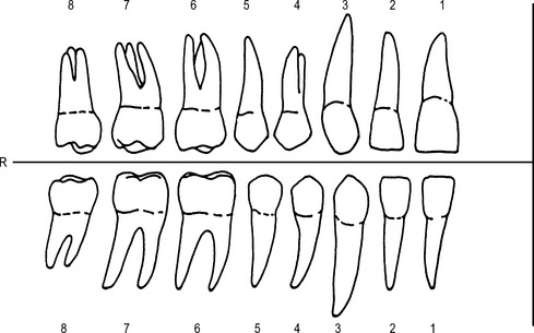
Structure
Types of teeth

 .
.


Lacrimal bones
Vomer
Articulations
With the maxillae, sphenoid, ethmoid, and palatine bones and the cartilage of the nasal septum.
Palatine bones


Zygomatic bones
Hyoid bone
Position
Lies in the front of the neck, above the thyroid cartilage of the larynx at the root of the tongue.
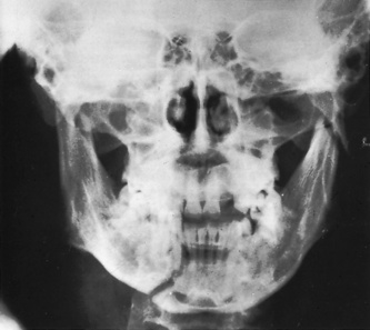
Radiographic appearances of the facial bones (Figs 10.51, 10.52 and 10.53)
Temporomandibular joint (Fig. 10.54)
Type
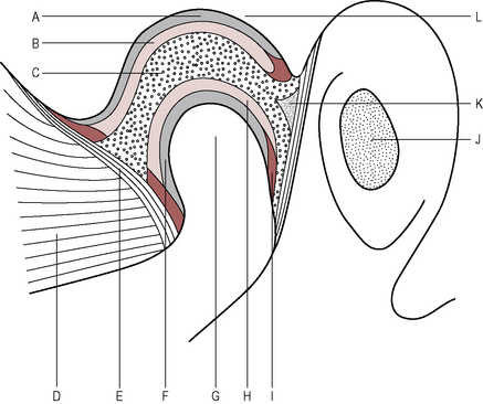
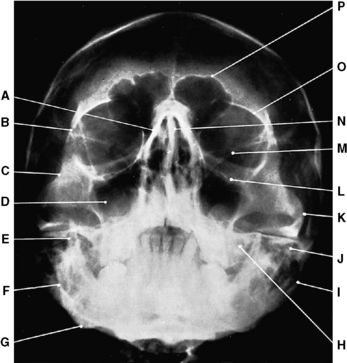
Fig. 10.51 Facial bones: occipitomental projection.
E– Coronoid process of mandible
H– Petrous part of temporal bone
(From Bryan 1996.)
Synovial membrane
Lines the fibrous capsule. The membrane secretes synovial fluid, which lubricates the joint.
Movements
Depression by the lateral pterygoid muscle.
Elevation by the temporalis, masseter and medial pterygoid muscles.
Protrusion by the lateral and medial pterygoid muscles.
Retraction by the temporalis muscle.
Lateral movement by the medial and lateral pterygoid muscles.
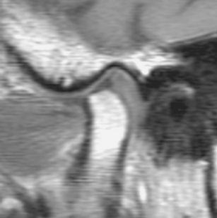
Radiographic appearances of the temporomandibular joint (Figs 10.55, 10.56, 10.57 and 10.58)
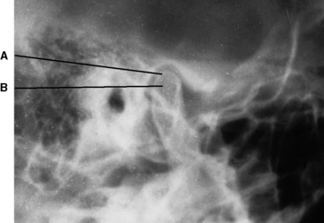
Fig. 10.55 Tem- poromandibular joint: lateral oblique projection with mouth closed.
A – Mandibular fossa of temporal bone
(From Bryan 1996.)
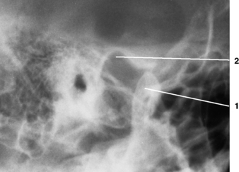
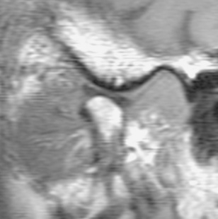
Fig. 10.56 Tem- poromandibular joint: lateral oblique projection with mouth open.
2 – Mandibular fossa of temporal bone.
(From Bryan 1996.)