The salivary glands
Investigations
For most patients, the history is the most useful single guide to the diagnosis. The presenting symptom is most often of a mass in the salivary gland (see Box 8.1).
The duct orifices should be inspected intraorally, preferably prior to palpation, which should be performed bimanually with a gloved finger in the mouth. Note if there is xerostomia or any stigmata of connective tissue disease. Examine the oropharynx for any deep lobe extensions. Measure the mass and note its exact position within the gland identified. Grade facial nerve function, e.g by the House–Brackmann scale,1 as a baseline and to monitor for future change. Examine the cervical lymph nodes and finally the external auditory meatus, to ensure no direct spread of a parotid neoplasm.
Imaging
Sialography is considered the most appropriate and sensitive in assessing ductal pathology but is increasingly being replaced by magnetic resonance (MR) sialography and ultrasound.2 A sialagogue is first used to stimulate saliva production to identify the duct. This is cannulated and contrast injected under fluoroscopic control to ensure adequate filling. Plain X-rays are taken to identify filling defects, delays in emptying and extravasation. Digital subtraction sialography removes the pre-contrast image from the post-contrast image to give improved identification of filling defects within the duct system. The disadvantages of sialography are that it is invasive, it can fail if the duct cannot be cannulated and it cannot be performed in the setting of acute salivary gland infection.
Ultrasound (US) is increasingly the imaging modality of choice for the initial investigation of salivary glands.3 It is cheap, widely available and avoids the use of ionising radiation. It is excellent at imaging superficial structures such as the parotid and submandibular glands. An advantage of US is that it can allow simultaneous and guided fine-needle aspiration cytology (FNAC). US will pick up 90% of salivary gland stones and can characterise salivary gland tumours in great detail, but the technique is highly operator dependent. US will not identify the facial nerve or bone erosion, and the mandible impedes visualisation of the deep parotid lobe.

Recent studies suggest that for clinically benign superficial lobe parotid tumours, there is no benefit in additional imaging after good-quality US examination4 and that the move towards surgical management of benign tumours with extracapsular dissection means that the identification of the facial nerve is less of an intraoperative issue. Brennan et al.4 studied 37 such patients and found that US imaging was sufficient as a single modality before surgery in 34 patients. In the three patients that had ultrasound features suggestive of malignancy (benign cytology and subsequent benign tissue pathology), the preoperative review of CT and MRI imaging did not change the surgical management plan. This study suggests that US is adequate to guide surgical management for superficial tumours with benign features.
Computed tomography (CT) is generally more accessible and cheaper than MRI but images can be distorted by dental artefacts (Fig. 8.1). It is superior to MRI in evaluation of bony cortical involvement in malignant disease. Non-contrast images will identify sialolithiasis within sialadenitis in the benign setting. CT is also useful in imaging the thorax and, where necessary, the abdomen in the case of metastatic disease from and to the salivary glands. However, its disadvantage is the relatively high dose of ionising radiation involved. Cone-beam CT has, however, similar radiation to sialography and is an emerging variant.5 MRI gives better defined soft-tissue images than CT as well as better imaging of the facial nerve and tumours in the deep lobe, clearer definition of anatomical relations to cranial nerves and perineural spread, and is thus superior in planning a surgical approach to the tumour.2 Low signal intensity on T2-weighted images and post-contrast ill-defined margins of a parotid tumour are highly suggestive of malignancy.6 Where identification of the nerve is particularly important, the use of new MRI techniques, such as gradient recalled acquisition in the steady state (GRASS) and balance turbo field echo (BTFE) sequences, may improve definition.3
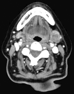
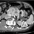
Figure 8.1 An axial CT scan showing a 1.5-cm lesion in the left submandibular gland. The lesion was reported as having the appearances of a benign tumour. The histological result was a carcinoma ex-pleomorphic adenoma.
For suspected malignant tumours a combination of both MRI and CT is often performed to allow precise tumour localisation and identification of bone invasion, perineural spread or distant metastases.2
Fine-needle aspiration cytology
The ideal management of patients with salivary masses is in the context of a one-stop clinic where the clinician is able to perform outpatient-based ultrasound-guided FNAC, with a cytology technician present in the clinic to confirm the presence of cellular material within the sample.7 An inadequate aspirate can be repeated at the same clinic appointment, and on-site microscopy can reduce the non-diagnostic rate to less than 1%.8
Salivary gland FNAC aims to distinguish neoplastic from non-neoplastic disease and, more importantly, also benign from malignant neoplasms. Clearly, knowing that a salivary gland lesion is malignant aids surgical planning. A systematic review9 of the accuracy of FNAC quotes positive predictive values from 16 studies, including 1782 cases of FNAC with histological concordance. In the case of a ‘benign’ FNAC result, the final histological diagnosis was confirmed as benign in 95% of cases. In malignancy the concordance was 93%. A recent systematic review10 of ultrasound-guided core biopsy found it to be more accurate than FNAC in determining benign from malignant pathology (sensitivity 92%, specificity 100% in the five studies included), compared with reports of equivalent 72% sensitivity and 100% specificity from ultrasound-guided FNAC.11
The take-home message is therefore that a positive result from either cytology technique is strongly suggestive of a malignancy, but FNAC yields more false-negative results for malignancy than core biopsy. The improved sensitivity of core biopsy is to be expected as far more tissue is obtained, but the investigation is more painful for patients and has a higher risk of haematoma formation. In the case of non-diagnostic initial cytology, repeat FNAC of salivary gland neoplasms has been shown to improve sensitivity from 70% to 84%.12 However, this still does not reach the sensitivity of core biopsy. If there is clinical or radiological suspicion of malignancy and in a situation where the cytology could alter the surgical treatment plan for an individual patient, then a core biopsy should be recommended following a negative initial FNAC.
Sialendoscopy
Sialendoscopy is the use of low-diameter optical endoscopes in the diagnosis and treatment of Stenson’s and Wharton’s pathology. It is most applicable to patients who have symptoms of salivary gland swelling on eating, indicative of a stone or stenosis.13 Diagnostic sialendoscopy may distinguish between stones, stenosis, mucous plugs or debris within the duct, all of which may present in a similar manner. It has also been used in the treatment of childhood sialolithiasis, juvenile recurrent parotitis14 and autoimmune disease, to dilate Stenson’s duct in patients with Sjögren’s syndrome.15 Interventional sialendoscopes incorporate a working channel, through which Dormia baskets, guide wires, laser fibres and balloons can be passed (see below).
Non-neoplastic disease of the salivary glands
Acute viral inflammation
The commonest cause of acute viral parotitis is mumps, caused by the paromyxovirus. The combined measles, mumps and rubella (MMR) vaccine was introduced throughout the UK in 1988 as a single dose, with a double-dose vaccine replacing this in 1996. The incidence of mumps was reduced by vaccination, but there has been a UK epidemic over the past decade, with a peak of 43 000 cases of mumps in 2005. Whereas mumps most commonly occurs in 4- to 6-year-olds, this rise was due to increasing numbers of adolescents getting the disease, too old to have received the vaccine prior to its routine introduction, or having only received the single-dose vaccine16 and then attending university, the ideal semi-closed environment to allow the virus to spread. The diagnosis is clinical but can be confirmed by serology, demonstrating antibodies to mumps S and V antigens. It can also be diagnosed by saliva testing for immunoglobulins. Polymerase chain reaction (PCR) can be used to further assay any negative samples.16 Malaise, fever and loss of appetite, followed by acute bilateral parotid enlargement, are typical presentations. The parotitis may be unilateral. About 30% of cases present as swelling in the submandibular and sublingual glands. Systemic complications include meningitis, encephalitis, hepatitis, carditis, orchitis and hearing loss. Treatment is symptomatic. Other viruses may mimic mumps: coxsackie A, enteric cytopathic human orphan (ECHO) virus, influenza A and cytomegalovirus.
Acute suppurative sialadenitis
Bacterial salivary gland infections are relatively uncommon and most frequently occur in the parotid gland. Traditionally, postoperative patients were deemed most at risk. With improved perioperative care and hydration this is now less common. However, the presentation of unilateral parotid enlargement with cellulitis in the dehydrated elderly patient still occurs. Pus may be expressed from the duct orifice and should be swabbed for microbiology. The commonest causative agent is Staphylococcus aureus. Haemophilus influenza is also common, but up to half of microbial isolates may be anaerobic, usually Gram-negative bacilli (Prevotella, Porphyromonas and Fusobacterium spp.).17 Intravenous antibiotic treatment should cover both aerobic and anaerobic organisms, and rehydration is important. Occasionally, abscess formation can accompany the presentation. If suspected, an ultrasound scan will confirm the presence of pus and therefore the need for incision and drainage. Acute bacterial salivary gland infections may also occur with duct calculi.
Chronic inflammatory conditions
Atypical tuberculosis
This is now an increasingly common condition affecting children, usually between the ages of 2 and 5, being rare after the age of 12.18 Although there are 13 ‘atypical’ strains of Mycobacterium, Mycobacterium avium intracellulare is the commonest, probably transmitted through contact with soil. The patient has painless lesions over either the parotid or submandibular glands, but is otherwise well. The infection is actually within the periglandular lymph nodes and any of the cervical lymph nodes can be affected. As the lymph nodes enlarge, pus may form and progression affects the skin, causing a typical discoloration before breakdown occurs. Whilst combination antibiotics are favoured by many (clarithromycin, ethambutol and rifampicin), some paediatric surgeons feel the diagnosis is so obvious clinically that surgical excision of all involved nodes is preferable, before the skin changes occur.18 There is also doubt as to the advantage, if any, conferred by antituberculous therapy.19 Where the salivary glands are involved, surgery requires gland excision. Wide local excision is advocated over an initial incisional biopsy to confirm the diagnosis. If an incisional biopsy is performed there is a risk of causing a chronic sinus and subsequent definitive surgery is less likely to control the disease.18 Left untreated the natural history of the disease is to break through the skin, discharging as a sinus on a chronic basis, before ‘burning out’. The end result is scarring of the overlying skin.
Cat-scratch disease
This is a granulomatous disease affecting the periglandular lymph nodes in the parotid and submandibular regions. The disease is caused by the Gram-negative bacterium Bartonella henselae and transmitted through a bite or scratch from a domestic cat. The cat flea is the vector for spread between animals and possibly also to humans. Serological testing of IgG and IgM, by immunofluorescence antibody testing, at presentation, and a rising titre at 10–14 days20 will confirm the diagnosis. It presents as a discoloured mass progressing to cervical lymphadenopathy, with 25% suffering with low-grade pyrexia. Swelling of the parotid gland may also occur. It may also manifest as Parinaud’s oculoglandular syndrome or granulomatous conjunctivitis in association with ipsilateral pre-auricular lymphadenopathy. Treatment is supportive, although occasionally surgery may be considered for persistent, enlarging, tender lymphadenopathy.21 Antibiotics have no proven role in disease resolution. A single randomised trial showed an improvement in lymphadenopathy, as measured by ultrasonography, at 30 days with a 5-day course of azithromycin, but no benefit over placebo at 2 and 4 months.22
Actinomycosis
The Gram-positive anaerobe, Actinomyces israelii, causes painless hard masses in the neck that may overlie the salivary glands. The infection may involve the mandible, especially in the presence of osteoradionecrosis.23 Necrosis and multiple sinus tracts often occur within the lesions. Treatment consists of surgical debridement or excision with long courses of antibiotics.
Sarcoidosis
Sarcoidosis is a multisystem inflammatory disease characterised by non-caseating granulomata. It is thought to be mediated by Th1 lymphocytes, and interleukin (IL)-17A has been implicated in granuloma formation.24 The disease most commonly affects young adults. Any organ can be involved but the lungs are the most commonly affected and extrapulmonary disease is usually associated with lung disease.25 The salivary glands, usually the parotids, are involved in 10–30% of patients, with non-tender, diffuse swelling of the glands. Intraoral nodules may also occur. Rarely, Heerfordt-Waldenstrom’s syndrome occurs, with uveitis, parotitis, fever and facial nerve palsy. Imaging of the parotid glands may show multiple non-cavitating masses. FNAC is useful in excluding malignancy. The diagnosis may be suggested via a chest radiograph, demonstrating mediastinal lymphadenopethy with or without pulmonary infiltrates, in association with a raised serum angiotensin-converting enzyme level. Serum levels of IL-18 are also elevated.26 Ultimately, a tissue diagnosis of non-caseating granulomata is required. This can be performed by biopsy of the salivary glands, intraoral sublabial biopsy, or a mediastinoscopy and lymph node biopsy. Treatment most frequently consists of systemic steroids or steroid-sparing immunosuppressants.
Sjögren’s syndrome
Sjögren’s syndrome (SS) is the most common autoimmune disease to affect the salivary glands. It may be primary or secondary to another connective tissue disorder. The American–European Consensus27 lists six diagnostic criteria:
3. Objective eye signs (Schirmer test < 5 mm in 5 minutes, Rose Bengal score).
4. Histopathology of minor salivary glands showing focal lymphocytic sialadenitis. The most accessible and reliable area to biopsy is the sublabial minor salivary gland tissue in the lower lip.
5. Salivary gland involvement demonstrated on objective testing: reduced whole salivary flow, diffuse sialectasis, reduced scintigraphy uptake.
The disease is most common in women aged 40–60 years, typically with dry, gritty eyes and a dry mouth. Reduced salivary function also affects swallowing and voice function, as well as leaving the patient susceptible to dental caries. Patients may be referred to an otolaryngologist with symptoms of recurrent salivary gland swelling, usually the parotids. Other systemic features of primary SS include arthritis, skin vasculitis and pulmonary fibrosis. Treatment involves saliva and tear replacement substitutes. Pilocarpine is used to improve any residual salivary gland tissue. Systemic therapy is delivered by a rheumatologist and may consist of non-steroidal anti-inflammatories for arthritis and immunomodulatory medications or steroids for more severe SS symptoms. Patients with SS are up to 16 times more likely to develop mucosa-associated lymphoid tissue (MALT) lymphoma.28 Any persistent swelling in the gland of a patient with SS should raise the possibility of lymphoma, prompting the standard work-up for a salivary gland neoplasm with imaging and FNAC. FNAC, being less accurate in diagnosing lymphoma, is chiefly used here to exclude other neoplasms.
Human immunodeficiency virus
Generalised parotid gland enlargement and xerostomia may be a presenting feature of undiagnosed human immunodeficiency virus (HIV) infection. The pathogenesis remains unclear, as the symptoms may not be related to direct infection of the salivary glands.29 Associated cervical lymphadenopathy may also feature. Lymphoepithelial cysts may occur, most commonly in the parotid and bilateral in nature. Conservative management of HIV-associated cysts is advocated.30
Sialolithiasis
Unlike renal calculi, metabolic disorders do not seem to predispose patients to salivary stones. Calculi tend to be composed of calcium carbonates and calcium phosphates along with glycoproteins and mucopolysaccharides.31 Theories for stone formation include the occurrence of intracellular microcalculi, which form a nidus when excreted into the duct,32 or mucous plugs of saliva may act as the nidus. Stones may either be single or multiple. Huoh and Eisele33 retrospectively looked at the aetiological factors in 153 patients with stone disease in California over nearly 10 years. In 82% the stone disease was submandibular. The patient population had a higher use of diuretics and there was a positive smoking history in 44% of patients.
Treatment
A further option for particularly large salivary gland stones is extracorporeal shock wave lithotripsy. First introduced in 1986, this uses pressure to fragment stones, making them easier to be flushed out of the gland in saliva. The technique is minimally invasive and painless with few side-effects. Long-term follow-up studies show good results for selected patients34 and it can now be used electively before sialendoscopic techniques are undertaken. Results are better with smaller calculi and with parotid than submandibular disease.35
Interventional sialendoscopy
The ability to remove salivary duct calculi with this method is dependent on the size and position of the stone. Calculi affecting the parotid or submandibular duct can be managed by interventional sialendoscopy (IS) alone when less than 3 and 4 mm, respectively.13 When larger than these sizes, combined treatment with IS, transoral excision, laser fragmentation, lithotripsy or external surgical approach may be considered. IS undoubtedly offers a genuine conservative approach to the management of salivary gland duct calculi and stenosis but the treatment tends to be concentrated in specialist centres. However, Bowen et al.36 have reported their preliminary experience in a newly developed sialendoscopy service. They report 36 procedures: 17 for calculi, 16 for recurrent sialadenitis and three for Sjögren’s syndrome. Symptoms fully resolved in 26. Thirteen stones were successfully removed, five purely by endoscopy with a mean size of 3.7 mm and eight by a combined endoscopic and external approach, with a mean size of 10.6 mm. One patient had a salivary fistula as a complication.

As mentioned, another use of sialendoscopy is to aid conservative surgical excision of larger salivary calculi, usually in the parotid. The standard treatment of such stones in previous times would have been a parotidectomy. Karavidas et al.37 describe 70 patients treated with a combined endoscopic and conservative external excision of parotid duct calculi. The stone is identified within the parotid duct by sialendoscopy and ultrasound. Either a pre-auricular incision or an incision directly over the stone is made to remove it. No facial nerve weakness or salivary fistulae were noted in this series.
Non-inflammatory conditions
Sialadenosis or sialosis is a non-inflammatory, non-neoplastic, non-painful, bilateral swelling of the major salivary glands and is usually most clinically apparent in the parotid glands. Histologically it is characterised by acinar cell hypertrophy and atrophy of striated ducts associated with oedema of interstitial connective tissue. There are a number of factors associated with this condition, including some drugs (particularly antihypertensive medications, sympathomimetics, antithyroid agents and phenothiazines), endocrine disorders and nutritional disorders.38 Treatment of the underlying pathology is the mainstay of treatment. Surgical management should only be considered for failure of medical management coupled with gross cosmetic concerns.
Post-radiotherapy xerostomia
Radiotherapy to the head and neck region results in a chronic long-term dry mouth and is universally recognised by patients as one of the major determinants on quality of life after such treatment. Radiotherapy to the salivary glands results in decreased salivary flow. There is emerging evidence from randomised controlled studies supporting the use of intensity modulated radiotherapy (IMRT) in head and neck oncology,39 in allowing a reduced dose of radiation to the parotid glands resulting in an improved salivary flow. Whilst amifostine and pilocarpine have both been used to improve salivary flow, their use has never replaced the humble bottle of water in relieving the troublesome symptoms of a chronic dry mouth.
Neoplastic disease

The likelihood of a neoplasm being malignant increases with decreasing size of the affected gland. In a large series, 25% of parotid neoplasms were malignant, 43% of submandibular neoplasms and 82% of minor salivary gland neoplasms.41 The chance of a sublingual gland neoplasm being malignant is said to approach 100%.
epithelial or non-epithelial in origin. Most (70%) salivary gland tumours are found in the parotid gland, with 8% in the submandibular glands and 22% in the minor glands.40
Benign epithelial neoplasms
In a large population study, the incidence of benign salivary gland neoplasms has been found to be 6.2–7.2 per 100 000 population over a 20-year period, 1988–2007. Of 916 neoplasms diagnosed in this study, 71% were pleomorphic adenomas, 22% Warthin’s tumours, 2.4% basal cell adenomas and 1.0% each were oncocytomas, monomorphic adenomas and myoepitheliomas.42
The aetiology of benign salivary gland disease is difficult to define, given the heterogeneity of the tumours. Environmental and genetic factors have been proposed. The strongest link seems to be radiation exposure. The survivor populations in Nagasaki and Hiroshima exposed to radiation after the atomic bombs at the end of the Second World War had an increased relative risk of 3.5 for benign and 11 for malignant salivary neoplasm.43 Smoking too has been implicated in the development of Warthin’s tumours,44 perhaps because smokers have six times higher mitochondrial DNA damage in alveolar macrophage samples than do non-smokers.
The World Health Organisation (2005)45 classify benign salivary gland tumours into 13 subtypes (Table 8.1). Because of the epithelial and myoepithelial tissue components of salivary glands, the tumours are a heterogeneous group that can be defined according to their dominant tissue type (epithelial or myoepithelial) or can be described as mixed.
Pleomorphic adenoma
Pleomorphic adenoma (PA) is the most common tumour type occurring in the salivary glands. It is most frequently found in the parotid gland. Tumours present as slow-growing painless masses. PAs are of mixed origin and the epithelial, myoepithelial and mesenchymal components demonstrate huge cell variation, architecture and morphology. Surgical excision is the preferred management due to the small but definite potential for malignant transformation. The anecdotal opinion on the risk of malignant transformation is of the order of 1% per annum. Surgical management of pleomorphic adenoma is controversial because of the issue of capsular rupture and tumour spillage causing tumour recurrence. Zbären and Stauffer46 analysed the histological features of the capsule of pleomorphic adenomas and found that in 73% of the tumours there were features such as incomplete capsule (33%), capsule penetration (26%), tumour pseudopodia (40%) and satellite nodules present (13%).
Warthin’s tumour
These are also known as papillary cystadenoma, lymphomatosum and cystic papillary adenoma adenolymphoma. After PAs, Warthin’s tumours are the most common benign neoplasms of the salivary glands, making up 10–15% of epithelial lesions. They are found exclusively in the parotid glands. Ten per cent are bilateral and there is an association with smoking. Recent evidence questions whether Warthin’s tumours are truly parotid neoplasms, or rather are a disease of the periparotid lymph nodes. Specific components of lymph nodes were consistently identified within a series of Warthin’s tumours, leading the authors to conclude that they are tumour-like lesions rather than true adenomata.47 Where appropriate, the management of these tumours is surgical. There are rare reported cases of malignant transformation. In surgically unsuitable patients, a conservative approach will be supported by appropriate cytological characterisation.
Management of benign epithelial neoplasms

George and McGurk48 state that there is no increase in tumour recurrence when tumours are dissected off the nerve in the manner described above. McGurk has championed the technique of extracapsular dissection in the UK. A pre-auricular incision is made and a standard skin flap raised over the parotid fascia. A cruciate incision is then made over the tumour through the fascia. Blunt dissection and careful division of tissues proceeds around the tumour, with meticulous haemostasis. Branches of the nerve are thus identified where necessary, but the nerve is not formally located. The tumour is therefore excised with minimal surrounding tissue but with an intact tumour capsule.
Over a 10-year period (1999–2009),48 156 benign parotid tumours were excised using this technique. With a mean follow-up of 3 years 8 months, no recurrences have been noted. The rates of temporary and permanent facial nerve weakness were 3% and 1%, respectively. Previously,49 between 1952 and 1992, a series of 662 clinically benign, less than 4-cm tumours were treated by either extracapsular dissection (502) or superficial parotidectomy (159). At 15-year follow-up the recurrence rate of benign tumours was 1.7% and 1.8%, respectively. In those tumours that ultimately turned out to be malignant (32), the cancer-specific 10-year survival was extremely high and no difference between the surgical techniques was found (100% vs. 98%).
Recurrent benign epithelial neoplasms
In reality this is almost always recurrent PA. Patients present any number of years following initial surgery, most commonly parotidectomy. The presentation is usually of one or more painless nodules in the parotid ‘bed’. Pain, rapid growth or facial nerve palsy should alert the clinician to the possibility of carcinoma developing from a pleomorphic adenoma, which occurred in as many as 16% of one series.50 It is important to investigate recurrent disease in the same manner as primary neoplasms, with cytology and radiology. For uninodular disease, the distinction between recurrent tumour and a neuroma is important. For multinodular disease, the diagnosis of recurrent PA is usually clinically obvious. Surgical excision is the treatment of choice. Local excision of a single nodule is rarely feasible. A complete superficial parotidectomy with facial nerve preservation and excision of the overlying scar is therefore the minimum required for lateral disease. Total parotidectomy with or without nerve sacrifice or even radical/extended parotidectomy may be considered for more extensive recurrences or for when the presence of carcinoma is considered likely. Postoperative radiotherapy should be considered, with the aim of improving local control. The dose is usually lower than for head and neck carcinoma, in the region of 50 Gy. As PA is a benign disease, the risk of radiation side-effects and the risk of radiation-induced neoplasms should be discussed with the patient, especially if they are young.
Benign non-epithelial neoplasms
Haemangiomas most commonly affect the parotid gland in children. Parental concern is understandable as the tumours have a characteristic rapid growth phase in the first 6 months. The diagnosis is entirely clinical and reassurance can be provided that natural resolution will ensue between 1 and 2 years. Oral propranolol may have a place in treatment.51 These should be distinguished from vascular malformations, which are usually present at birth and may require surgical excision from the major salivary glands. Lipomas may also affect the major salivary glands.
Malignant epithelial neoplasms
The most recent WHO classification of malignant salivary gland neoplasm includes 24 subtypes45 (Table 8.1). The majority, 60–70%, of patients will have either mucoepidermoid carcinoma, adenoid cystic carcinoma, acinic cell carcinoma or polymorphous low-grade adenocarcinoma. Signs that suggest a malignant tumour in the salivary gland include facial or hypoglossal nerve involvement, pain, a sudden increase in size of a pre-existing salivary gland tumour and associated cervical lymphadenopathy. Jones et al.52 analysed a series of 741 salivary gland tumours over 30 years. Of the 260 malignant neoplasms, 33% were mucoepidermoid carcinoma and 24% were adenoid cystic. The USA Surveillance, Epidemiology and End Results Program identified 6391 malignant salivary neoplasms (1.195/100 000 person-years) from 1992 to 2006. Squamous cell and mucoepidermoid carcinomas were the commonest male variants, with mucoepidermoid, acinic cell and adenoid cystic carcinomas most common among females. As with benign tumours of the salivary glands, radiation exposure has been implicated in the risk of developing malignant tumours.
Mucoepidermoid carcinoma
Mucoepidermoid carcinoma (MEC) is the most common malignant neoplasm of the salivary glands in both adults and children but has the most favourable outcome. Half of these tumours present in the major glands, most commonly in the parotid gland (45%). Histological grading of this tumour in particular has prognostic value. The majority of tumours are low or intermediate grade, are treated surgically and have a good prognosis. Those that are high grade have an increased metastatic potential with a reduced cure rate following both surgical and radiotherapy management.53
Polymorphous low-grade adenocarcinoma
This tumour was described in 198454 and, since then, an increasing number have been diagnosed. Confusion sometimes occurs histologically with PA and ACC and many polymorphous low-grade adenocarcinoma (PMLA) will have been previously misdiagnosed. PMLA is the second most common minor salivary gland tumour, mainly affecting the palate. Treatment involves wide local surgical resection and carries an excellent prognosis. Local control failures can occur after as long as 10 years, hence the rationale for long-term follow-up.55
Carcinoma ex-pleomorphic adenoma
These tumours are those in which a new malignancy has arisen in a previous PA. They represent 12% of malignant salivary gland tumours and frequently present as a long-standing mass that has recently increased in size. Wide local surgical excision with a neck dissection is the treatment of choice followed by postoperative radiotherapy. The subtypes are non-invasive, minimally invasive and invasive, with non-invasive and minimally invasive having a better prognosis. Invasive disease has an aggressive course, with 25–50% patients developing one or more recurrences.56
Management of epithelial malignancies
A different approach to treatment exists between low-grade tumours and high-grade tumours, but also between tumours less than 4 cm and those greater than 4 cm. In general, low-grade, low-stage tumours (T1–2) may be treated by complete surgical excision alone.57 Many are initially excised completely as presumed benign tumours, only for the final histology to be revealed. For all other tumours, postoperative radiotherapy improves locoregional control. High-grade tumours, on the other hand, exhibit aggressive growth patterns and may merit more aggressive treatment. The ‘4-cm rule’ is often quoted, implying any tumour over this size has a reduced rate of locoregional control and worse survival outcomes.58
Advanced stage (T3–4), high-grade parotid tumours should be treated by at least a total parotidectomy, with preservation of the facial nerve where appropriate.59 Any tumour with clinical or radiological cervical lymphadenopathy should have a simultaneous modified radical neck dissection, levels I–V. For advanced tumours with clinical N0 neck disease, the options are to treat the neck electively with either a selective neck dissection levels II–V, elective adjuvant radiotherapy after salivary gland excision or to watch and wait. The evidence supports active treatment to the neck in these patients over observation.59 Clinically high-grade submandibular malignancies should be treated with radical excision incorporating a 2-cm cuff of normal tissue, sacrificing the overlying facial nerve branches. Clinically N0 disease should undergo simultaneous level I and IIa neck dissection. Any clinically positive nodal disease should be treated by at least a modified radical neck dissection.60
Malignant non-epithelial neoplasm
By far the most common of these neoplasms is lymphoma: 5% of extranodal lymphomas affect the major salivary glands and almost 50% of head and neck lymphomas involve the major salivary glands. The mucosal associated lymphoid tissue (MALT) type of non-Hodgkin’s lymphoma predominates. Presentation is usually early and diagnosis and imaging will determine treatment, whether surgical or with chemotherapy.61 MALT lymphoma may occur bilaterally and metachronously. Surgical management in isolated disease may be considered.
Metastatic disease to the major salivary glands
The parotid gland is the major salivary gland most frequently involved by metastatic disease (80–90%), the commonest source being cutaneous squamous cell carcinoma (CSCC) of the head and neck. This is thus rare in the northern hemisphere, but sun exposure in Australia and New Zealand has led to metastatic CSCC being the commonest malignant neoplasm to affect the parotid. In a series of 232 patients treated in Sydney62 for malignant parotid neoplasms, 54 were primary parotid cancers, 101 were metastatic CSCC, 69 were metastatic melanoma and eight were other metastases. The parotid gland has a rich lymphatic network that drains the temple area and the cheek region. Any tumour in this area is likely to metastasise to the parotid region and subsequently to the upper cervical lymph nodes. There are approximately 15–20 periparotid lymph nodes and approximately four to five lymph nodes in the deeper portion of the parotid gland. The majority of patients with metastatic parotid disease present after the primary CSCC has been treated, usually up to 12 months, but the interval can be up to 2–3 years. Immunosuppressed patients, in particular transplant recipients, are especially prone to metastatic CSSC.
Metastases from infraclavicular primary sites are rare.63 The commonest primary sites are the lung, breast and kidney, although metastases from the stomach, bladder and prostate have also been described. Seifert et al.64 looked at 108 cases of metastases to the salivary glands between 1965 and 1985. They found 21 cases from infraclavicular primary sites: seven from lung, six renal, six breast, one colonic and one uterus. Whenever an infraclavicular primary site is suspected, either clinically or cytologically, then radiological imaging should cover the neck, thorax, abdomen and pelvis. This should be with a CT or CT-PET scan.
Surgical principles
Partial parotidectomy
The term superficial parotidectomy is commonly used by surgeons and implies removal of all parotid tissue lateral to the facial nerve. In reality, for the most common pathology, benign neoplasms, all that is required is removal of the tumour with a cuff of normal tissue surrounding it. As discussed above, this can be in the form of an extracapsular dissection, but more commonly surgeons perform a formal partial parotidectomy with identification of the facial nerve. Frequently, a benign tumour may extend between branches of the nerve, therefore entering the deep lobe. For this reason, the preferred terminology is a ‘partial superficial parotidectomy with or without deep lobe dissection’.65
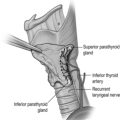
In the neck, dissection proceeds down to the sternocleidomastoid muscle where the great auricular nerve is found. This nerve can then be dissected out superiorly up to the gland in order to preserve it. It has anterior and posterior branches. The anterior branch always needs to be sacrificed in this approach, but the posterior branch can be preserved, maintaining sensory function to the ear lobe (Fig. 8.2).
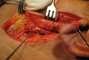
Three landmarks can be used to identify the main trunk of the nerve: the tragal ‘pointer’ – part of the cartilage of the external auditory canal. The nerve lies 1 cm deep and inferior to this (less in cadaveric dissections66). Secondly, the tympanomastoid suture line lies between the tympanic ring of bone that forms the bony external auditory canal and the mastoid bone. This suture line leads to the stylomastoid foramen and hence the nerve. Palpating this suture line gives a reliable impression of where the nerve will lie.66 Thirdly, the nerve will lie 1 cm deep to the posterior belly of digastric. In reality, a broad dissection and the use of all three of the landmarks will allow identification of the nerve.
Once the main trunk of the nerve is identified (Fig. 8.3), an artery clip is inserted on top of the nerve and opened so the nerve can be seen and the parotid tissue divided superficial to it. This action splits open the gland, vastly improving exposure to the nerve. This pattern continues to expose the bifurcation of the nerve. Care must be taken to orientate the dissection so as not to breach the surgical margin of normal tissue around the tumour.
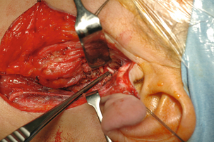
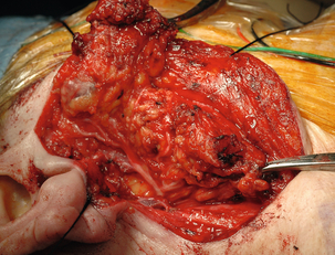
For an inferiorly placed tumour one of the middle branches of the nerve can be followed out to the periphery. The lower branches are then followed out one by one, from superior to inferior, so the tumour and gland are retracted inferiorly before being completely excised. For a larger tumour, all the branches are followed out to the periphery one by one, often starting superiorly and going inferiorly in a similar manner (Fig. 8.4).
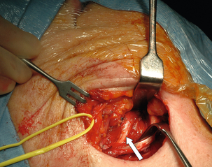
Submandibular gland surgery
Unlike parotid surgery, this is more commonly performed for inflammatory conditions of the gland than neoplasms. In a single large series of submandibular gland excisions, 75% were performed for non-neoplastic disease and 15% for neoplastic disease.67 The distinct difference between the two indications for surgery lies in the surgical dissection of the deep investing layer of cervical fascia.
The patient is positioned supine with a head ring and shoulder support. The incision is marked in a skin crease at least two finger breadths inferior to the lower border of the mandible. This is because the mandibular branch of the facial nerve can lie anything up to 1.2 cm below the mandible as it courses over the gland.68
If surgery is being performed for neoplastic disease then the above dissection would compromise oncological clearance. The nerve is usually sacrificed for malignant neoplasms,60 but is preserved for benign neoplastic disease. The correct dissection to preserve the nerve is to leave the investing fascia over the gland intact. A subplatysmal plane is raised up to the inferior border of the mandible. The mandibular nerve is then located on the fascia and completely mobilised. It can be reliably located just inferior to the angle of the mandible. There is often more than one branch. As the nerve is released posteriorly towards the parotid gland, the cervical branch may also be located and preserved. The nerve should be protected by retracting it superiorly out of the operating field using a vascular sling.
Removal of the gland involves dissection and release of the gland from both bellies of the digastric muscles. Deep to the posterior belly, the facial artery and vein are located. These should be ligated and divided, releasing the gland. The artery and vein also should be ligated and divided as they pass over the inferior border of the mandible. Keeping the ligature long on the distal vascular stump also facilitates the retraction of the mandibular nerve. The gland must then be dissected out superiorly under the mandible. Once the superficial lobe is mobile the mylohyoid muscle is approached and retracted anteriorly. This gives access to the deep lobe. Lying deep to the gland are the lingual nerve superiorly and hypoglossal nerve inferiorly, on the hyoglossus muscle. A small secretorimotor, parasympathetic nerve branch leaves the lingual nerve to enter the submandibular ganglion and then gland. A vessel always accompanies this nerve branch, and must be cauterised and divided to allow mobilisation of the gland with preservation of the lingual nerve (Fig. 8.5). The final stage is ligation and division of the submandibular salivary duct.
Surgical complications
Facial nerve palsy
The risk to the facial nerve may vary with the extent of the surgery, experience of the surgeon, pathology and with recurrent disease. For partial or superficial lateral parotid surgery the risk is in the region of 25% for a temporary palsy (literature range 18–65%) and between 1% and 6% for a permanent palsy, with the mandibular nerve most commonly affected.48,69 The use of the facial nerve monitor or stimulator is becoming more common. However, reports suggest that they do not reduce the rate of facial nerve palsies.70 For submandibular gland excision, temporary mandibular nerve paralysis has been reported in up to 36% of cases.71 The risk of a permanent mandibular nerve palsy has been reported to be as high as 8%.72
Frey’s syndrome
This is sweating, erythema or warmth over the parotid bed area whilst eating. It is thought to be due to parasympathetic nerve fibres from the auriculotemporal nerve re-anastomosing with sweat glands following parotidectomy. The reported incidence is up to 63%,73 but may be much higher when objectively tested for with electrogustometry or starch–iodine testing. There are several surgical methods to reduce Frey’s syndrome: sternocleidomastoid muscle flap insertion, SMAS flap, dermal fillers and fat graft. Postoperatively injecting the area with botulinum A is a method frequently employed to reduce symptoms.
References
1. Henstrom, D.K., Skilbeck, C.J., Weinberg, J., et al, Good correlation between original and modified House Brackmann facial grading systems. Laryngoscope. 2011;121(1):47–50. 21120826
2. Yousem, D.M., Kraut, M.A., Chalian, A.A., Major salivary gland imaging. Radiology. 2000;216(1):19–29. 10887223
3. Burke, C.J., Thomas, R.H., Howlett, D., Imaging the major salivary glands. Br J Oral Maxillofac Surg. 2011;49(4):261–269. 20381221
4. Brennan, P.A., Herd, M.K., Howlett, D.C., et al, Is ultrasound alone sufficient for imaging superficial lobe benign parotid tumours before surgery? Br J Oral Maxillofac Surg. 2012;50(4):333–337. 21371794
5. Jadu, F., Yaffe, M.J., Lam, E.W.N., A comparative study of the effective radiation doses from cone beam computed tomography and plain radiography for sialography. Dentomaxillofac Radiol. 2010;39(5):257–263. 20587648
6. Christe, A., Waldherr, C., Hallett, R., et al, MR imaging of parotid tumors: typical lesion characteristics in MR imaging improve discrimination between benign and malignant disease. AJNR Am J Neuroradiol. 2011;32(7):1202–1207. 21724574
7. NICE. Guidance on cancer services. Improving outcomes in head and neck cancers – the manual. London: National Institute for Clinical Excellence; 2004.
8. Eisele, D.W., Sherman, M.E., Koch, W.M., et al, Utility of immediate on-site cytopathological procurement and evaluation in fine needle aspiration biopsy of head and neck masses. Laryngoscope. 1992;102(12, Pt 1):1328–1330. 1280752
9. Colella, G., Cannavale, R., Flamminio, F., et al, Fine-needle aspiration cytology of salivary gland lesions: a systematic review. J Oral Maxillofac Surg. 2010;68(9):2146–2153. 20580145
10. Schmidt, R.L., Hall, B.J., Layfield, L.J., A systematic review and meta-analysis of the diagnostic accuracy of ultrasound-guided core needle biopsy for salivary gland lesions. Am J Clin Pathol. 2011;136(4):516–526. 21917673
11. Cho, H.W., Kim, J., Choi, J., et al, Sonographically guided fine-needle aspiration biopsy of major salivary gland masses: a review of 245 cases. AJR Am J Roentgenol. 2011;196(5):1160–1163. 21512086
12. Brennan, P.A., Davies, B., Poller, D., et al, Fine needle aspiration cytology (FNAC) of salivary gland tumours: repeat aspiration provides further information in cases with an unclear initial cytological diagnosis. Br J Oral Maxillofac Surg. 2010;48(1):26–29. 19233526
13. Chossegros, C., Faure, F., Marchal, F. Sialadenitis and sialadenosis – interventional sialendoscopy. In: Bradley P.J., Guntinus-Lichius O., eds. Salivary gland disorders and diseases: diagnosis and management. Stuttgart: Thieme, 2011. [Chapter 15].
14. Martins-Carvalho, C., Plouin-Gaudon, I., Quenin, S., et al, Pediatric sialendoscopy: a 5-year experience at a single institution. Arch Otolaryngol Head Neck Surg. 2010;136(1):33–36. 20083775
15. Shacham, R., Puterman, M.B., Ohana, N., et al, Endoscopic treatment of salivary glands affected by autoimmune diseases. J Oral Maxillofac Surg. 2011;69(2):476–481. 21145154
16. Kay, D., Roche, M., Atkinson, J., et al, Mumps outbreaks in four universities in the North West of England: prevention, detection and response. Vaccine. 2011;29(22):3883–3887. 21447315
17. Brook, I., Aerobic and anaerobic microbiology of suppurative sialadenitis. J Med Microbiol. 2002;51(6):526–529. 12018662
18. Fraser, L., Moore, P., Kubba, H., Atypical mycobacterial infection of the head and neck in children: a 5-year retrospective review. Otolaryngol Head Neck Surg. 2008;138(3):311–314. 18312877
19. Clarke, J.E., Nontuberculous lymphadenopathy in children: using the evidence to plan optimal management. Adv Exp Med Biol 2011; 719:117–121. 22125039
20. Ridder, G.J., Boedeker, C.C., Technau-Ihling, K., et al, Cat-scratch disease: otolaryngologic manifestations and management. Otolaryngol Head Neck Surg. 2005;132(3):353–358. 15746844
21. Munson, P.D., Boyce, T.G., Salomao, D.R., et al, Cat-scratch disease of the head and neck in a pediatric population: surgical indications and outcomes. Otolaryngol Head Neck Surg. 2008;139(3):358–363. 18722212
22. Bass, J.W., Freitas, B.C., Freitas, A.D., et al, Prospective randomized double blind placebo-controlled evaluation of azithromycin for treatment of cat-scratch disease. Pediatr Infect Dis J. 1998;17(6):447–452. 9655532
23. Hansen, T., Kunkel, M., Springer, E., et al, Actinomycosis of the jaws – histopathological study of 45 patients shows significant involvement in bisphosphonate-associated osteonecrosis and infected osteoradionecrosis. Virchows Arch. 2007;451(6):1009–1017. 17952459
24. Ten Berge, B., Paats, M., Bergen, I., et al, Increased IL-17A expression in granulomas and in circulating memory T cells in sarcoidosis. Rheumatology (Oxford). 2012;51(1):37–46. 22075064
25. Knopf, A., Bas, M., Chaker, A., et al, Rheumatic disorders affecting the head and neck: underestimated diseases. Rheumatology. 2011;50(11):2029–2034. 21865282
26. Liu, D., Yao, Y., Cui, W., et al, The association between interleukin-18 and pulmonary sarcoidosis: a meta-analysis. Scand J Clin Lab Invest. 2010;70(6):428–432. 20653400
27. Vitali, C., Bombardieri, S., Jonsson, R., et al, Classification criteria for Sjogren’s syndrome: a revised version of the European criteria proposed by the American–European Consensus Group. Ann Rheum Dis. 2002;61(6):554–558. 12006334
28. Theander, E., Henriksson, G., Ljungberg, O., et al, Lymphoma and other malignancies in primary Sjogren’s syndrome: a cohort study on cancer incidence and lymphoma predictors. Ann Rheum Dis. 2006;65(6):796–803. 16284097
29. Schiodt, M., Greenspan, D., Levy, J.A., et al, Does HIV cause salivary gland disease? AIDS. 1989;3(12):819–822. 2517203
30. Bradley, P.J. Cystic salivary gland tumours including cystic neoplasms. In: Bradley P.J., Guntinas-Lichius O., eds. Salivary gland disorders and diseases: diagnosis and management. Stuttgart: Thieme, 2011. [Chapter 17].
31. Marchal, F., Dulguerov, P., Lehmann, W., Interventional sialendoscopy. N Engl J Med. 1999;341(16):1242–1243. 10523164
32. Marchal, F., Kurt, A.M., Dulguerov, P., et al, Retrograde theory in sialolithiasis formation. Arch Otolaryngol Head Neck Surg. 2001;127(1):66–68. 11177017
33. Huoh, K.C., Eisele, D.W., Etiologic factors in sialolithiasis. Otolaryngol Head Neck Surg. 2011;145(6):935–939. 21753035
34. Schmitz, S., Zengel, P., Alvir, I., et al, Long-term evaluation of extracorporeal shock wave lithotripsy in the treatment of salivary stones. J Laryngol Otol. 2008;122(1):65–71. 17466089
35. Escudier, M.P., Brown, J.E., Putcha, V., et al, Factors influencing the outcome of extracorporeal shock wave lithotripsy in the management of salivary calculi. Laryngoscope. 2010;120(8):1545–1549. 20641082
36. Bowen, M.A., Tauzin, M., Kluka, E.A., et al, Diagnostic and interventional sialendoscopy: a preliminary experience. Laryngoscope. 2011;121(2):299–303. 21271577
37. Karavidas, K., Nahlieli, O., Fritsch, M., et al, Minimal surgery for parotid stones: a 7-year endoscopic experience. Int J Oral Maxillofac Surg. 2010;39(1):1–4. 19897340
38. Pape, S.A., MacLeod, R.I., McLean, N.R., et al, Sialadenosis of the salivary glands. Br J Plast Surg. 1995;48(6):419–422. 7551515
39. Nutting, C.M., Morden, J.P., Harrington, K.J., et al, Parotid-sparing intensity modulated versus conventional radiotherapy in head and neck cancer (PARSPORT): a phase 3 multicentre randomised controlled trial. Lancet Oncol. 2011;12(2):127–136. 21236730
40. Eveson, J.W., Cawson, R.A., Salivary gland tumours. A review of 2410 cases with particular reference to histological types, site, age and sex distribution. J Pathol. 1985;146(1):51–58. 4009321
41. Spiro, R.H., Salivary neoplasms: overview of a 35-year experience with 2,807 patients. Head Neck Surg. 1986;8(3):177–184. 3744850
42. Grant, D.G., Bradley, P.T. Epidemiology of benign salivary gland neoplasms. In: Bradley P.J., Guntinas-Lichius O., eds. Salivary gland disorders and diseases: diagnosis and management. Thieme: Stuttgart, 2011. [Chapter 18].
43. Takeichi, N., Hirose, F., Yamamoto, H., et al, Salivary gland tumors in atomic bomb survivors, Hiroshima, Japan. II. Pathologic study and supplementary epidemiologic observations. Cancer. 1983;52(2):377–385. 6305475
44. Sadetzki, S., Oberman, B., Mandelzweig, L., et al, Smoking and risk of parotid gland tumors: a nationwide case-control study. Cancer. 2008;112(9):1974–1982. 18361448
45. Barnes, L. Pathology and genetics of head and neck tumours. Lyon: IARC Press; 2005.
46. Zbären, P., Stauffer, E., Pleomorphic adenoma of the parotid gland: histopathologic analysis of the capsular characteristics of 218 tumors. Head Neck. 2007;29(8):751–757. 17252593
47. Teymoortash, A., Werner, J.A., Moll, R., Is Warthin’s tumour of the parotid gland a lymph node disease? Histopathology. 2011;59(1):143–145. 21771031
48. George, K.S., McGurk, M., Extracapsular dissection – minimal resection for benign parotid tumours. Br J Oral Maxillofac Surg. 2011;49(6):451–454. 21215499
49. McGurk, M., Thomas, B.L., Renehan, A.G., Extracapsular dissection for clinically benign parotid lumps: reduced morbidity without oncological compromise. Br J Cancer. 2003;89(9):1610–1613. 14583757
50. Makeieff, M., Pelliccia, P., Letois, F., et al, Recurrent pleomorphic adenoma: results of surgical treatment. Ann Surg Oncol. 2010;17(12):3308–3313. 20645014
51. Kupeli, S., Use of propranolol for infantile hemangiomas. Pediatr Hematol Oncol. 2012;29(3):293–298. 22303875
52. Jones, A.V., Craig, G.T., Speight, P.M., et al, The range and demographics of salivary gland tumours diagnosed in a UK population. Oral Oncol. 2008;44(4):407–417. 17825603
53. Emerick, K.S., Fabian, R.L., Deschler, D.G., Clinical presentation, management, and outcome of high-grade mucoepidermoid carcinoma of the parotid gland. Otolaryngol Head Neck Surg. 2007;136(5):783–787. 17478216
54. Evans, H.L., Batsakis, J.G., Polymorphous low-grade adenocarcinoma of minor salivary glands. A study of 14 cases of a distinctive neoplasm. Cancer. 1984;53(4):935–942. 6692293
55. Paleri, V., Robinson, M., Bradley, P., Polymorphous low-grade adenocarcinoma of the head and neck. Curr Opin Otolaryngol Head Neck Surg. 2008;16(2):163–169. 18327037
56. Olsen, K.D., Lewis, J.E., Carcinoma ex pleomorphic adenoma: a clinicopathologic review. Head Neck. 2001;23(9):705–712. 11505478
57. Vander Poorten, V., Bradley, P.J., Takes, R.P., et al, Diagnosis and management of parotid carcinoma with a special focus on recent advances in molecular biology. Head Neck. 2012;34(3):429–440. 21618326
58. Speight, P.M., Barrett, A.W., Salivary gland tumours. Oral Dis. 2002;8(5):229–240. 12363107
59. Jeannon, J.P., Calman, F., Gleeson, M., et al, Management of advanced parotid cancer. A systematic review. Eur J Surg Oncol. 2009;35(9):908–915. 19027261
60. Roland, N.J., Paleri V. Head and neck cancer: multidisciplinary management guidelines. London: ENT UK; 2011. [Chapter 27].
61. Carbone, A., Gloghini, A., Ferlito, A., Pathological features of lymphoid proliferations of the salivary glands: lymphoepithelial sialadenitis versus low-grade B-cell lymphoma of the malt type. Ann Otol Rhinol Laryngol. 2000;109(12, Pt 1):1170–1175. 11130833
62. Bron, L.P., Traynor, S.J., McNeil, E.B., et al, Primary and metastatic cancer of the parotid: comparison of clinical behavior in 232 cases. Laryngoscope. 2003;113(6):1070–1075. 12782825
63. O’Hara, J.T., Paleri, V. Metastases to the major salivary glands from non-head and neck primary malignancies. In: Bradley P.J., Guntinus-Lichius O., eds. Salivary gland disorders and diseases: diagnosis and management. Stuttgart: Thieme, 2011. [Chapter 37].
64. Seifert, G., Hennings, K., Caselitz, J., Metastatic tumors to the parotid and submandibular glands – analysis and differential diagnosis of 108 cases. Pathol Res Pract. 1986;181(6):684–692. 3562340
65. Tweedie, D.J., Jacob, A., Surgery of the parotid gland: evolution of techniques, nomenclature and a revised classification system. Clin Otolaryngol. 2009;34(4):303–308. 19673976
66. Rea, P.M., McGarry, G., Shaw-Dunn, J., The precision of four commonly used surgical landmarks for locating the facial nerve in anterograde parotidectomy in humans. Ann Anat. 2010;192(1):27–32. 19883997
67. Gallina, E., Gallo, O., Boccuzzi, S., et al, Analysis of 185 submandibular gland excisions. Acta Otorhinolaryngol Belg. 1990;44(1):7–10. 2239263
68. Ziarah, H.A., Atkinson, M.E., The surgical anatomy of the mandibular distribution of the facial nerve. Br J Oral Surg. 1981;19(3):159–170. 6945120
69. Guntinas-Lichius, O., Klussmann, J.P., Wittekindt, C., et al, Parotidectomy for benign parotid disease at a university teaching hospital: outcome of 963 operations. Laryngoscope. 2006;116(4):534–540. 16585855
70. Reilly, J., Myssiorek, D., Facial nerve stimulation and postparotidectomy facial paresis. Otolaryngol Head Neck Surg. 2003;128(4):530–533. 12707656
71. Smith, W.P., Peters, W.J., Markus, A.F., Submandibular gland surgery: an audit of clinical findings, pathology and postoperative morbidity. Ann R Coll Surg Engl. 1993;75(3):164–167. 8323209
72. Ichimura, K., Nibu, K., Tanaka, T., Nerve paralysis after surgery in the submandibular triangle: review of University of Tokyo Hospital experience. Head Neck. 1997;19(1):48–53. 9030945
73. Koch, M., Zenk, J., Iro, H., Long-term results of morbidity after parotid gland surgery in benign disease. Laryngoscope. 2010;120(4):724–730. 20205175
74. Berini-Aytes, L., Gay-Escoda, C., Morbidity associated with removal of the submandibular gland. J Craniomaxillofac Surg. 1992;20(5):216–219. 1328305