[level-membership-for-orthopaedics-category]
CHAPTER 16 The Patella in Medial Unicompartmental Knee Arthroplasty
 The premise behind Oxford criteria is that UKA is utilized to treat anteromedial osteoarthritis. When these criteria are present, other unnecessary contraindications such as age, weight, and the status of the patellofemoral articulation can be safely ignored without affecting the outcome of Oxford UKA.


Introduction
Unicompartmental knee arthroplasty (UKA) or partial knee replacement represents the ultimate minimally invasive procedure for treating degenerative joint disease of the knee in the appropriately indicated patient. The conservative nature of the procedure stems from the bone conservation, the preservation and correction of normal ligamentous structures, and the restoration of normal knee kinematics. These outcomes cannot be achieved with any total knee arthroplasty (TKA). By incorporating a fully congruent, meniscal bearing, the Oxford UKA (Biomet, Inc., Warsaw, IN) has demonstrated some of the lowest wear rates seen with any knee implant in both retrieval studies and RSA studies, averaging 0.02 mm per year in both.1,2 Thus, wear does not appear to be a limiting factor in the indications for or contraindications against UKA using this device. The fully congruent articulation provides a larger contact area and reduces the contact stresses, leading to significantly decreased wear when compared to fixed bearing, noncongruent designs. Other design-specific differences, such as an inset, spherical femoral component, may specifically address the patellofemoral joint problems seen with many fixed-bearing UKA designs. The long-term outcomes that have been published using this device make it one of the most successful devices available today for the treatment of medial compartmental disease of the knee and therefore make this device the logical choice for UKA. Survivorship rates out to 15 years and 20 years have been 95% and 92%, respectively, in an independent series.3
The indications for UKA have been debated widely over the past several decades. Classical indications, when followed closely, have included only a very small percentage of degenerative knees. The Kozinn and Scott criteria limit the utilization of UKA to those patients older than 60 years, weighing less than 82 kg, and exclude active individuals. Additionally, these classical indications exclude cumulative deformity of more than 15°, any fixed flexion contracture of more than 5°, and more than minimal arthritic changes in the patellofemoral joint.4 With these indications, only between 2% and 12% of arthritic knees would meet the criteria for UKA.5,6 The so-called Oxford criteria are somewhat more liberal and encompass an anatomic and radiographic philosophy for inclusion or exclusion of patients. The premise behind these expanded criteria is that UKA is utilized to treat a specific disease entity: anteromedial osteoarthritis. Anteromedial disease is defined as the combination of full-thickness cartilage loss in the medial compartment, a correctible intra-articular varus deformity, and preserved lateral joint space seen with a valgus stress radiograph. By definition, these criteria are only present when the ligaments are functionally intact and the disease is confined to the anterior one third or two thirds of the tibial plateau. When these criteria are present, other unnecessary contraindications such as age, weight, and the status of the patellofemoral articulation can be safely ignored without affecting the outcome of Oxford UKA.
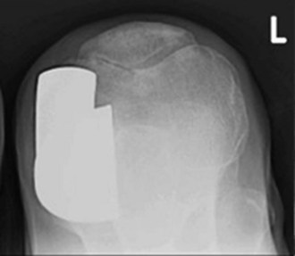
In the United States, perhaps the biggest area of debate is the status of the patellofemoral joint. Concern over the status of this joint in an otherwise suitable candidate for UKA is also unwarranted. It is this author’s opinion that the correction of varus deformity with preservation of the anterior cruciate ligament has a protective effect on the patellofemoral joint, even if there is significant arthritic disease present. Despite ignoring patellofemoral disease, the long-term results of Oxford UKA have not been affected by progression of patellofemoral disease or anterior knee pain (Fig. 16–1).
Results
To date we have performed over 1500 medial Oxford UKAs. At 6 years, the cumulative survivorship is better than 98%. We reported on 318 medial compartmental UKAs in 268 patients using the aforementioned Oxford indications.7 Isolated medial pain was present preoperatively in 211 knees (67%). Anterior knee pain was described in 20 knees (6%) and lateral pain described in 9 knees (3%). Posterior knee pain was described in 9 knees (3%), and in 65 knees (21%) the pain was described as global in nature. Follow-up averaged 8 months (range, 6 weeks to 28 months). There were six revisions in this group for an early survivorship of 98.1%. Survivorship in knees with preexisting radiographic signs of patellofemoral joint disease was no different than in those knees without patellofemoral disease. In fact, there was a trend toward a higher average Knee Society pain score in knees with preexisting patellofemoral joint disease (p = .052). The preoperative site of pain was not a significant predictor of survivorship or failure with the device, nor did preoperative location of pain influence postoperative knee scores.
Discussion
Kuipers et al. recently described factors associated with reduced early survivorship using the Oxford Phase 3 device.8 They noted that young age (<60 years) was the only variable associated with early revision, but that preoperative patellofemoral arthritis was not associated with decreased implant survivorship and the status of the patellofemoral joint should not be considered as an indication or contraindication when using this device. Beard et al. studied a consecutive series of 100 Oxford UKAs, specifically evaluating the role of preoperative anterior knee pain and patellofemoral arthritis on the outcomes.9 More than half the patients reported anterior knee pain preoperatively and yet no patients were revised at 2 years for patellofemoral pain or progression. Furthermore, 54% of patients had degenerative changes in the patellofemoral joint and, again, no patient failed from progression. Similar to our findings, the preoperative status of the patellofemoral joint did not affect the outcomes, unless bone-on-bone lateral facet disease was present with grooving and subluxation. In these cases, Beard et al. reported lower knee scores in this subset of patients. This severe lateral patellofemoral disease, while not associated with failure, did have a negative effect on outcomes, and its presence should be seen as a relative contraindication to medial UKA.
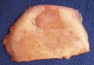
It appears that the mobile bearing, with its inset femoral component, may be more tolerant to patellofemoral disease than a polyradial/polycentric femoral component used in fixed-bearing UKA. Berger et al. described a rapid increase in second-decade patellofemoral symptoms with a fixed-bearing UKA design. At 10 years only 1.6% of patients were thought to have anterior knee pain, and this rose to 10% at 15 years.10 At 15 years, survivorship of this fixed-bearing design began to decline, predominantly due to patellofemoral progression, dropping from 98% at 10 years to 95.7% at 15 years.10 This is remarkably different than the second-decade survivorship seen with the medial mobile-bearing device. The Oxford group followed 824 consecutive UKAs performed between 1998 and 2005. Full-thickness cartilage loss was observed in the trochlea in 13% of knees and full-thickness loss in any area of the patellofemoral joint was seen in 16%. The severity of patellofemoral disease did not influence outcome in this series.11 It is hoped that the early success in the United States will mirror the excellent long-term results seen in other series. At 6 years, survivorship of better than 98%, despite ignoring the status of the patellofemoral joint, is being reported. The data clearly demonstrate that not only should anterior knee pain and patellofemoral disease be ignored, but that its presence preoperatively has no effect on outcome or failure (Fig. 16–2).
1 Kendrick BJ, Longino D, Pandit H, et al. Polyethylene wear in Oxford unicompartmental knee replacement: a retrieval study of 47 bearings. J Bone Joint Surg [Br]. 2010;92:367-373.
2 Price AJ, Short A, Kellett C, et al. Ten-year in vivo wear measurement of a fully congruent mobile bearing unicompartmental knee arthroplasty. J Bone Joint Surg [Br]. 2005;87:1493-1497.
3 Price AJ, Waite JC, Svard U. Long-term clinical results of the medial Oxford unicompartmental knee arthroplasty. Clin Orthop Relat Res. 2005;435:171-180.
4 Kozinn SC, Scott R. Unicondylar knee arthroplasty. J Bone Joint Surg [Am]. 1989;71:145-150.
5 Ritter MA, Faris PM, Thong AE, et al. Intra-operative findings of varus osteoarthritis of the knee: an analysis of pre-operative alignment in potential candidates for unicompartmental arthroplasty. J Bone Joint Surg [Am]. 2004;86:43-47.
6 Laskin RS. Unicompartmental knee replacement: some unanswered questions. Clin Orthop Relat Res. 2001;392:267-271.
7 Berend KR, Lombardi AVJr, Adams JB. Obesity, young age, patellofemoral disease, and anterior knee pain: identifying the unicondylar arthroplasty patient in the United States. Orthopedics. 2007;30(Suppl 5):19-23.
8 Kuipers BM, Kollen BJ, Bots PC, et al. Factors associated with reduced early survival in the Oxford phase III medial unicompartmental knee replacement. Knee. 2010;17:48-52.
9 Beard DJ, Pandit H, Gill HS, et al. The influence of the presence and severity of pre-existing patellofemoral degenerative changes on the outcome of the Oxford medial unicompartmental knee replacement. J Bone Joint Surg [Br]. 2007;89:1597-1601.
10 Berger RA, Meneghini RM, Sheinkop MB, et al. The progression of patellofemoral arthritis after medial unicompartmental replacement: results at 11 to 15 years. Clin Orthop Relat Res. 2004;428:92-99.
11 Beard DJ, Pandit H, Ostlere S, et al. Pre-operative clinical and radiological assessment of the patellofemoral joint in unicompartmental knee replacement and its influence on outcome. J Bone Joint Surg [Br]. 2007;89:1602-1607.
[/level-membership-for-orthopaedics-category][not-level-membership-for-orthopaedics-category]
CHAPTER 16 The Patella in Medial Unicompartmental Knee Arthroplasty
The premise behind Oxford criteria is that UKA is utilized to treat anteromedial osteoarthritis. When these criteria are present, other unnecessary contraindications such as age, weight, and the status of the patellofemoral articulation can be safely ignored without affecting the outcome of Oxford UKA.Introduction
Unicompartmental knee arthroplasty (UKA) or partial knee replacement represents the ultimate minimally invasive procedure for treating degenerative joint disease of the knee in the appropriately indicated patient. The conservative nature of the procedure stems from the bone conservation, the preservation and correction of normal ligamentous structures, and the restoration of normal knee kinematics. These outcomes cannot be achieved with any total knee arthroplasty (TKA). By incorporating a fully congruent, meniscal bearing, the Oxford UKA (Biomet, Inc., Warsaw, IN) has demonstrated some of the lowest wear rates seen with any knee implant in both retrieval studies and RSA studies, averaging 0.02 mm per year in both.1,2 Thus, wear does not appear to be a limiting factor in the indications for or contraindications against UKA using this device. The fully congruent articulation provides a larger contact area and reduces the contact stresses, leading to significantly decreased wear when compared to fixed bearing, noncongruent designs. Other design-specific differences, such as an inset, spherical femoral component, may specifically address the patellofemoral joint problems seen with many fixed-bearing UKA designs. The long-term outcomes that have been published using this device make it one of the most successful devices available today for the treatment of medial compartmental disease of the knee and therefore make this device the logical choice for UKA. Survivorship rates out to 15 years and 20 years have been 95% and 92%, respectively, in an independent series.3
The indications for UKA have been debated widely over the past several decades. Classical indications, when followed closely, have included only a very small percentage of degenerative knees. The Kozinn and Scott criteria limit the utilization of UKA to those patients older than 60 years, weighing less than 82 kg, and exclude active individuals. Additionally, these classical indications exclude cumulative deformity of more than 15°, any fixed flexion contracture of more than 5°, and more than minimal arthritic changes in the patellofemoral joint.4 With these indications, only between 2% and 12% of arthritic knees would meet the criteria for UKA.5,6 The so-called Oxford criteria are somewhat more liberal and encompass an anatomic and radiographic philosophy for inclusion or exclusion of patients. The premise behind these expanded criteria is that UKA is utilized to treat a specific disease entity: anteromedial osteoarthritis. Anteromedial disease is defined as the combination of full-thickness cartilage loss in the medial compartment, a correctible intra-articular varus deformity, and preserved lateral joint space seen with a valgus stress radiograph. By definition, these criteria are only present when the ligaments are functionally intact and the disease is confined to the anterior one third or two thirds of the tibial plateau. When these criteria are present, other unnecessary contraindications such as age, weight, and the status of the patellofemoral articulation can be safely ignored without affecting the outcome of Oxford UKA.
[/not-level-membership-for-orthopaedics-category]
