The neurological examination: Peripheral nervous system

Published on 12/04/2015 by admin

Filed under Neurology

Last modified 22/04/2025

Print this page

rate 1 star rate 2 star rate 3 star rate 4 star rate 5 star
Your rating: none, Average: 0 (0 votes)

This article have been viewed 3138 times

4 The neurological examination

Peripheral nervous system

Examination of the peripheral nervous system assumes a stylised approach when adopting the traditional method. This means following a set pattern of: observation and inspection; tone; power; reflexes; sensation; coordination; and gait. This ignores what was stated earlier, namely that the consultation starts long before the patient enters the consultation room.

The first part of testing of the peripheral nervous system is observation, namely observing how the patient gets out of the chair and walks into the room. Difficulty rising from the chair may suggest Parkinson’s disease. Not swinging one arm when walking may suggest either pyramidal disease, such as a stroke, or may also suggest Parkinson’s disease. Spastic posturing (flexed upper limb and extended lower limb) with circumduction of affected lower limb suggests upper motor neurone damage, such as stroke. Stooped posture, shuffling gait with festination, hesitancy and inertia of gait initiation are almost diagnostic of Parkinson’s disease, and are apparent long before the consultation formally commences.

Ataxic, wide-based gait is suggestive of cerebellar disease. Wide-based gait is far less common than one would think. The width between the feet is usually wider than the shoulders of the patient and gives the impression that the patient is drunk—hence the statement that the patient walks like a drunken sailor. Wide-based gait provides stability of stance, and sailors used to moving ships often walk with such wide-based gait to maintain stability on a ship.

Before undergoing the formal peripheral assessment it is worth testing for grasp reflexes. This is done by distracting the patient, possibly by engaging in ‘small talk’, and while doing so sliding the hand out, pushing up against the patient’s palm and fingers. Grasp reflex may be as subtle as feeling the fingers of the patient flexing downwards towards the examiner’s sliding fingers. A positive grasp reflex is indicative of contralateral frontal lobe (upper motor neurone) damage. Other frontal lobe signs may include palmar–mental response. This is evoked by applying a noxious stimulus (such as scratching) to the palm of the patient’s hand and observing the movement of the chin (mentalis muscle) on the same side as the scratched hand. This again suggests contralateral frontal lobe damage.

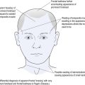
The glabellar tap is elicited by tapping the index finger on the patient’s forehead. It is best achieved by holding the hand above the forehead and tapping the forehead without coming front-on as this movement, equivocal to menace (as described when testing visual fields in Ch 2), may itself evoke a blink response. When testing the glabellar tap the normal response allows up to three blinks. More than three blinks represent a positive response. When testing glabellar tap it is worth reappraising the patient for ptosis, as attention is focused on the eyelids.

Much of the above actually tests central nervous system function, but it is best examined at this point in patient assessment. This uses the peripheral to test central competence.

While the approach adopted has been compartmentalised, this has been done for ease of understanding. An additional consideration has been to instil time-efficient consultation methods: all busy doctors are ‘time poor’ and anything that can improve clinical acumen and save time is of value.

Tone

Tone is tested by a number of different methods. Initial resistance, followed by a feeling of giving way, is called ‘clasp knife’ as it is similar to opening the blade of a pocketknife. This is found with spasticity in upper motor neurone damage.

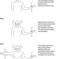
If Parkinson’s disease is considered, then the initial testing of tone by moving the hand up and down at the wrist should be normal early in the disease process. The patient is asked to move their head from side to side, pushing the ear into the shoulder with each turn, at which time the tone increases with ‘cogwheeling’ and ‘lead pipe’ rigidity. This is pathognomic of early Parkinson’s disease.

If the patient is examined soon after the onset of a stroke, the hemiplegic limbs may be totally flaccid and floppy. After longstanding upper motor neurone damage, the hyper-tonicity may progress to clonus. This is evoked by short, sharp joint movement, such as sudden sharp dorsiflexion of the foot, similar to the initial provocative jerking movement. The same may be caused by jerking the patella bone towards the foot in the extended knee. This may evoke repeated movement of the knee bone up and down in a similar fashion to that seen at the foot. Clonus may be evoked at other joints, such as the wrist, but lower limb clonus is more common.

Cerebellar damage does not cause increased tone which is often decreased, causing pendular movements on testing. This is often difficult for the inexperienced to discern. While increased or decreased tone is relatively easy to identify, often the subtleties of tone are not that easy to identify for the doctor not attuned to the neurological examination.

Power

Various conditions affect different muscles. Upper motor neurone weakness, as with stroke, affects the antigravity muscles. Most inexperienced doctors have difficulty remembering which muscles constitute the antigravity muscles (see Fig 4.1).

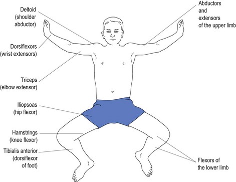
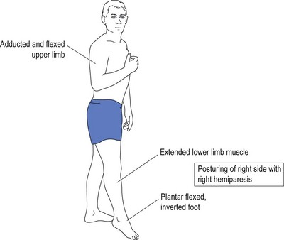
It is because of weakness of the antigravity muscles that the patient with upper motor neurone damage presents with flexion of the upper limbs and extension of the lower limbs. With weakness of the antigravity muscles, the opposing muscles exert their effect causing posturing without counter effect by opposing muscles (see Fig 4.2).

To make it simpler for the doctor, it is easier to remember just one group of muscles, such as the extensors in the upper limbs. It is even easier to remember just one upper limb muscle, such as the triceps, which is an extensor muscle. This will reinforce extensors in the upper limbs and the opposite, namely the flexors with lower limbs. Alternatively, the doctor may choose to remember just a single flexor in the lower limb, such as the hip flexor, the iliopsoas.

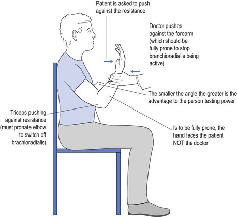
It is obvious that a 60 kg female may have trouble overcoming the strength of a 100 kg male who is engaged in very physical work. One way to overcome this is to use mechanical advantage to test power. An example of this might be to test triceps power by flexing the elbow much more than the traditional 90° (see Fig 4.3).

Power is graded on a five-point scale:

Some examiners try to add further sophistication (for example 4+ or 4−) but this is less universally accepted.

Endocrine disorders, such as excessive exposure to steroids, may cause proximal weakness. Some myopathies specifically target certain muscles, such as facio-scapular humerus muscles. Other diseases, like Charcot-Marie-Tooth, may cause wasting of calf muscles, giving the appearance of inverted champagne bottles in the lower limbs. It helps to remember some of these patterns when performing the neurological examination.

Weakness may be restricted to particular innervated muscles with mononeuritic disease. An example of this is specific weakness of abductor pollicis brevis (APB), flexor pollicis brevis (FPB), opponens pollicis and the lateral two lumbricals with median nerve entrapment and carpal tunnel disease.

Reflexes

Upper motor neurone weakness causes hyperreflexia. By far the most common cause of hyperreflexia is anxiety with excess adrenaline. When reflexes are particularly brisk, the tapping of a reflex with a tendon hammer may be sufficient to provoke clonus, as described above.

The basis of the deep tendon reflex is to stretch receptors within the muscle. This causes the muscle to contract, thereby resulting in the jerk of the joint being tested. The way that clonus is provoked is as a consequence of increased tone within the muscle. The single jerk that normally occurs when tapping a reflex is enough to cause another jerk to happen because of the hypertonicity of the tight muscle. This is repeated until the size of the resultant movement is too small to evoke a further jerk response.

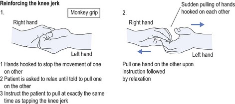
Some anxious patients are too ‘uptight’ to allow the free movement/jerk of the muscle. The patient needs to relax, and the best way to encourage this is to distract the patient. Ask the patient to adopt the finger clasp known as a ‘monkey grip’ (see Fig 4.4). The patient is asked to close the eyes and relax, and to only pull on the grip when told to do so. This instruction is given as the reflex is tapped. Some neurologists ask the patient to clench the teeth at the exact same time as tapping the reflex. This is another method of distraction.

It is more difficult to test upper limb deep tendon reflexes in a patient who is stressed (and uptight) and who is ‘splinting’ their muscles. To test the right upper limb deep tendon reflexes, the patient is given an object to hold in the left hand. The patient is asked to squeeze the object at exactly the same time as the deep tendon is tapped with the hammer, or more correctly just as the swing of the hammer starts. The sides are reversed when testing left upper limb reflexes. Again, clenching the teeth tightly at the same time as swinging the tendon hammer to tap the reflex is an alternative method of distraction. This is often best achieved with the patient’s eyes closed so they cannot anticipate the tap of the hammer.

Deep tendon reflexes represent specific spinal root levels:

It is difficult to be sure that the tendon hammer can be appropriately targeted at the biceps jerk. To assist with this and ensure that the focus of stimulus is correctly directed at the biceps tendon in the elbow, the doctor places a thumb over the biceps tendon at the elbow and hits their thumb, thereby ensuring that the immediate jerk is evoked over the tendon.

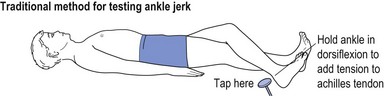
Ankle jerks are difficult to elicit if one is not sure how to do it. If the patient is lying down then the foot is drawn across the opposite shin and the Achilles tendon is tapped (see Fig 4.5).

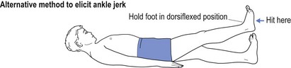
An alternative method is to dorsiflex the ankle joint and then tap the sole of the foot (see Fig 4.6).

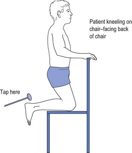
If both these methods fail, then the patient may be asked to kneel on a chair while the Achilles tendon is tapped (see Fig 4.7). This way the patient is distracted by kneeling on the chair and cannot predict when the ankle jerk will be tested, thus reducing the propensity of ‘splinting’.

The Babinski response is often misunderstood. Most people focus on the great toe, but for subtle response the examiner should focus on the little toe. Rather than scratch the sole of the foot, my preference is to stimulate the lateral border of the foot and see if the little toe moves medially (joining the other toes as they all move in a downward direction) or moves laterally (away from the other toes) in a splaying motion as the great toe moves up. Upper motor neurone lesions cause the toes to splay, followed by upward movement of the great toe if it is sufficiently positive. The easiest way to understand this is to consider that the Babinski response is the foot either opening or closing, the equivalent of a fist. When a hand closes to make a fist all the fingers come together and the last action is the thumb wrapping around the front of the fist. Equating the thumb with the great toe, the thumb is the last digit of the hand to move—as is the great toe with the Babinski response.

The role of other reflexes, such as the cremasteric reflex (L1), abdominal reflexes (T6–T12) or adductor reflexes of the lower limb (L2), are all relevant to the neurological examination but they are subtle tests generally extraneous to the needs or role of the general practitioner.

Sensation

While neurologists test light touch, temperature, pain, position (proprioception) sense, vibration sense and two-point discrimination, the routine neurological examination by the general practitioner may be limited to light touch (which travels together with pain and temperature) in the lateral spinothalamic tracts of the spinal cord and joint position (which travels with vibration sense) in the posterior columns of the cord.

It is easier to work from the area of impaired sensation towards the area of increased sensation. As sensory loss usually starts in the periphery, it follows that light touch stimulation should start at the feet or hands and move centrally up the limbs to the torso until the patient reports increased perception of the stimulus.

Fingers are as useful as cotton wool to test light touch as we know how heavily we place our fingers, and hence we can better gauge the stimulus applied when testing light touch. If there is greater need for subtlety when testing light touch, then personal preference is to use a small paintbrush because the handle of the small brush lends itself to tighter manipulation and control than does holding a wisp of cotton wool.

While many advocate using a pin or needle to test pain, it is difficult to apply consistent pressure with a pin to be sure that the patient is responding to the same stimulus each time. A simple tool that allows consistent and acceptable stimulation is a serrated edge haberdashery tracing wheel rolled from the foot, or hand, up the limb until a change in perception is recognised. As stated earlier, pain travels a similar pathway to light touch and hence it needs to be tested only if the history dictates a need for more accurate sensory demarcation, which may be better defined with painful stimulation.

Similarly, temperature perception accompanies light touch and pain pathways, and thus only needs testing in more subtle cases. Use of a cold metal object, such as a tuning fork, may be adequate for the crude testing of temperature perception and follows a similar pattern, moving from the periphery, centrally. The patient may report the cold metal suddenly feels colder, thereby establishing a level if there is ‘glove and stocking’ hyposensitivity as occurs with peripheral neuropathy.

For vibration sense testing, the tuning fork should vibrate more slowly than is the case for testing hearing. A 128 Hz cycle frequency, the C note one octave below middle C on the piano, is the ideal tuning fork to test vibration sense. Vibration sense accompanies joint position sense (proprioception) in the posterior columns of the spinal cord. Testing vibration sense is a comparative test in which the patient compares perception of vibration to areas thought to be normal. Again it behoves the examiner to move from the periphery. Obviously the doctor needs to have an appropriate tuning fork to test vibration sense and this is not always the case. Joint position testing also evaluates posterior column integrity.

Joint position should be tested moving from the periphery centrally, namely the great toe before the ankle, before the knee, before the hip or, in the upper limb, finger, before the wrist, before the elbow, before the shoulder. The great toe is held between index finger and thumb (held on the sides of the toe rather than the top and bottom). The reason for holding the sides is to avoid additional position sense cues, as may be provided by pushing up and down with pressure on the top or bottom of the toe. A similar approach can be adopted with other joints if the patient cannot identify upward/downward movement of the joint, starting at the periphery, namely great toe or little finger.
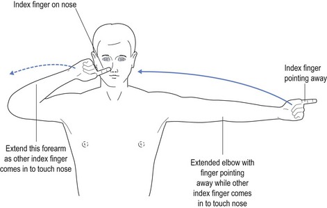
It is important to remember that spinothalamic sensation (pain, light touch and temperature) travels up two to three segments above where clinical examination suggests the level to be. It then crosses to the other side of the spinal cord at this level two to three segments above where sensory changes are detected. This means a right-sided spinal cord lesion at this level will result in altered pain, light touch and temperature perception on the left from about two to three segments below the lesion. Thus, a level at T10, the umbilicus, on clinical testing suggests a spinal cord lesion at T7 or T8.

Alternatively, posterior column sensation (such as joint position sense, vibration sense and possibly two-point discrimination) travels up the posterior columns of the spinal cord to the brainstem where they cross over in the medullar oblongata. It follows that spinal cord lesion on the right side of the cord, affecting the right side posterior columns, will result in loss of joint position sense and vibration sense on the right side (the same side as the lesion). Appreciation of these points is important when evaluating hemicord lesions, as may occur with Brown-Séquard’s syndrome.