The maxillary antra
Introduction
The maxillary antra, because of their close proximity to the upper teeth, are the most important of the paranasal sinuses in dentistry. Nowadays the imaging modalities of choice to investigate possible disease within the antra are computed tomography (CT), cone beam CT (CBCT) and magnetic resonance (MR) (see Chapters 16 and 18). However, as the antra are often imaged on conventional dental radiographs, as well as on certain skull views, clinicians still need to know:
• The anatomy of the antra, including their shape, size, normal variations and related structures.
• How the antra are represented normally on conventional dental and skull radiographs and how these radiographs are assessed.
Similar radiographic changes can be seen in the other paranasal sinuses – frontal, ethmoidal and sphenoidal. They are of less clinical relevance in dentistry and are discussed only briefly.
Normal anatomy
The main anatomical parts of the antra (see Fig. 31.1) can be divided into:
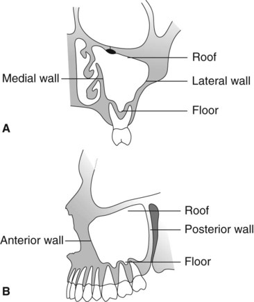
• A roof or upper border, bounded by the orbit
• A medial wall, bounded by the nasal cavity
• A posterior wall, related to the pterygopalatine fossa
• A lateral wall, related to the zygoma and cheek
• An anterior wall, related to the cheek
• A floor, related to the apices of the upper posterior teeth.
Normal appearance of the antra on conventional radiographs
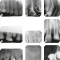
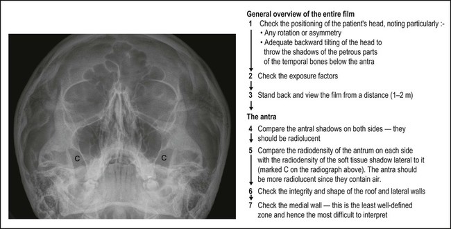
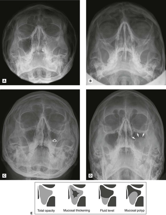
An antrum appears radiographically as a radiolucent cavity in the maxilla, with well-defined, dense, corticated radiopaque margins or walls. In general, the larger the cavity the more radiolucent it will appear. The internal bony septa and blood vessel canals in the walls all produce their own shadows. The thin epithelial lining is not normally seen. The different parts of the antra shown on conventional dental and skull radiographs are summarized in Table 31.1. Typical normal radiographic appearances are shown in Figs 31.2–31.4. In addition, a suggested systematic approach to viewing the antra on the 0° OM is shown in Fig. 31.4.
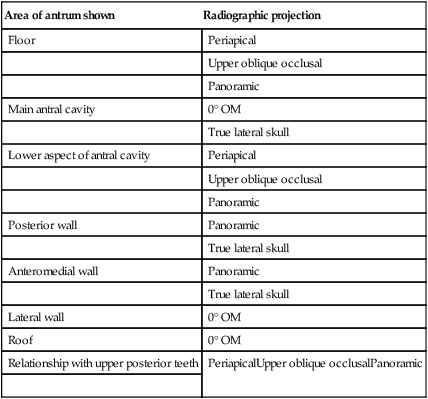
Table 31.1
Summary of the different parts of the antra shown on conventional dental and skull radiographs
| Area of antrum shown | Radiographic projection |
| Floor | Periapical |
| Upper oblique occlusal | |
| Panoramic | |
| Main antral cavity | 0° OM |
| True lateral skull | |
| Lower aspect of antral cavity | Periapical |
| Upper oblique occlusal | |
| Panoramic | |
| Posterior wall | Panoramic |
| True lateral skull | |
| Anteromedial wall | Panoramic |
| True lateral skull | |
| Lateral wall | 0° OM |
| Roof | 0° OM |
| Relationship with upper posterior teeth | PeriapicalUpper oblique occlusalPanoramic |
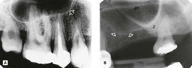
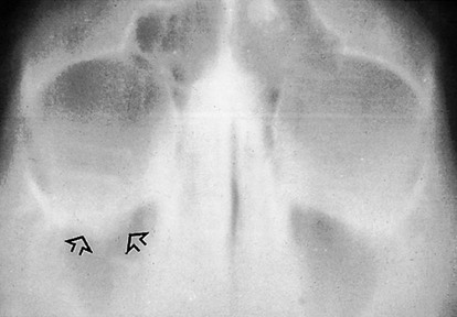
 showing the usual appearance of the floor (arrowed) and base of the antral cavity in relation to the upper posterior teeth in a dentate adult. B Periapical of
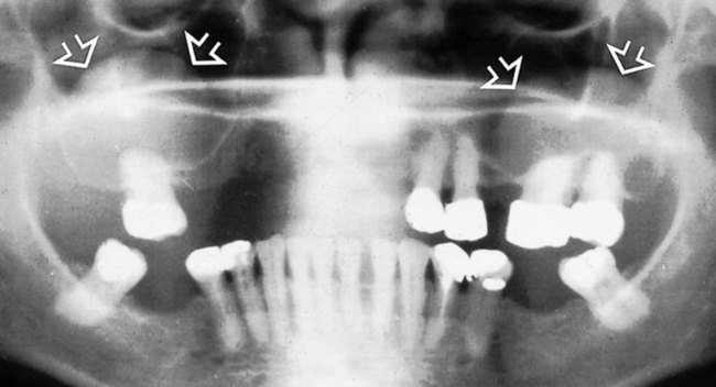
  of a partially dentate adult showing various normal anatomical structures. These include the floor of the antrum (open white arrows), the hard palate/floor of the nasal cavity (open black arrows) and the zygomatic buttress (Z).
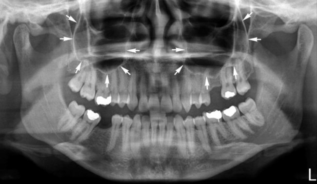
Antral disease
The major pathological conditions that can affect the antra, directly or indirectly, include:
These various disease entities can result in the following radiological changes:
• Total or partial opacity/obliteration of the main antral cavity
• Alteration in the integrity of the antral walls, including discontinuity as a result of a fracture or destruction by an intrinsic or extrinsic tumour
• Alteration in the antral outline, including expansion or compression caused by an intrinsic or extrinsic lesion or disease process
Investigation and appearance of disease within the antra
• Computed tomography (CT) – currently recommended by the Royal College of Radiologists in the UK
• Multidirectional (spiral) tomography
Appearances of antral diseases using various imaging modalities are shown in Figs 31.5–31.23.
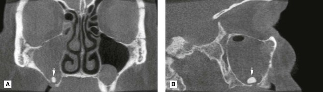
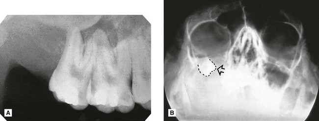
 showing a discontinuity of the antral floor (arrowed) following the extraction of the molar tooth that resulted in a clinical oro-antral communication. Note this break in continuity of the floor is not always evident in cases of oro-antral communication.
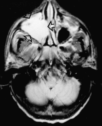
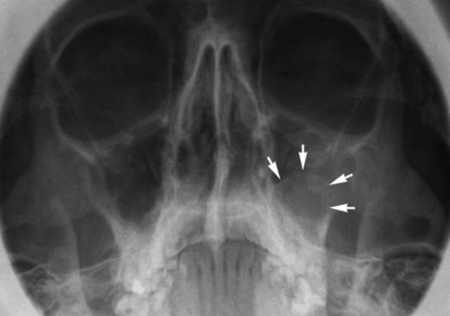
 which has displaced the antral floor.
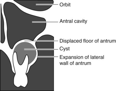
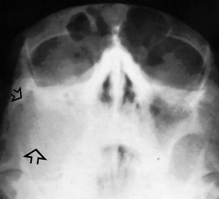
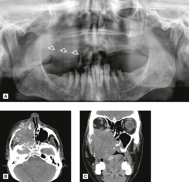
 A Periapical of the upper right posterior teeth. Note the lack of antral floor outline and absence of
  B Occipitomental of the same patient showing total opacity of the right antral region with no evidence of the lateral antral margin. The displaced upper wisdom tooth is evident underneath the orbit (outlined and arrowed).
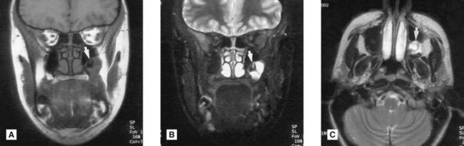
Infection/inflammation
Acute sinusitis
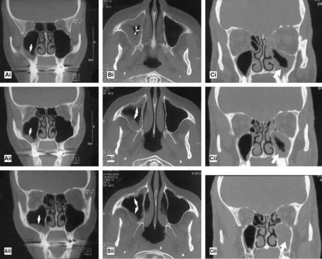
Chronic sinusitis (Figs 31.5–31.9)
Main radiological features
• Total/partial opacity within the antral cavity
• Fluid level – usually in the base of antral cavity with a characteristic meniscus shape
• Round, domed opacity produced by a mucosal polyp
• Occasionally an increase in the bony antral walls
• Typical inflammatory changes if infected teeth are involved – this may lead to resorption and remodelling to produce the appearance described as antral halo (see also Ch. 21 and Fig. 31.8).
Trauma
Oro-antral communication
Main radiographic features
• Break in the continuity of the floor may be evident (see Fig. 31.10) – however, the diagnosis of an oro-antral communication is made clinically, not radiographically, since the defect in the floor of the antrum may not be evident on the two-dimensional radiograph
• Characteristic features of acute or chronic sinusitis owing to the ingress of bacteria
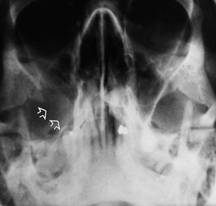
• Evidence of the displaced root or tooth – a second view of the antrum with the head in a different position or CBCT may be required to ascertain the exact location of the displaced object (see Fig. 31.5).
Fractures of the maxillofacial skeleton
Fractures are discussed in detail in Chapter 29 and only a brief summary is shown below.
Main radiographic features
• Break in the continuity of one or more of the antral walls depending on the type of fracture.
• Total opacity or fluid level within the antral cavity caused by haemorrhage (see Figs 31.11 and 31.12).
• Features of sinusitis if subsequent infection develops (this is in fact surprisingly rare).
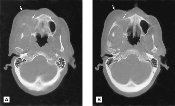
• The orbital blow-out fracture classically produces a tear-drop-shaped opacity in the upper part of the antrum, the hanging drop appearance, caused by herniation of the orbital contents downwards into the antrum following collapse of the antral roof (see Fig. 31.13). The infraorbital margin remains intact. See also Fig. 29.30.
Cysts
The more important cysts that can affect the antra include:
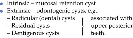
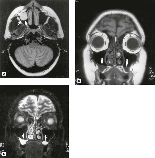
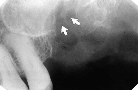
Mucosal retention cyst
Main radiographic features (see Figs 31.7 and 31.9B)
Odontogenic cysts
These cysts are extrinsic to the antra developing in the alveolar bone beneath the antral floor.
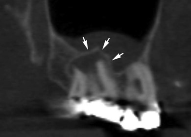
Main radiographic features of a small cyst
• Round, dome-shaped opacity in the base of the antrum with a well-defined, radiopaque corticated margin to the edge of the meniscus, i.e. the odontogenic cyst has a bony margin and so can be differentiated from the soft tissue mucosal retention cyst or antral polyp (see Figs 31.15 and 31.16)
Main radiographic features of a large cyst
• Total opacity of the antral region owing to complete compression of the antral cavity
• Sometimes displacement of the associated tooth (see Figs 31.17 and 31.18).
Note: Subsequent marsupialization or decompression of the cyst will usually result in the reformation of the antral cavity.
Tumours
Benign intrinsic tumours
These are all rare but can include:
• Papilloma – this produces, radiographically, a well-defined, non-specific, soft tissue opacity within the antrum
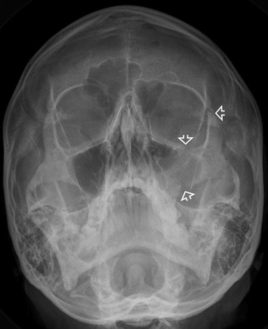
• Osteoma – this produces, radiographically, a well-defined, round or lobulated, homogeneous, densely opaque mass within the antrum. The osteoma is most common in the frontal sinus.
Malignant intrinsic tumours
Main radiographic features of a large well-established lesion
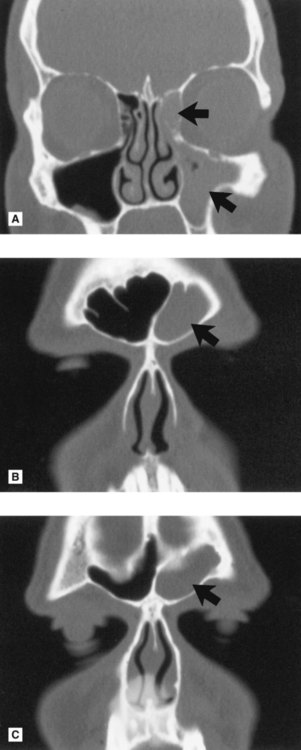
• Total opacity of the antral cavity (see Fig. 31.19) – in the absence of symptoms suggesting infection, or a history of trauma, a totally opaque antrum is a cause for serious concern and further investigation is necessary.
• Destruction of one or more of the antral walls (see Fig. 31.20) and invasion of surrounding hard and soft tissues – hence the need for CT
Other paranasal air sinuses
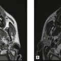
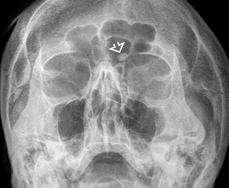
As mentioned earlier, the frontal, ethmoidal and sphenoidal air sinuses are of limited importance in routine dentistry. Many of the conditions described in relation to the maxillary antra can affect these paranasal sinuses and produce similar effects. Occasionally routine skull radiography reveals unusual abnormalities, such as the ivory osteoma in the frontal sinus (see Fig. 31.23). But generally the investigation of choice for the paranasal air sinuses is computed tomography (CT), as shown in Fig. 31.24.
To access the self assessment questions for this chapter please go to www.whaitesessentialsdentalradiography.com