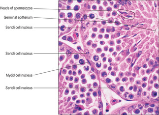
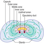
The male reproductive system

Published on 02/03/2015 by admin

Filed under Basic Science

Last modified 02/03/2015

Print this page

rate 1 star rate 2 star rate 3 star rate 4 star rate 5 star
Your rating: none, Average: 0 (0 votes)

This article have been viewed 1748 times

The male reproductive system comprises paired testes, associated glands, ducts and the penis (Fig. 15.1). The testes are the male gonads and are the site of production of spermatozoa (the male gametes). Each spermatozoon (in humans) has 23 chromosomes containing the haploid amount of DNA. Additionally, testes produce and secrete a group of steroid hormones, the male sex hormones (androgens), of which testosterone is the main type. The glands that are associated with the male reproductive system are the paired seminal vesicles, the prostate and paired bulbourethral glands. All these glands contribute fluid secretions which support spermatozoa and form semen. The ducts form the reproductive tract and transport spermatozoa and secretions from the testes and glands to the urethra. The urethra is shared by the reproductive and urinary systems and it passes from the bladder through the prostate and penis and opens at a meatus at the glans penis. The urethra transports urine from the urinary bladder as it is emptied at micturition. The penis is also the organ of copulation able to deliver spermatozoa, in semen, into the vagina of the female reproductive tract.

Testes

Each testis is an organ suspended within a pouch of skin known as the scrotum. The testes thus lie outside the abdominal cavity and this ensures that spermatogenesis (the formation of spermatozoa) occurs at a temperature slightly below body temperature, a requirement for successful gamete production. Each testis is surrounded by a tough connective tissue covering known as the tunica albuginea. Connective tissue septa from the tunica albuginea penetrate into the substance of each testis and divide it into lobules. One septum forms a wedge which penetrates the posterior surface of each testis.

Seminiferous tubules

Located within the lobules of each testis are coiled seminiferous tubules which are the sites of spermatogenesis. Seminiferous tubules are lined by several layers of epithelial cells forming a germinal (seminiferous) epithelium (Fig. 15.2). There are two types of cell in the germinal epithelium of adults: spermatogenic cells, which are in layers, and Sertoli cells, which form a single layer and are supportive of the spermatogenic cells (see below). The germinal epithelium lies on a basement membrane and is surrounded by a thin layer of loose connective tissue. This connective tissue supports blood vessels (Fig. 15.2), lymph capillaries, nerves and Leydig (interstitial) cells (Fig. 15.3) which secrete androgens (see below). Myoid cells (Fig. 15.4) are closely adjacent to seminiferous tubules and are capable of contraction, thus helping to move the spermatozoa along tubules and away from where they develop.
After puberty, spermatogenesis begins as spermatogenic cells undergo meiosis and differentiation in a cycle of events that result in the production of spermatozoa, a process which takes about 65days in humans. The stages of spermatogenesis take different lengths of time to complete and, as a result, not all the stages are present in the layers of cells in any one region of the germinal epithelium. The appearance of seminiferous tubules depends on the stages the spermatogenic cells have reached in forming spermatozoa and on the angle at which they are sectioned (Figs 15.2 and 15.5).

Spermatogenic cells

The formation of spermatozoa (spermatogenesis) may be divided into three stages: mitosis, meiosis and spermiogenesis. These stages begin at about puberty and continue throughout life.

Mitosis. In this stage, stem cells (spermatogonia) at the base of the germinal epithelium undergo regular cycles of mitosis and produce new diploid cells, i.e. cells containing the normal number of chromosomes (46 in humans, comprising 22 homologous pairs and two sex chromosomes). Some of the new cells formed by mitosis remain in the basal layer as spermatogonia, others move towards the lumen and enter meiosis.
Meiosis. At the start of this phase the cells begin to enlarge and are known as primary spermatocytes. They undergo an S phase when they replicate the DNA in their chromosomes prior to cell division (Chapter 2). They then enter the prophase of the first division in meiosis (Fig. 15.6), a process which lasts about 20days. During this time, chromosomes begin to coil and condense (and stain densely) and homologous pairs of chromosomes move close together and exchange lengths of DNA. This exchange introduces genetic variation to the gametes produced.
The first meiotic division of meiosis (also known as the reduction division) continues into metaphase. Each pair of homologous chromosomes in each primary spermatocyte line up together on the same part of the equator of the spindle of the metaphase plate, prior to cell division. (This contrasts with mitosis, where each chromosome attaches to a different part of the equator of the spindle.) At this stage, the chromosomes are at their most condensed and densely stained and are readily identified in histological sections (Fig. 15.7). The paired chromosomes then move away from each other in anaphase and telophase. The result of this reduction division is the production of two secondary spermatocytes each with only half the original number of chromosomes (i.e. 23 in humans). (This reduction division introduces further genetic variation to the new cells as maternally and paternally derived chromosomes in the homologous pairs are randomly arranged on the spindle, and therefore randomly distributed between the two offspring cells.)
Separation of cytoplasm during meiosis is incomplete and spermatids derived from one spermatogonium are partially joined together. This is thought to aid synchrony of their progression through the next stage (spermiogenesis). Groups of newly formed, related spermatids appear as clusters of small, spherical cells near the luminal surface of seminiferous tubules (Fig. 15.6).

Spermiogenesis. During this stage, spermatids differentiate and each forms one spermatozoon. Approximately 200 million spermatozoa are produced daily by the testes in humans. During spermiogenesis the spherical spermatids change in form and shed cytoplasm as they become highly differentiated spermatozoa characterised by a head, middle region and tail (Fig. 15.8). As spermatozoa develop, the head regions remain in the epithelium but the tail regions extend into the lumen of the seminiferous tubule (Fig. 15.5). Eventually, spermatozoa pass into the lumen and fluid secreted by Sertoli cells (see below) aids their transport away from the seminiferous tubules.
The head of each spermatozoon contains the nucleus in which the DNA in the chromosomes is condensed and stains densely with bases such as haematoxylin (Figs 15.4, 15.5 and 15.6). At one end of the nucleus of a developing spermatozoon a head ‘cap’ develops known as the acrosome (Fig. 15.8). The contents of the acrosome include hydrolytic enzymes that take part in the acrosome reaction, which is the process by which the head of a spermatozoon penetrates the cells and structures surrounding the oocyte prior to fertilisation (Chapter 16

Buy Membership for Basic Science Category to continue reading. Learn more here