The eyes and visual system

Published on 09/04/2015 by admin

Filed under Neurology

Last modified 22/04/2025

Print this page

rate 1 star rate 2 star rate 3 star rate 4 star rate 5 star
Your rating: none, Average: 0 (0 votes)

This article have been viewed 1839 times

The eyes and visual system

An examination of the eyes and the visual system can prove helpful in patients with no visual or ocular symptoms. This section will first describe examination of the eye generally, then examination of the pupils, visual function, acuity, visual fields and fundoscopy. Eye movements are discussed on pages 1819.

General examination of the eye

Ptosis is common and is often missed (Table 1). Partial ptosis is usually associated with unilateral overactivity of the frontalis. Ptosis is not a feature of facial nerve palsy (with a facial nerve palsy the eye does not close).

Table 1 Ptosis

Ptosis Causes Features
Neurogenic

Neuromuscular junction Myasthenia gravis Variable ptosis that fatigues, may be associated diplopia and facial weakness Myopathic Myopathies, especially myotonic dystrophy Usually symmetrical. Features of associated muscle disease Mechanical Aponeurotic dehiscence (common in the elderly) The tarsal plate is separated from the levator muscle

Exophthalmos is usually a feature of hyperthyroidism but may indicate orbital disease. Enophthalmos is a feature of Horner’s syndrome. Lid retraction is seen in hyperthyroidism and in rare upper brain stem lesions.

Remember false eyes can be cosmetically effective – a pitfall in exams.

Examination of the pupils

First look at the pupils. Are they the same size? Examination then aims to assess the afferent (optic nerve) and the efferent (parasympathetic via 3rd nerve constricting and sympathetic dilating) elements of the pupillary reflexes.

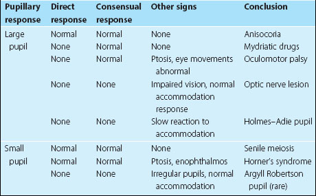
Ask the patient to look into the distance and shine a light twice in each eye in turn. First, look at the response in the eye into which you are shining the torch (the direct response), and then at the response in the other eye (the consensual response). Then ask the patient to look at your finger held 15 cm from the patient’s face and look at the pupils for brisk constriction – the accommodation response. If no response is obtained from shining a light into the eye, but there is a normal response on accommodation, this is called an afferent pupillary defect and indicates significant optic nerve disease. Other abnormalities are summarized in Table 2.

When a bright light is shone into the eye with an intact optic nerve, there is a brisk constriction of both pupils. When an optic nerve is partly damaged, the constriction to the same stimulus is less brisk. If the light is moved rapidly from a normal eye to one with an abnormal optic nerve, the pupil of the damaged eye continues to dilate. This is because the relatively weaker stimulus to constriction transmitted by the impaired optic nerve does not overcome the relaxation following a powerful stimulation on the other side. This is called a relative afferent pupillary defect.

Examination of visual fields

The visual fields are described from the patient’s point of view. When looking out of the right eye, the right half of the vision is the temporal field and the left the nasal field, and vice versa from the left eye.

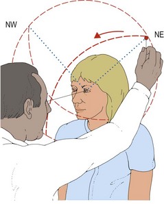
The visual fields are examined by covering one of the patient’s eyes, asking the patient to fixate on your opposite eye and then bringing an object in from the periphery to find the edge of the patient’s vision (Fig. 1). This is done slightly differently depending on the type of object at which the patient is asked to look. The visual field for a large moving object is normally much larger than for a small red object. It is easiest to screen the visual fields using a crude stimulus, for example a wiggling finger, and then refine any defect with a smaller object, usually a small, red hat-pin head.

More detailed examination

Keeping the red pin in a plane midway between yourself and the patient, bring the pin in from the same directions, as described above, starting outside your own field for the red object. Ask the patient to tell you when the object is seen as red.

Ask the patient to compare the red colour each side of the point of fixation. Test for the blind spot. This is in the temporal field about 15° from the point of fixation. This should be quite difficult to find and be the same size as your own blind spot. If patients complain of holes in their visual field, it is easiest to ask them to find them for you. Holes in the central vision are called scotomas, and are described according to their shape and position.

Examination of the visual field can be recorded using manual or automated perimeters (e.g. Goldman or Humphrey perimeters).

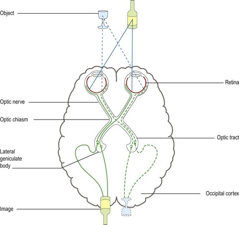
The location of a lesion producing a field defect can be deduced from the anatomy and organization of the visual system (Fig. 2; Table 3) (see p. 56).

Table 3 Field defects

  Site of lesion Possible associations
Unilateral visual field loss Optic nerve or retina Loss of pupillary responses, reduced acuity
Bitemporal hemianopia Optic chiasm Pituitary abnormalities, hypersecretion with or without hypopituitarism
Superior homonymous quadrantanopia Temporal lobe Hemisensory loss, mild hemianopia
Inferior homonymous quadrantanopia Parietal lobe Sensory agnosias (see p. 13)
Congruent homonymous hemianopia or quadrantanopia Occipital lobe Patients often have limited awareness of the defect
Incongruent homonymous hemianopia Optic tract  

Any field defect affecting only one eye must be due to a lesion anterior to the optic chiasm. This can result from retinal disease or optic nerve disease.

Field defects affecting both eyes can indicate bilateral ocular or optic nerve disease. They usually occur with lesions at the chiasm, which produce bitemporal hemianopias, or in the optic tract, optic radiation or occipital lobe when they result in homonymous hemianopias or quadrantanopias. A homonymous field defect is one where the loss from both eyes overlaps such that there is a defect in binocular vision. These defects can be identical in both eyes (congruent) or differ (incongruent). The macula can be spared, which is important functionally and needs to be assessed specifically, though this finding is of limited localizing value.

Occipital lobe lesions are associated with highly congruent fields.