Chapter 15
The Diencephalon
Although it is considered by some investigators to be part of the brainstem, the diencephalon is treated here as a portion of the forebrain. The diencephalon includes the dorsal thalamus, hypothalamus, ventral thalamus, and epithalamus, and it is situated between the telencephalon and the brainstem. In general, the diencephalon is the main processing center for information destined to reach the cerebral cortex from all ascending sensory pathways (except those related to olfaction). The right and left halves of the diencephalon, for the most part, contain symmetrically distributed cell groups separated by the space of the third ventricle.
OVERVIEW
The dorsal thalamus, or thalamus as it is commonly called, is the largest of the four principal subdivisions of the diencephalon and consists of pools of neurons that collectively project to nearly all areas of the cerebral cortex. Some of the thalamic nuclei receive somatosensory, visual, or auditory input and transmit this information to the appropriate area of the cerebral cortex. Other thalamic nuclei receive input from subcortical motor areas and project to those parts of the overlying cortex that influence the successful execution of a motor act. A few thalamic nuclei receive a more diffuse input and accordingly relate in a more diffuse way to widespread areas of the cortex.
The hypothalamus is also composed of multiple nuclear subdivisions and is connected primarily to portions of the forebrain, brainstem, and spinal cord. This part of the diencephalon is involved in the control of visceromotor (autonomic) functions. In this respect, the hypothalamus regulates functions that are “automatically” adjusted (such as blood pressure and body temperature) without our being aware of the change. In contrast, conscious sensation and some aspects of motor control are mediated by the dorsal thalamus.
The ventral thalamus and epithalamus are the smallest subdivisions of the diencephalon. The ventral thalamus includes the subthalamic nucleus, which is linked to the basal nuclei of the forebrain and functions in the motor sphere; lesions in the subthalamus give rise to characteristic involuntary movement disorders. The epithalamus is functionally related to the limbic system.
DEVELOPMENT OF THE DIENCEPHALON
The cell groups that give rise to the diencephalon form in the caudomedial portion of the prosencephalon, bordering on the space that will become the third ventricle. The developing brain at this level consists initially of a roof plate and the two alar plates; it lacks a well-defined floor plate and basal plates.
A shallow groove appears in the wall of the third ventricle and extends rostrally from the developing cerebral aqueduct to the ventral edge of the interventricular foramen (Fig. 15-1A, B). This groove, the hypothalamic sulcus, divides the alar plate into a superior (dorsal) area, the future dorsal thalamus, and an inferior (ventral) portion, the future hypothalamus. The third ventricle, which is the vertically oriented, midline space of the diencephalon, is continuous with the paired lateral ventricles via the interventricular foramina (foramina of Monro) (Figs. 15-1B and 15-2). The dorsal thalamus on each side of the third ventricle increases rapidly in size and, in about 80% of patients, will partially fuse across the space of the third ventricle to form the massa intermedia, or interthalamic adhesion (Figs. 15-2 to 15-4). The epithalamus develops from the caudal portion of the roof plate (Fig. 15-1B). By the seventh week, a small thickening of the roof plate forms. It gradually increases in size and evaginates to form the epiphysis, which develops into the pineal gland of the adult (Figs. 15-2 to 15-4). The portion of the roof plate immediately rostral to the epiphysis gives rise to the habenula, a small thickening in which the habenular nuclei will develop (Figs. 15-2 and 15-4).
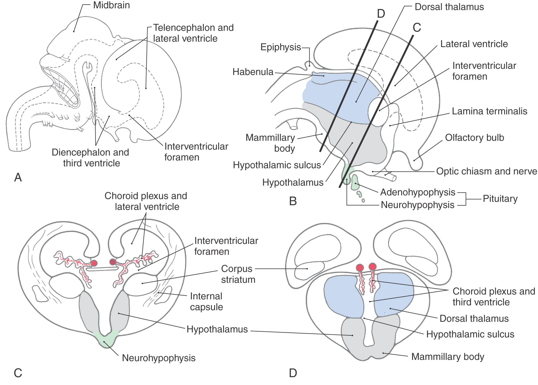
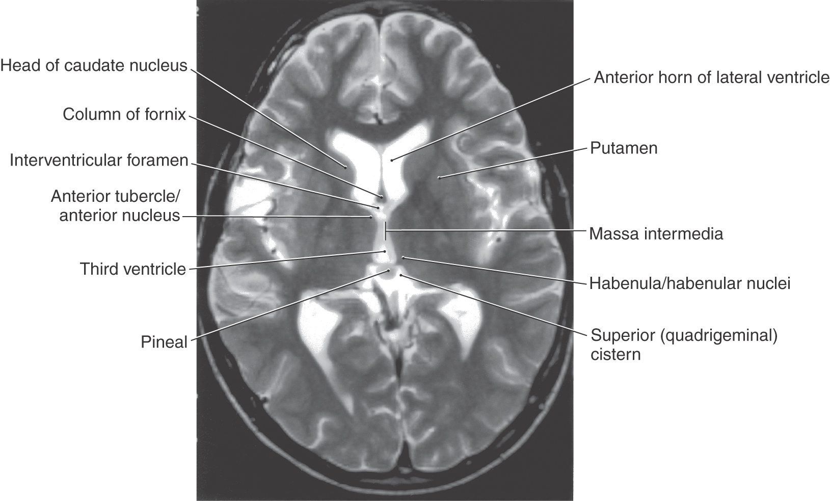
Figure 15-2. Axial T2-weighted magnetic resonance image showing the interventricular foramen, the massa intermedia, the area of the habenula, and the pineal in the superior cistern. The interventricular foramen is the space (containing a small portion of choroid plexus) located between the column of the fornix and the anterior tubercle of the thalamus. The major nuclei of the dorsal thalamus in an axial plane are shown in Figure 15-10.
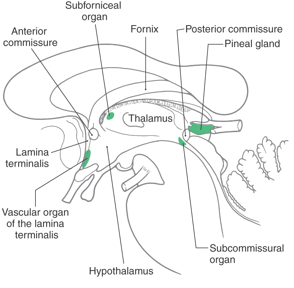
Figure 15-3. Midsagittal view showing the locations of circumventricular organs.
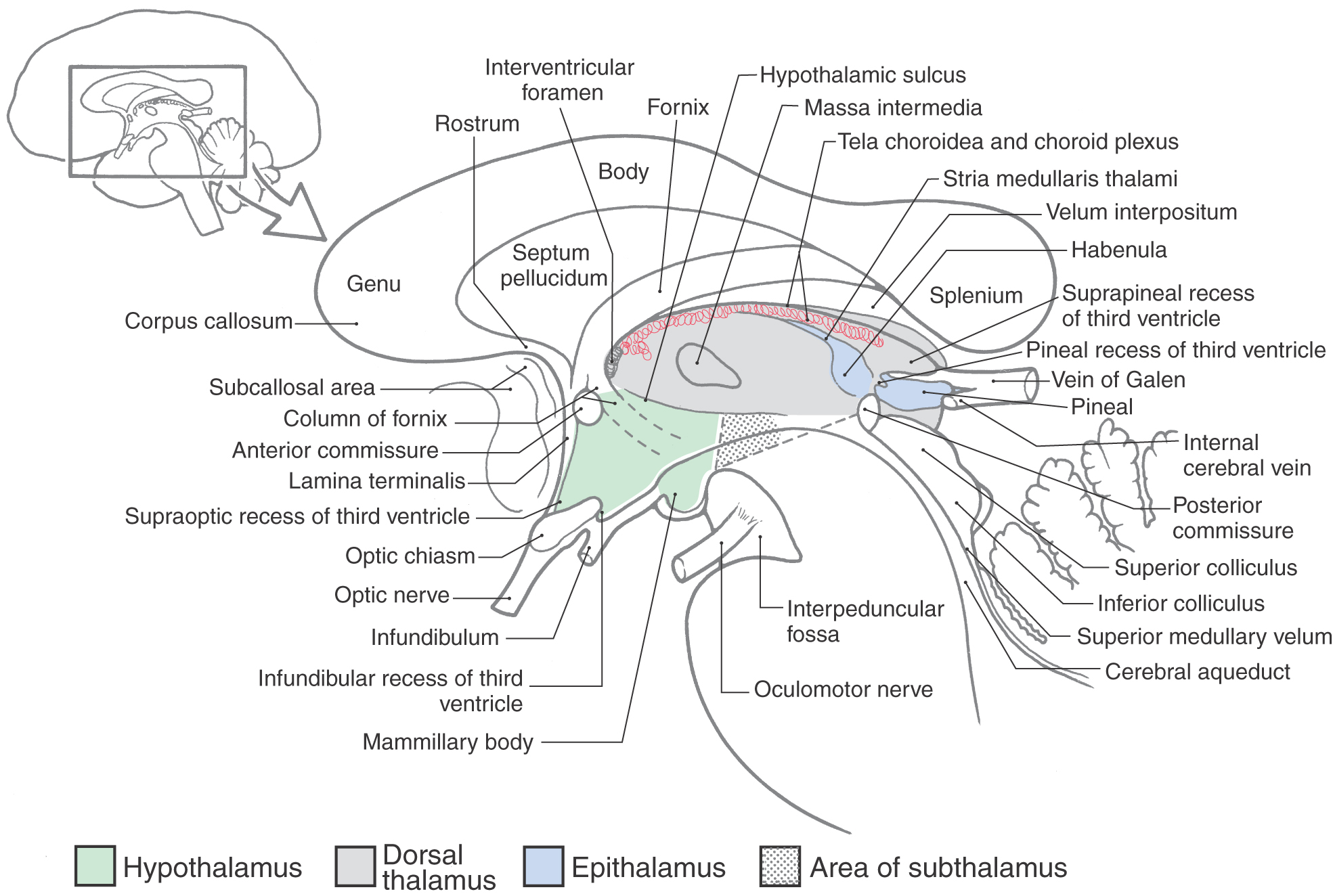
Figure 15-4. Midsagittal view of the diencephalon and closely related structures. This is a drawing of the specimen shown in Figure 15-6.
Just anterior to the habenular region, the roof plate epithelium and adjacent pia mater give rise to the choroid plexus of the third ventricle, which, in the adult, remains suspended from the roof of this space (Fig. 15-1B-D). This choroid plexus is continuous through the interventricular foramina with that of the lateral ventricles. Elsewhere, in locations around the perimeter of the third ventricle, specialized patches of ependyma lie on the midline and form unpaired structures called the circumventricular organs. These structures include the subfornical organ, the organum vasculosum of the lamina terminalis, the subcommissural organ, and the pineal gland (Fig. 15-3). These cellular regions are characterized by the presence of fenestrated capillaries, which implies an absence of the blood-brain barrier. These structures are thought to release metabolites and neuropeptides into the cerebrospinal fluid or into the cerebrovascular system.
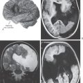
The development of the pituitary gland during the third week is linked to that of the diencephalon (Fig. 15-1B, C). A downward extension of the floor of the third ventricle, the infundibulum, meets the Rathke pouch, an upward outpocketing of the stomodeum, the primitive oral cavity. By the end of the second month, the Rathke pouch loses its connection with the developing oral cavity but maintains its attachment to the infundibulum. As development continues, the Rathke pouch gives rise to the anterior lobe (adenohypophysis) and pars intermedia of the pituitary gland, whereas the infundibulum differentiates into the posterior lobe of the pituitary gland, or neurohypophysis (Fig. 15-1B; see Chapter 30). A craniopharyngioma (Rathke pouch tumor) can arise from a portion of the Rathke pouch that fails to undergo proper migration and apposition to the infundibulum. These tumors mimic lesions of the pituitary gland and may cause visual problems, diabetes insipidus, and increased intracranial pressure.
BASIC ORGANIZATION
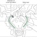
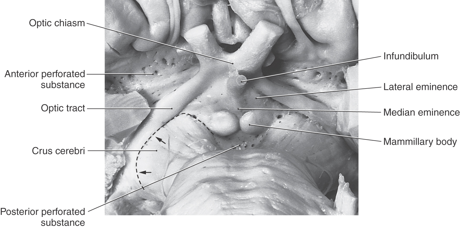
The junction between the diencephalon and midbrain lies along a line extending from the posterior commissure to the caudal edge of the mammillary body on the medial aspect of the hemisphere (Fig. 15-4). On the surface of the hemisphere, this interface is represented by a line starting at the caudal aspect of the mammillary body, extending anterolaterally over the edge of the crus cerebri, and following the caudal edge of the optic tract (Fig. 15-5). The boundary between the diencephalon and surrounding telencephalon is less distinct and is represented laterally by the internal capsule and rostrally by the interventricular foramen, lamina terminalis, and optic chiasm (Fig. 15-4).
The cavity of the diencephalon, the third ventricle, is a narrow, vertically oriented midline space located between the paired dorsal thalami and hypothalami of the two sides (Figs. 15-6 and 15-7). In addition to its connections with the lateral ventricles and the cerebral aqueduct, the third ventricle has small evaginations or recesses associated with the optic chiasm (supraoptic recess), the infundibulum (infundibular recess), and the pineal gland (pineal and suprapineal recesses) (Figs. 15-1 and 15-4).
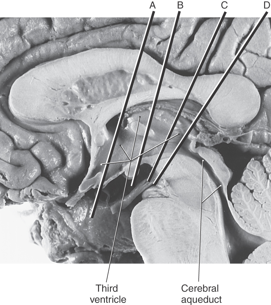
Figure 15-6. Midsagittal view of the diencephalon. This view correlates with the drawing in Figure 15-3. Lines A through D indicate the planes of the stained sections in Figure 15-7.
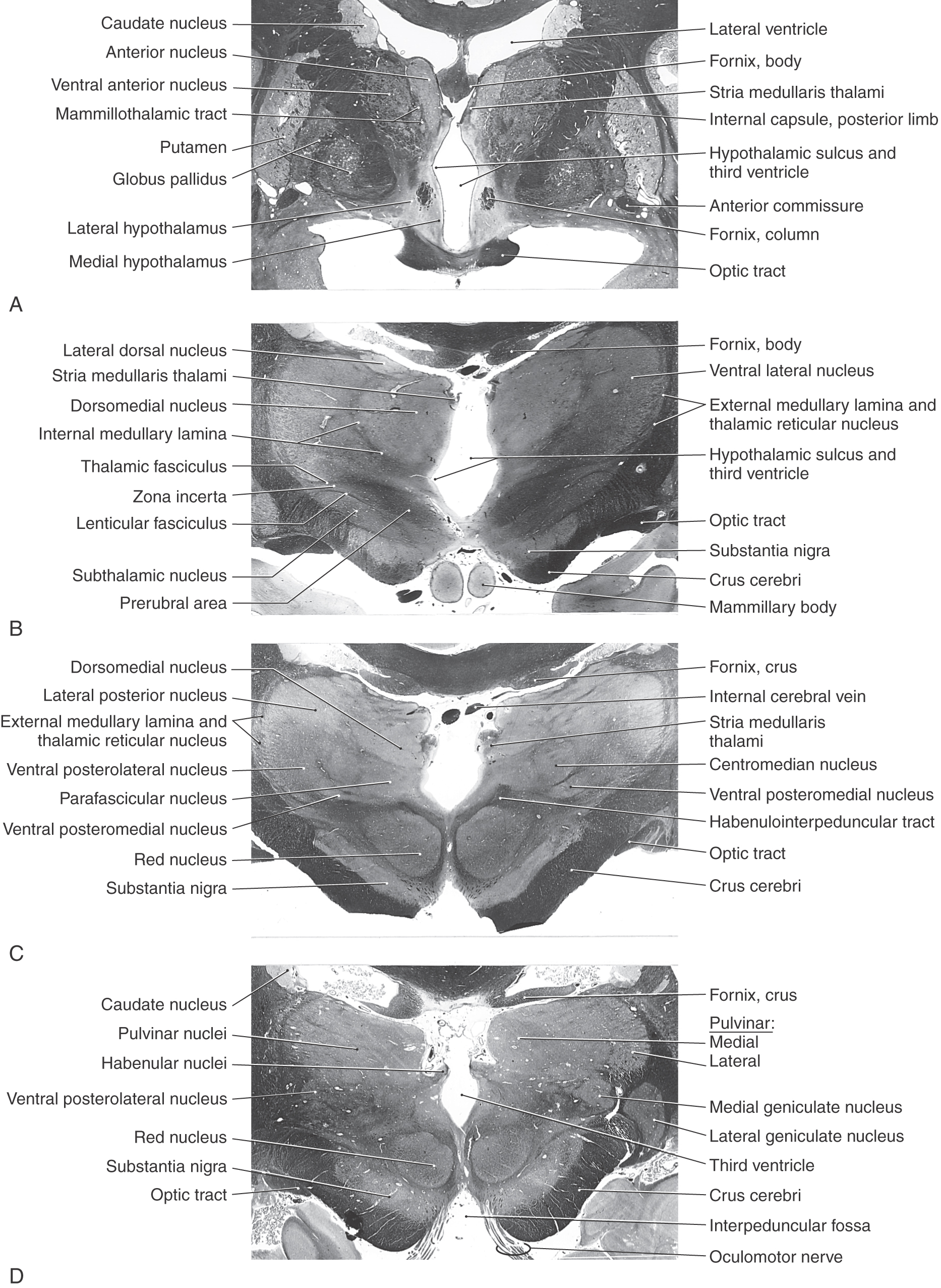
Figure 15-7. Four levels of the forebrain from rostral (A) to caudal (D) showing the internal structure of the hemisphere with emphasis on the diencephalon. These levels correlate with those shown in Figure 15-6 and with the planes represented in the exploded view in Figure 15-10. Weil stain.
All four diencephalic subdivisions can be approximated in a midsagittal section of the forebrain (Figs. 15-4 and 15-6). The dorsal thalamus is located superior to the hypothalamic sulcus and extends from the interventricular foramen caudally to the level of the splenium of the corpus callosum. The hypothalamus lies inferior to the hypothalamic sulcus and is bordered rostrally by the lamina terminalis and caudally by a line that extends from the posterior aspect of the mammillary body superiorly to intersect with the hypothalamic sulcus. The only diencephalic structures visible on the inferior surface of the hemisphere are those related to the hypothalamus, including the optic chiasm, infundibulum, medial and lateral eminences, and mammillary bodies (Fig. 15-5). The ventral thalamus (subthalamus) does not border on the ventricle; rather, it occupies a position caudal to the hypothalamus, rostral to the diencephalon-midbrain junction, and lateral to the midline (Figs. 15-4 and 15-7B). Epithalamic structures are located posteriorly and caudally, in close apposition to the posterior commissure, and include the pineal gland, the habenular nuclei, and the main afferent bundle of these nuclei, the stria medullaris thalami.
DORSAL THALAMUS (THALAMUS)
The dorsal thalamus (or thalamus) (Figs. 15-4 and 15-7 to 15-9) is a massive collection of neuronal cell groups that participate in a widely diverse array of functions involving motor, sensory, and limbic systems. It receives a variety of ascending inputs and projects, via thalamocortical fibers to various cortical areas or gyri, and receives reciprocal connections, via corticothalamic fibers, from those cortical targets to which it sends projections. As a result, the thalamus is often regarded as the functional “gateway” to the cerebral cortex.
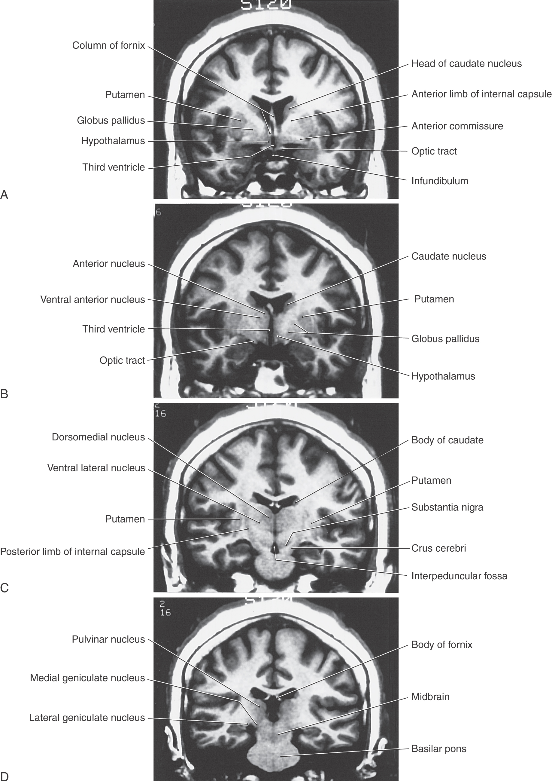
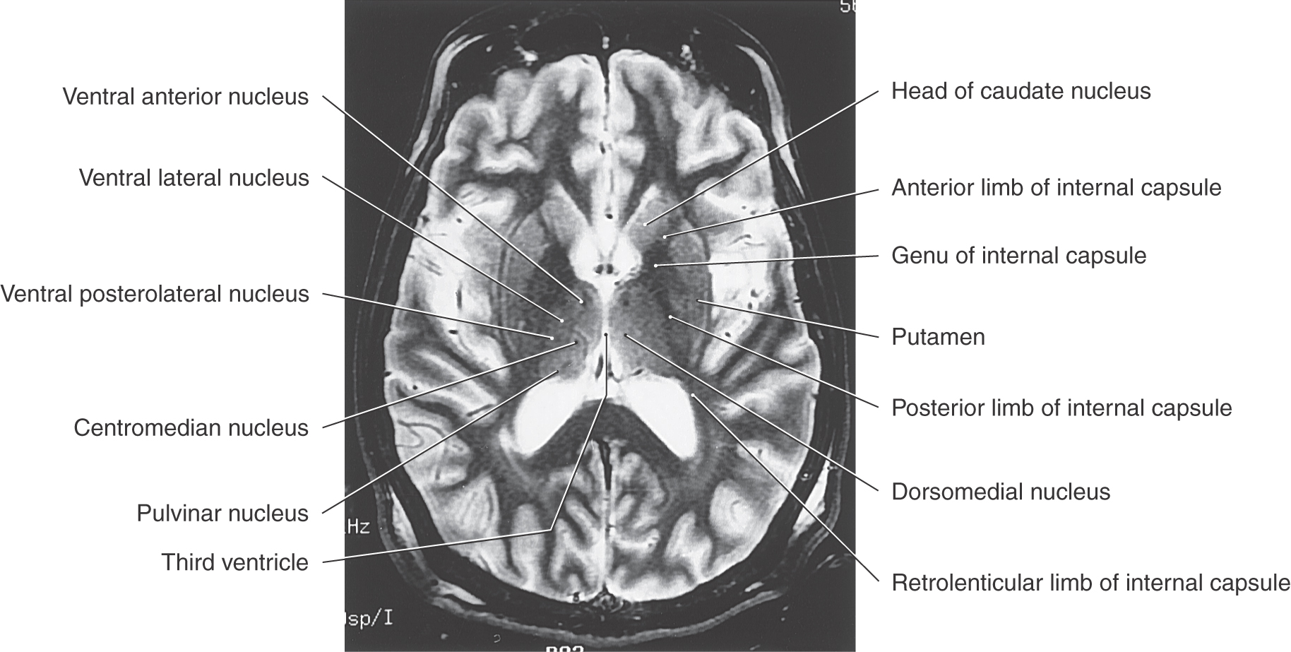
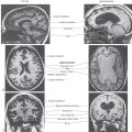
Figure 15-9. T2-weighted magnetic resonance image of the cerebral hemisphere in the axial plane. Emphasis in labeling is on diencephalic structures. Compare with Figure 15-12.
The thalamus is covered on its lateral aspect by a layer of myelinated axons, the external medullary lamina, which includes fibers that enter or leave the subcortical white matter (Fig. 15-7B, C). Within the external medullary lamina are clusters of neurons that form the thalamic reticular nucleus. The medial surface of the thalamus borders the third ventricle, and the external medullary lamina and thalamic reticular nucleus blend with the thalamic fasciculus and zona incerta, respectively, to form an interface between dorsal and ventral thalami (Fig. 15-7B).
An internal medullary lamina, also consisting of myelinated fibers, extends into the substance of the thalamus, where it forms partitions or boundaries that divide the thalamus into its principal cell groups (Figs. 15-9 and 15-10): the anterior, medial, lateral, and intralaminar nuclear groups. The last cell group is located in the portion of the internal medullary lamina that separates the lateral and medial nuclear groups. In addition, there are midline thalamic nuclei located just superior to the hypothalamic sulcus.
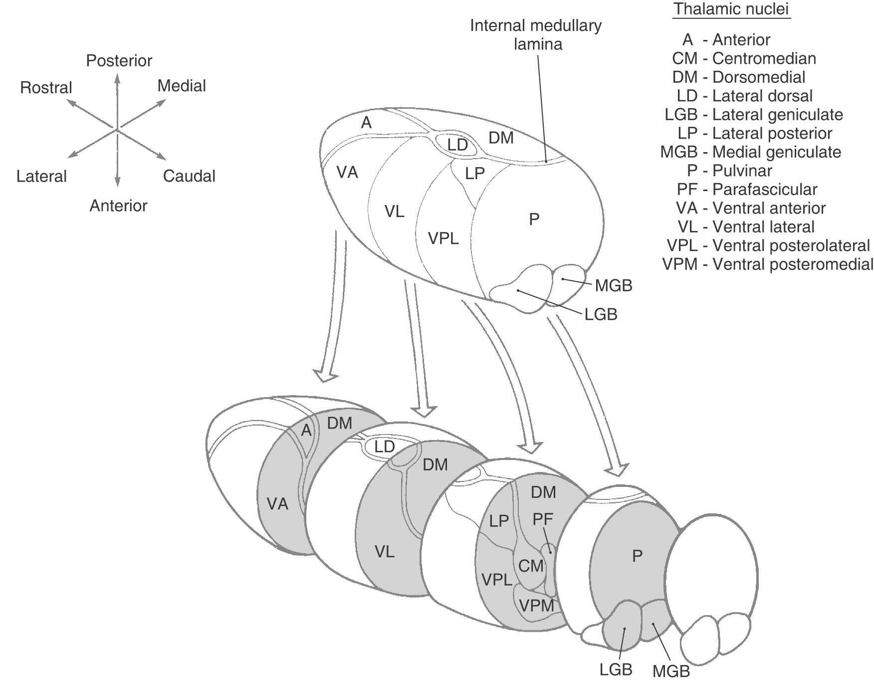
Figure 15-10. Exploded view of the dorsal thalamus illustrating the organization of thalamic nuclei. Compare with Figures 15-6 and 15-8.
Finally, attached to the caudolateral portion of the thalamus are the medial and lateral geniculate bodies (and their correspondingly named subjacent nuclei) (Figs. 15-7D, 15-8D, and 15-10). Although considered here as components of the lateral nuclear group, the geniculate nuclei are sometimes considered as a separate part of the thalamus, the metathalamus.
Anterior Thalamic Nuclei
The anterior nucleus forms a prominent wedge on the rostral aspect of the dorsal thalamus just caudolateral to the interventricular foramen; this wedge is the anterior thalamic tubercle. Internal to the anterior tubercle is a large principal nucleus and two smaller nuclei that collectively form the anterior nucleus of the thalamus (Figs. 15-7A, 15-8B, 15-9, and 15-10). Rostrally, the internal medullary lamina divides to partially encapsulate the anterior nucleus. The cells of this nucleus receive dense limbic-related projections from (1) the mammillary nuclei via the mammillothalamic tract and (2) the medial temporal lobe (hippocampus) via the fornix. The output of this nucleus is primarily directed to the cingulate gyrus through the anterior limb of the internal capsule (Fig. 15-11; see p. 205). The anterior nucleus is an important synaptic station in the Papez circuit, which is related to emotion and memory acquisition.
Medial Thalamic Nuclei
This region of the dorsal thalamus comprises the dorsomedial nucleus (Figs. 15-8C and 15-9). This expansive group of neuronal cell bodies is composed of large parvicellular (located caudally) and magnocellular (located rostrally) parts and a small paralaminar part adjacent to the internal medullary lamina (Figs. 15-7B-D and 15-10). The two larger portions are linked to parts of the frontal and temporal lobes and to the amygdaloid complex (Fig. 15-11). Cells of the paralaminar subdivision receive input from the frontal lobe and substantia nigra and may play a role in the control of eye movement.
Lateral Thalamic Nuclei
This large collection of thalamic neurons is grouped into dorsal and ventral tiers. The relatively small group of dorsal tier nuclei includes the lateral dorsal and lateral posterior nuclei along with the much larger pulvinar nucleus (pulvinar) (Figs. 15-7B-D, 15-8D, 15-9, and 15-10). The connections of the lateral dorsal and lateral posterior nuclei are formed with the cingulate gyrus and parietal lobe, respectively (Fig. 15-10). The large pulvinar nucleus consists of anterior, medial, lateral, and inferior subdivisions. The inferior division receives input from the superior colliculus and projects to the visual association cortex. Other portions of the pulvinar project to areas of the temporal, parietal, and frontal lobes that are especially concerned with visual function and eye movements (Fig. 15-11).
The large ventral tier of the lateral group consists of three separate nuclei (Figs. 15-7, 15-8B, C, and 15-9). The ventral anterior nucleus (VA) and the slightly more caudal ventral lateral nucleus (VL) are important motor-related nuclei; the ventral posterior nucleus, consisting of ventral posterolateral (VPL) and ventral posteromedial (VPM) nuclei, convey somatosensory information to the cerebral cortex.
The VA is composed of a large parvocellular portion and a small magnocellular part. The former receives input from the medial segment of the globus pallidus, and the latter receives afferents from the reticular portion of substantia nigra. The efferent projections from the VA are diffuse and appear to include selected parts of the frontal lobe (Fig. 15-11).
The VL (Figs. 15-7B and 15-10) is also composed of three subdivisions: a pars oralis, a pars medialis, and a pars caudalis. The largest of these, the pars oralis, receives a dense projection from the internal segment of the ipsilateral globus pallidus; some of these afferents enter the caudal subdivision. In contrast, the pars caudalis subdivision of the VL receives its main input from the contralateral cerebellar nuclei. Consequently, pallidal and cerebellar projections are largely segregated within this nucleus. The output of the VL reflects its segregated input in that the oral and caudal parts project to largely separate areas of the frontal lobe (Fig. 15-11).
The larger and more laterally located VPL nucleus and the comparatively smaller and more medially located VPM nucleus both receive somatosensory input from the contralateral side of the body (Figs. 15-7C and 15-10). The medial lemniscus and spinothalamic fibers terminate in a somatotopic manner (cervical fibers medial, sacral fibers lateral) within the VPL, whereas trigeminothalamic fibers from the spinal trigeminal nucleus and the principal trigeminal sensory nucleus terminate in the VPM. Both the VPL and VPM project to the somatosensory cortex of the parietal lobe (Fig. 15-11).
A small group of cells called the ventral posterior inferior nucleus is situated ventrally between the VPL and VPM. These cells process vestibular input and project to lateral areas of the postcentral gyrus that are located in the depths of the central sulcus. Similarly, a small group of cells forming the rostral (oral) portion of the VPL receives cerebellar input and projects to the precentral gyrus of the frontal lobe; this nucleus probably represents a few cells that have been displaced from the slightly more rostrally located VL. This cell group is also called the ventral intermediate nucleus because of its location between the VL and VPL.
The medial (MGB) and lateral (LGB) geniculate nuclei are considered parts of the lateral thalamic nuclear group (Figs. 15-7D, 15-8D, and 15-10). The MGB receives ascending auditory input via the brachium of the inferior colliculus and projects to the primary auditory cortex in the temporal lobe. The LGB receives visual input from the retina via the optic tract and in turn projects to the primary visual cortex on the medial surface of the occipital lobe (Fig. 15-11).
Located in the posterior thalamus at about the level of the pulvinar and geniculate nuclei is a cluster of cell groups collectively called the posterior nuclear complex. This complex consists of the suprageniculate nucleus, the nucleus limitans, and the posterior nucleus. These nuclei are positioned superior to the medial geniculate and medial to the rostral pulvinar. The posterior nuclear complex receives and sends to the cortex nociceptive cutaneous input that is transmitted over somatosensory pathways.
Intralaminar Nuclei
Embedded within the internal medullary lamina are the discontinuous groups of neurons that form the intralaminar nuclei. These cells are characterized by their projections to the neostriatum and to other thalamic nuclei, along with diffuse projections to the cerebral cortex. Two of the most prominent cell groups are the centromedian and parafascicular nuclei (Figs. 15-9 and 15-10). The centromedian nucleus projects to the neostriatum and to motor areas of the cerebral cortex, whereas the parafascicular nucleus projects to rostral and lateral areas of the frontal lobe. Other intralaminar nuclei receive input from ascending pain pathways and project to the somatosensory and parietal cortex.
Midline Nuclei
The midline nuclei are the least understood components of the thalamus. The largest is the paratenial nucleus, which is located just ventral to the rostral portion of the stria medullaris thalami; other cells are associated with the interthalamic adhesion (massa intermedia). Although inputs are poorly defined, efferent fibers reach the amygdaloid complex and the anterior cingulate cortex, suggesting a role in the limbic system.
Thalamic Reticular Nucleus
The cells of this nucleus are situated within the external medullary lamina and between this lamina and the internal capsule (Fig. 15-7B, C). Axons of these cells project medially into the nuclei of the dorsal thalamus or to other parts of the reticular nucleus, but not into the cerebral cortex. Afferents are received from the cortex and from nuclei of the dorsal thalamus via collaterals of thalamocortical and corticothalamic axons. It appears that thalamic reticular neurons modulate, or gate, the responses of thalamic neurons to incoming cerebral cortical input.
Summary of Thalamic Organization
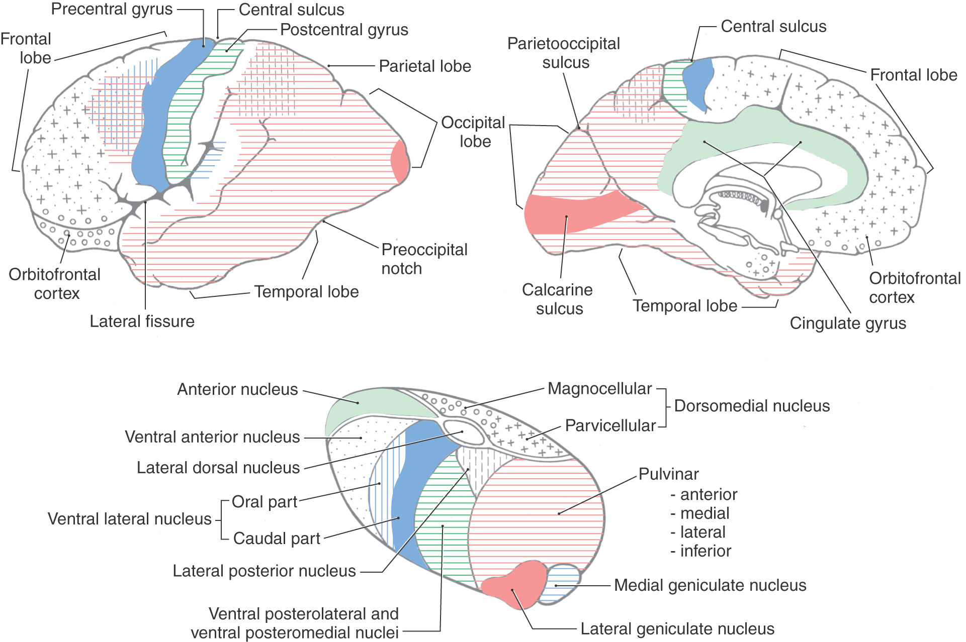
Each thalamic nucleus (with a few exceptions) gives rise to efferent projections (thalamocortical fibers) that target some portion of the cerebral cortex. That region of cortex then typically provides a reciprocal projection (corticothalamic fibers) that returns to the original thalamic nucleus. Figure 15-11 reviews the major thalamocortical relationships.
Some thalamic nuclei are primarily associated with a particular function and in turn with a specific gyrus (and functional area) of the cerebral cortex. The more important of these relationships are as follows: VL/motor/precentral gyrus and anterior paracentral gyrus; VPL/sensory for the body/postcentral gyrus and posterior paracentral gyrus; VPM/sensory for the face/postcentral gyrus; MGB/auditory/transverse temporal gyrus; LGB/vision/cortex on the calcarine sulcus. The anterior nucleus projects primarily to the cingulate gyrus and functions in the broad area of behavior.
The nuclei of the thalamus have been classified according to their connections as either relay nuclei or association nuclei. A relay nucleus is one that receives input predominantly from a single source, such as a sensory pathway or a cerebellar nucleus, or from the basal nuclei. The incoming neural information is processed and then sent to a localized region of sensory, motor, or limbic cortex. Relay nuclei include MGB, LGB, VPL, VPM, VL, VA, and the anterior thalamic nuclei. These nuclei do not merely relay neural signals; in fact, considerable neural processing also takes place in these nuclei. However, their position in a modality-specific pathway linking one particular source to one particular destination makes the word “relay” a useful designation. In contrast, an association nucleus receives input from a number of different structures or cortical regions and usually sends its output to more than one of the association areas of the cerebral cortex (i.e., areas that are neither sensory nor motor cortex; see Chapter 32). Association nuclei include dorsomedial, lateral dorsal, lateral posterior, and the nuclei of the pulvinar complex.
A thalamic nucleus can also be designated specific or nonspecific on the basis of thalamocortical signals generated in response to electrical stimulation delivered to a localized site in that thalamic nucleus. Focal electrical stimulation of a specific nucleus produces a rapidly conducted, sharply localized evoked response in the ipsilateral cerebral cortex. All relay nuclei and association nuclei are specific nuclei. Focal electrical stimulation of a nonspecific nucleus produces widespread activity in the cortex of both hemispheres, at a significantly longer time delay than with stimulation of a specific nucleus. It is thought that nonspecific nuclei play a role in modulating the excitability of large regions of cortex. Nonspecific nuclei include the midline nuclear group, the intralaminar nuclear group (such as the centromedian nucleus), and a portion of the VA.
INTERNAL CAPSULE
Axons pass between the diencephalon, particularly the dorsal thalamus, and the cerebral cortex in a fan-shaped mass of fibers, the internal capsule, that courses from the central core of the hemisphere into the brainstem (Figs. 15-7 and 15-12). Even though this structure consists mostly of axons that reciprocally link the thalamus and cerebral cortex, it also contains cortical efferent fibers that project to the brainstem (corticorubral, corticoreticular, corticonuclear-corticobulbar) or spinal cord (corticospinal).
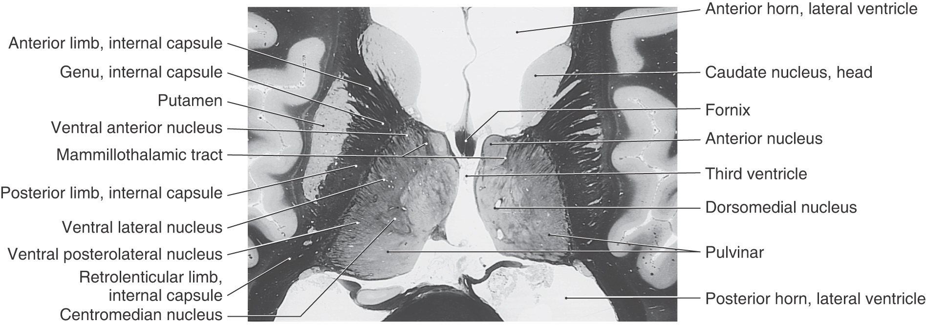
Figure 15-12. Axial view of the forebrain showing the relationship of the thalamus to the limbs of the internal capsule. Weil stain.
Although the internal capsule is described in detail in Chapter 16, it is summarized here because of its important relationship to the thalamus. As seen in axial section (Fig. 15-12), the internal capsule consists of an anterior limb, genu, posterior limb, and retrolenticular limb. The genu is located immediately lateral to the anterior thalamic nucleus, at about the same level as the interventricular foramen. The anterior limb extends rostrolateral from the genu and is insinuated between the caudate and lenticular nuclei. The posterior limb extends caudolateral from the genu and separates the thalamus from the globus pallidus. As its name implies, the retrolenticular limb is the white matter located immediately caudal to the lenticular nucleus (Latin retro-, for “behind”).
HYPOTHALAMUS
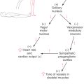
Unlike the thalamus, which is primarily related to somatic functions, the hypothalamus (Figs. 15-8A, B and 15-9) is mainly involved in visceromotor, viscerosensory, and endocrine activities. The hypothalamus and related limbic structures receive sensory input regarding the internal environment and in turn regulate the motor systems that modify the internal environment through four mechanisms. First, the hypothalamus is a principal modulator of autonomic nervous system function. Second, it is a viscerosensory transducer, containing neurons with specialized receptors capable of responding to changes in the temperature or osmolality of blood as well as to specific hormonal levels in the general circulation. Third, the hypothalamus regulates the activity of the anterior pituitary through the production of releasing factors (hormone-releasing hormones). Fourth, it performs an endocrine function by producing and releasing oxytocin and vasopressin into the general circulation within the posterior pituitary.
The hypothalamus can be divided into lateral, medial, and periventricular zones (Figs. 15-7A and 15-13). The lateral zone, often called the lateral hypothalamic area, extends the full rostrocaudal length of the hypothalamus and is separated from the medial zone by a line drawn through the fornix in the sagittal plane (Fig. 15-14C). The medial zone is divided from rostral to caudal into three regions: the chiasmatic, the tuberal, and the mammillary regions (Figs. 15-13 and 15-14). The periventricular zone includes the neurons that border the ependymal surfaces of the third ventricle.
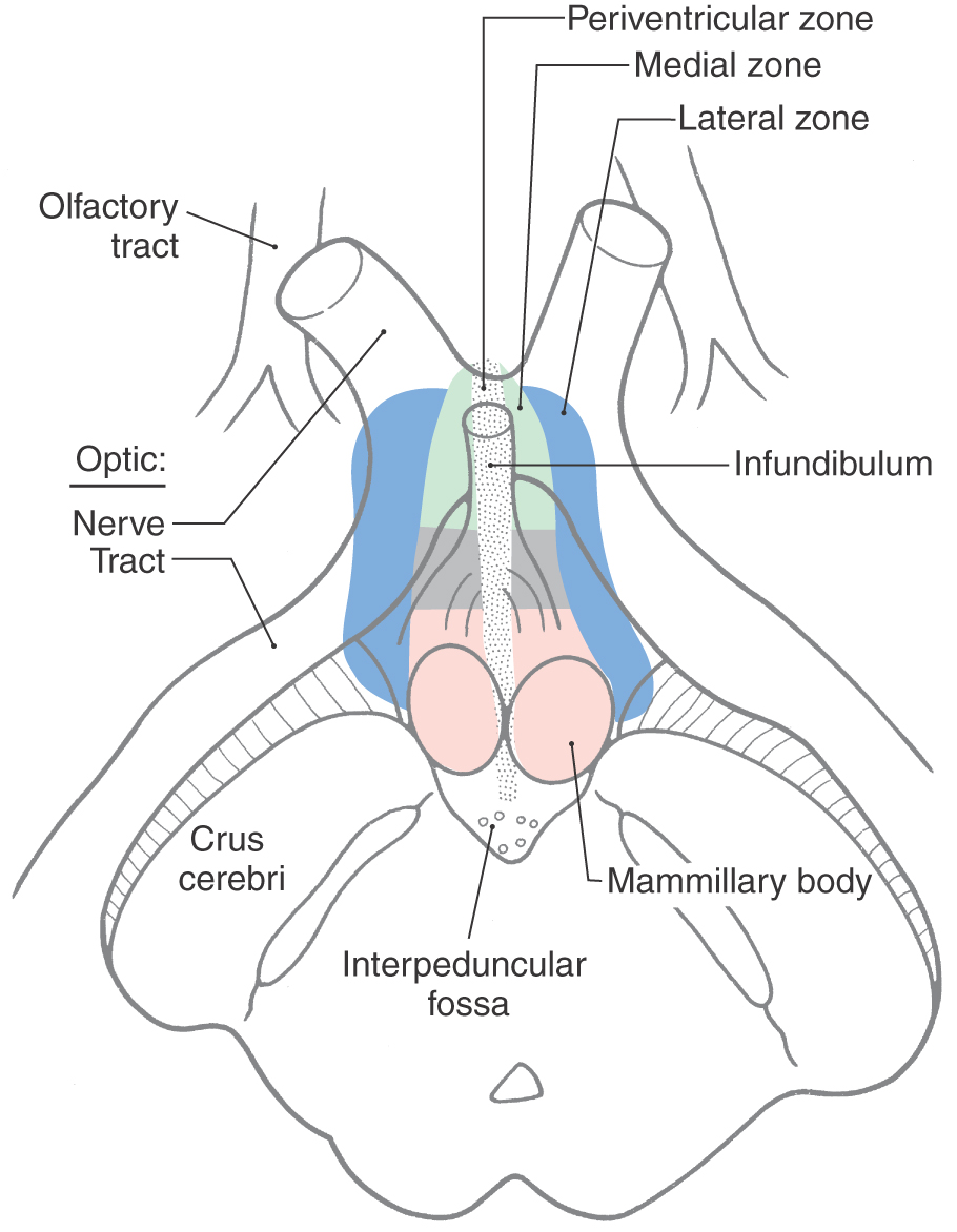
Figure 15-13. Anterior (ventral) view of the diencephalon illustrating the three zones of the hypothalamus as superimposed on external structures. The colors used for medial and lateral zones correlate with those in Figure 15-14.
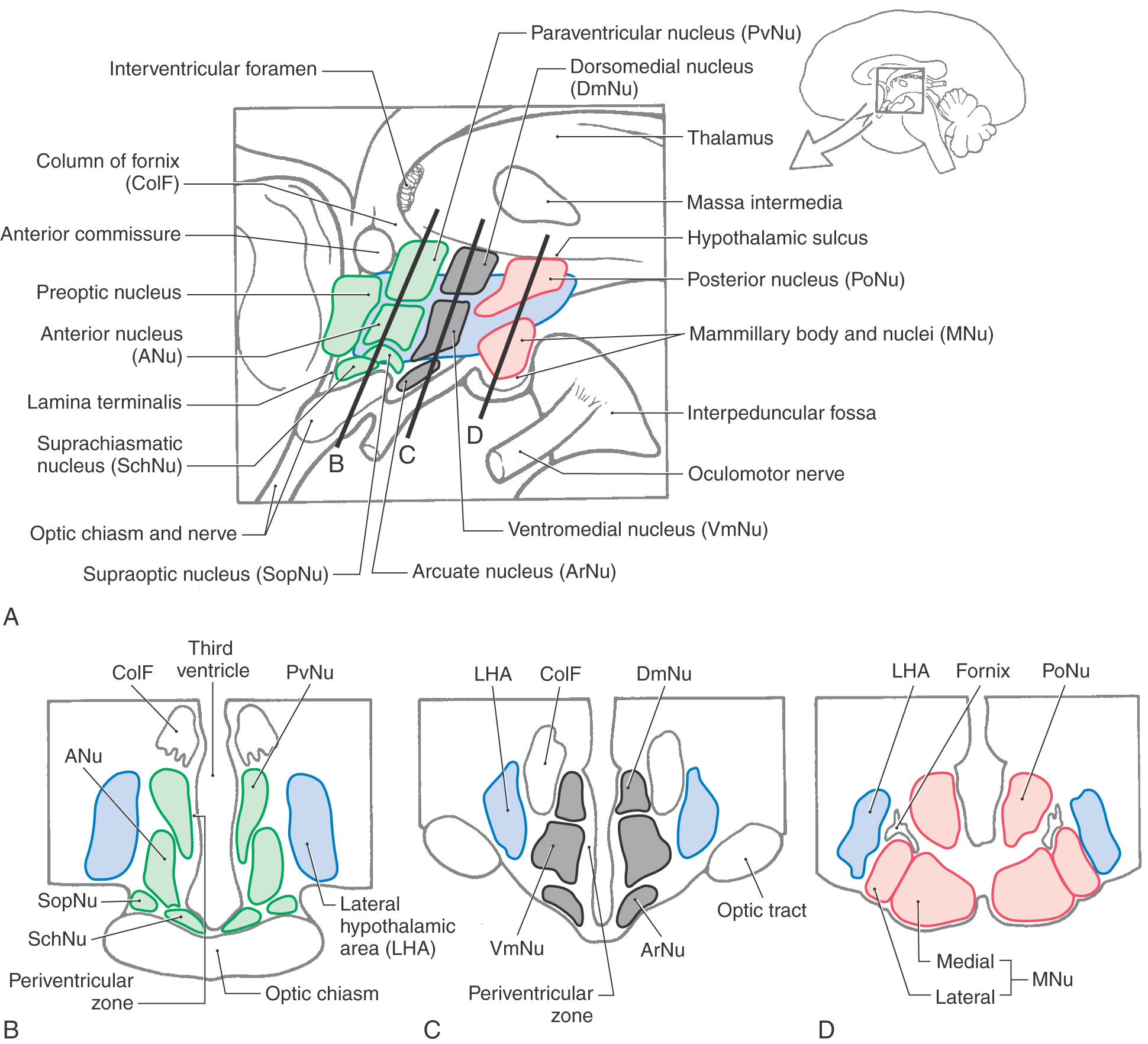
Figure 15-14. Midsagittal (A) and cross-sectional (B-D) views illustrating the nuclei of medial and lateral hypothalamic zones and the nuclei associated with chiasmatic (B), tuberal (C), and mammillary (D) regions. The colors used here correlate with those in Figure 15-13. (A Modified from Haymaker W, Anderson E, Nauta WJH: The Hypothalamus. Springfield, Ill, Charles C Thomas, 1969, with permission.)
Lateral Hypothalamic Zone
The lateral hypothalamic area (Fig. 15-14) is composed of diffuse clusters of neurons intermingled with longitudinally oriented axon bundles. The latter, which form the medial forebrain bundle, are diffusely organized in the human brain. No discrete named nuclei are present in this lateral area, although the supraoptic nucleus is considered by some authorities to be part of it. Cells of the lateral hypothalamic area are involved in cardiovascular function and in the regulation of food and water intake.
Medial Hypothalamic Zone
The medial hypothalamic zone contains discrete groups of neurons whose function and connections are established. Within the chiasmatic (anterior) region are five nuclei: the preoptic, supraoptic, paraventricular, anterior, and suprachiasmatic nuclei (Fig. 15-14A, B). Nuclei in the chiasmatic region are generally involved in regulating hormone release (preoptic, supraoptic, periventricular), cardiovascular function (anterior), circadian rhythms (suprachiasmatic), and body temperature and heat loss mechanisms (preoptic). In the tuberal region are the dorsomedial, ventromedial, and arcuate nuclei (Fig. 15-14A, C). The ventromedial nucleus is regarded as the food intake (satiety) center. Bilateral lesions of this hypothalamic region produce hyperphagia, a greatly increased food intake with resultant obesity. Cells of the arcuate nucleus deliver peptides to the portal vessels and, through these channels, to the anterior pituitary. Some of these peptides are releasing factors, which cause an increase in the secretion of specific hormones by the anterior pituitary, and some are inhibiting factors, which inhibit the secretion of specific hormones by the anterior pituitary.
At caudal levels, the mammillary region is composed of the posterior nucleus and the mammillary nuclei (Fig. 15-14A, D). In humans, the mammillary nuclei consist of a large medial nucleus and a small lateral nucleus. Although both of these nuclei receive input via the fornix, only the medial nucleus projects to the anterior thalamic nucleus through the mammillothalamic tract. This latter bundle traverses the internal medullary lamina as it enters the anterior nucleus (Figs. 15-7A and 15-12). The neurons of the posterior nucleus are involved in activities that include elevation of blood pressure, pupillary dilation, and shivering or body heat conservation. The mammillary nuclei are involved in the control of various reflexes associated with feeding as well as in mechanisms relating to memory formation.
Afferent Fiber Systems
Although many axonal systems extend into the hypothalamus, only four inputs are mentioned here (see Chapter 30). The fornix and the stria terminalis are two major afferent fiber bundles that reach the hypothalamus (Fig. 15-7). The fornix consists of axons that largely originate in the hippocampus, and the stria terminalis arises from neurons in the amygdaloid complex (see Fig. 16-18). Fibers comprising the ventral amygdalofugal bundle exit the amygdala and course through the substantia innominata to enter the hypothalamus and thalamus (see Fig. 16-18). As mentioned earlier, the medial forebrain bundle passes bidirectionally through the lateral hypothalamic region. This composite fiber bundle consists of ascending axons that originate in areas throughout the neuraxis and terminate in the hypothalamus and other axons that exit the hypothalamus to reach forebrain and brainstem targets.
Efferent Fibers
The hypothalamus is the source of a diverse array of efferent fibers (see Chapter 30). Several nuclei give rise to descending fibers that contribute to the dorsal longitudinal fasciculus and the medial forebrain bundle and to diffuse projections that pass into the tegmentum. These fiber systems project directly to numerous brainstem nuclei as well as to preganglionic sympathetic and parasympathetic neurons in the spinal cord. Other projections reach the thalamus and frontal cortex, and still others extend to the posterior pituitary or to the tuberohypophysial portal system for delivery of substances to the anterior pituitary.
VENTRAL THALAMUS (SUBTHALAMUS)
The ventral thalamus (also called subthalamus) includes the large subthalamic nucleus, the medially adjacent prerubral area (field H of Forel), and, posteriorly, the zona incerta (Figs. 15-4 and 15-7B). As the term ventral thalamus (or subthalamus) implies, these cell groups are located ventral (anterior) to the large expanse of the dorsal thalamus. The subthalamic nucleus is a lens-shaped cell group situated rostral and posterior to the substantia nigra and immediately inferior to a distinct myelinated fiber bundle, the lenticular fasciculus (Fig. 15-7B; see Chapter 26). The cells of the subthalamic nucleus receive input from motor areas of the cerebral cortex, project to the substantia nigra, and are reciprocally connected with the globus pallidus. The subthalamic nucleus can be affected by vascular lesions involving posteromedial branches of the posterior cerebral or posterior communicating arteries, which results in a characteristic clinical condition known as hemiballismus. Patients with this involuntary movement disorder exhibit rapid and forceful flailing movements, which usually involve the contralateral upper extremity. These movements can be very debilitating because the patient has no control over their initiation or duration.
The zona incerta is located superior to the subthalamic nucleus and is separated from it by the lenticular fasciculus (Fig. 15-7B). Superior to the zona incerta are the myelinated axons of the thalamic fasciculus. The zona incerta contains output neurons that project to a variety of locations, including the cerebral cortex, the superior colliculus, the pretectal region, and the basilar pons. Afferent projections arise from the motor cortex and as collaterals from the medial lemniscus.
The prerubral area (Forel’s field H) is located just rostral to the red nucleus and medial to the subthalamic nucleus (Fig. 15-7B). There are scattered neurons in this region, and traversing the prerubral area are fibers from the lenticular fasciculus (Forel’s field H2) that enter the thalamic fasciculus (Forel’s field H1).
EPITHALAMUS
The pineal gland, habenular nuclei, and stria medullaris thalami are the principal components of the epithalamus (Figs. 15-2, 15-4, and 15-15). The pineal gland consists of richly vascularized connective tissue containing glial cells and pinealocytes but no true neurons. Mammalian pinealocytes are related to the photoreceptor elements found in this gland in lower forms, such as amphibians. In humans, however, they remain only indirectly light sensitive and receive information concerning photic stimuli through a multisynaptic neural circuit.
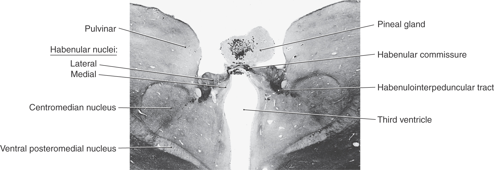
 Figure 15-15.
 Pinealocytes have club-like processes that are apposed to blood vessels but do not have direct synaptic contacts with central nervous system neurons. These cells synthesize melatonin from serotonin via enzymes that are sensitive to diurnal fluctuations in light. Levels of serotonin N-acetyltransferase increase during the night (in the absence of photic stimulation), and the synthesis of melatonin is enhanced. Exposure to light turns off the enzymatic activity, and melatonin production is diminished. Thus the production of melatonin by pinealocytes is rhythmic and calibrated to the 24-hour cycle of photic input to the retina. This is called a circadian rhythm.
Photic stimulation of pinealocytes occurs through an indirect route. Retinal ganglion cells project to the suprachiasmatic nucleus of the hypothalamus, which in turn influences neurons of the intermediolateral cell column in the spinal cord through descending connections. These preganglionic sympathetic neurons project to the superior cervical ganglion, which in turn innervates the pineal gland via postganglionic fibers that travel on branches of the internal carotid artery.
Pinealocytes also produce serotonin, norepinephrine, and neuroactive peptides, such as thyrotropin-releasing hormone, which are normally associated with the hypothalamus. These secretory products are released into the general circulation or the cerebrospinal fluid.
Pinealomas (tumors with large numbers of pinealocytes) are accompanied by depression of gonadal function and delayed puberty, whereas lesions that lead to the loss of pineal cells are associated with precocious puberty. This indicates that pineal secretory products exert an inhibitory influence on gonadal formation.
The habenular nuclei are located just anterior to the pineal gland and consist of a large lateral nucleus and a small medial nucleus (Figs. 15-2 and 15-15). Both nuclei contribute axons to the habenulointerpeduncular tract (fasciculus retroflexus), which terminates in the midbrain interpeduncular nucleus. The stria medullaris thalami, which arches over the medial aspect of the dorsal thalamus near the midline, conveys input to both habenular nuclei. The habenular commissure, a small bundle of fibers riding on the upper edge of the posterior commissure, connects the habenular regions of the two sides.
VASCULATURE OF THE DIENCEPHALON
The diencephalon is supplied by smaller vessels that branch from the various arteries making up the cerebral arterial circle (circle of Willis) and by larger arteries that originate from the proximal parts of the posterior cerebral artery (Figs. 15-16 and 15-17A, B). The hypothalamus and subthalamus are supplied by central (perforating or ganglionic) branches of the circle. Anterior parts of the hypothalamus are served by central branches (anteromedial group) arising from the anterior communicating artery and the A1 segment of the anterior cerebral artery and from branches of the proximal part of the posterior communicating artery. Caudal hypothalamic regions and the ventral thalamus are supplied by branches of the posteromedial group; these branches arise from the posterior communicating artery and the P1 segment of the posterior cerebral artery. The specific relationships of the anteromedial and posteromedial groups originating from the cerebral arterial circle can be reviewed in Chapter 8 (see Fig. 8-16).
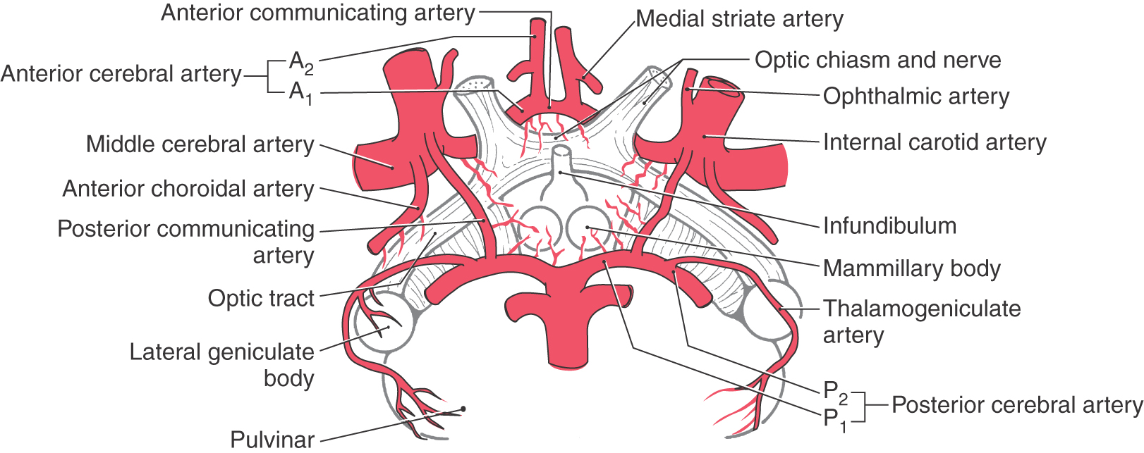
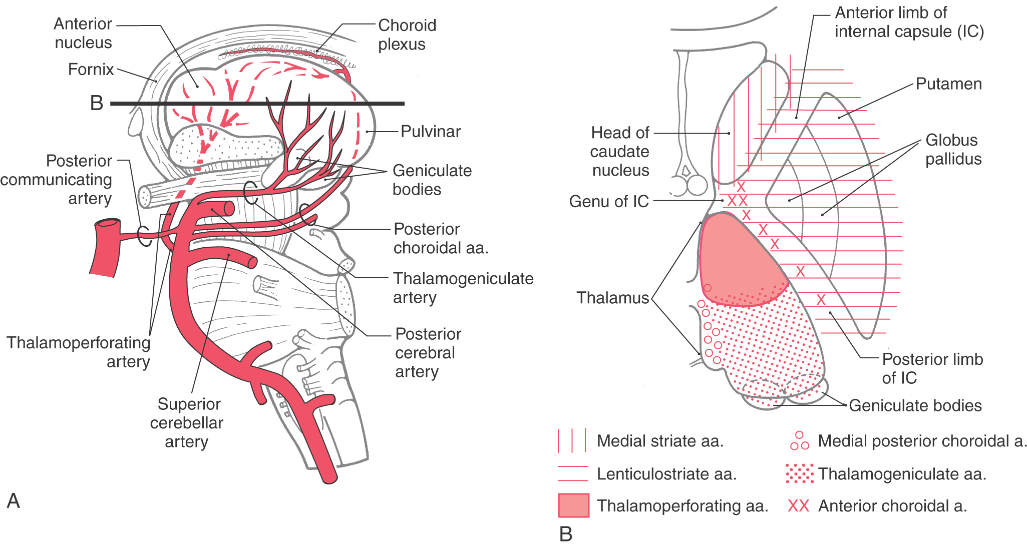
Some of the branches of the posteromedial group that arise from the P1 segment near the basilar bifurcation are called the thalamoperforating arteries. These vessels (of which there may be more than one on each side) penetrate deeply to supply rostral areas of the thalamus (Figs. 15-16 and 15-17A, B). If these vessels are occluded during surgery in this region, as can occur, for example, when an aneurysm of the basilar bifurcation is clipped, the patient can be rendered permanently comatose. Slightly more distal branches, which usually arise from the P2 segment, are the posterior choroidal and thalamogeniculate arteries. These arteries also supply portions of the diencephalon (Figs. 15-16 and 15-17A, B). A narrow portion of the caudal and medial thalamus bordering on the third ventricle is supplied by the medial posterior choroidal artery; the thalamogeniculate branches irrigate the caudal thalamus, including the pulvinar and the geniculate nuclei (Figs. 15-16 and 15-17B). In addition, branches of the medial posterior choroidal artery also serve the choroid plexus of the third ventricle.
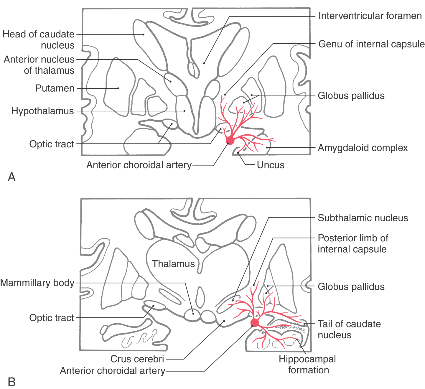
The anterior choroidal artery originates from the cerebral portion of the internal carotid artery and courses caudolaterally along the trajectory of the optic tract (Fig. 15-18A, B). This vessel serves important structures in this general area. It sends penetrating branches into the genu of the internal capsule and into the more inferior aspect of the posterior limb of the internal capsule (Fig. 15-18A, B). In addition, it serves the optic tract, inferior portions of the lenticular nucleus, the choroid plexus of the inferior horn of the lateral ventricle, much of the amygdala, the retrolenticular limb of the internal capsule, and large parts of the hippocampal formation. An occlusion of this vessel, an anterior choroidal artery syndrome, results in characteristic visual and motor deficits that reflect damage to the optic tract and the inferior portion of the posterior limb of the internal capsule (see Chapters 24 and 25).
Although the thalamus receives a blood supply largely separate from that of the internal capsule (Fig. 15-17B), vascular lesions in the thalamus may extend into the internal capsule or vice versa. Ischemic or hemorrhagic strokes in the hemisphere may result in contralateral hemiparesis in combination with hemianesthesia. These losses correlate with damage to corticospinal and thalamocortical fibers in the internal capsule. On the other hand, strokes involving the larger thalamic arteries, such as the thalamogeniculate artery, may result in total or dissociated sensory losses. These patients may subsequently experience persistent, intense pain (thalamic pain, Dejerine-Roussy syndrome).
Sources and Additional Reading
Alexander GE, Crutcher MD, DeLong MR. Basal ganglia–thalamocortical circuits: Parallel substrates for motor, oculomotor, “prefrontal” and “limbic” functions. In: Uylings HBM, Van Eden CG, DeBruin JPC, Corner MA, Feenstra MGP, eds. Progress in Brain Research, vol. 85, The Prefrontal Cortex: Its Structure, Function, and Pathology. Amsterdam: Elsevier Biomedical Press; 1990.
Burt AM. Textbook of Neuroanatomy. Philadelphia: WB Saunders; 1993.
Jones EG. The Thalamus. New York: Plenum Press; 1986.
Parent A. Carpenter’s Human Neuroanatomy. 9th ed Baltimore: Williams & Wilkins; 1996.
Walker AE. The Primate Thalamus. Chicago: University of Chicago Press; 1938.