The developing world

Published on 03/04/2015 by admin

Filed under Hematology, Oncology and Palliative Medicine

Last modified 22/04/2025

Print this page

rate 1 star rate 2 star rate 3 star rate 4 star rate 5 star
Your rating: none, Average: 0 (0 votes)

This article have been viewed 1050 times

49

The developing world

The term ‘developing world’ is used to describe the majority of tropical countries which are ‘hot, humid and poor’. An alternative term is the ‘less economically sound’ nations, as these countries are often advanced in human and cultural resources. Haematological practice is different to that in most developed countries. Genetic diseases such as the haemoglobinopathies and red cell enzymopathies are frequent in many tropical regions. Deficiency anaemia and haemolytic anaemia are often secondary to infections such as ancylostomiasis (hookworm) and malaria. Medical treatment regarded as routine in the developed countries is commonly unavailable. For instance, only about 20% of the world’s haemophiliac population has access to factor VIII replacement therapy.

With the ever-increasing availability of ‘exotic’ holidays and regular foreign travel within immigrant populations, doctors in the developed world are seeing more tropical diseases. In the patient with unexplained symptoms such as malaise and fever, or signs such as splenomegaly, a history of travel should not be overlooked.

Malaria

Malaria is a protozoal disease, the infectious agent being Plasmodium falciparum, P. vivax, P. ovale or P. malariae. Mortality from the disease has fallen over the last decade but it remains a serious health risk throughout the tropics and subtropics where insecticide resistance of anopheline mosquitoes and multiple drug resistance of malarial parasites make control and treatment challenging. According to the World Health Organization, there were 216 million cases of malaria and an estimated 655 000 deaths in 2010. Most deaths occur in children living in Africa where the disease accounts for 20% of all childhood mortality.

Pathogenesis

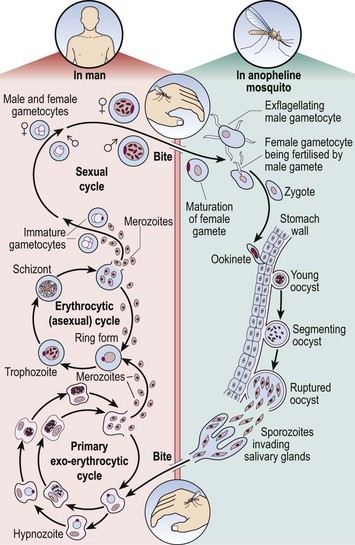
The life cycle of the malaria parasite is illustrated in Figure 49.1. When taking a meal of blood an infected mosquito initiates human infection by the inoculation of malarial sporozoites. These rapidly pass to the liver where they enter hepatocytes and divide. After several days, enormously increased numbers of parasites (merozoites) depart the liver and invade red cells. Here the merozoites develop via ring forms and trophozoites into schizonts. Rupture of the schizont releases 12–20 merozoites back into the blood, thus perpetuating the cycle. The duration of the blood cycle varies between malarial species, explaining the different periodicity of fever in each type. A further mosquito becomes infected when it feeds on blood containing gametocytes, the sexual form of the parasite.

Diagnosis

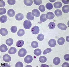
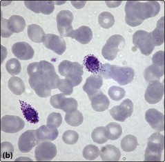
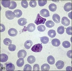
Although malarial parasites may be detected in normal blood films, their identification is generally easier in Leishmann or Giemsa stain at a higher pH. A thick film is best for detection and a thin film for determination of the species. Prolonged inspection of the film is sometimes necessary to spot malarial parasites as there can be a low level of parasitaemia. Where malaria is suspected on clinical grounds repeated samples may be needed to make or exclude the diagnosis. P. falciparum is often associated with higher parasite counts. Paradoxically, some very ill patients with malaria initially have no detectable parasites in the blood as there is sequestration of parasite-laden red cells in the tissues. It is beyond the scope of this book to make a detailed comparison of the four species but some typical appearances are shown in Figure 49.2. Supplementary methods of parasite detection include antigen and antibody detection and DNA-based techniques.

Clinical features

Malaria has a different clinical presentation in non-immune and immune patients.

Non-immune patient

The interval between the mosquito bite and the onset of symptoms is typically 1–2 weeks. Common symptoms are rigors, sweats, headache, vomiting, diarrhoea and muscle pains. P. vivax and P. ovale are classically associated with bouts of fever on alternate days and P. malariae on every third day. Possible clinical signs include a rising temperature, tachycardia, herpes labialis, jaundice, dehydration and splenomegaly. P. falciparum infection is the most dangerous form of malaria. The onset can be insidious and the fever has no particular pattern. Life-threatening complications such as cerebral malaria (with development of coma), acute renal failure and blackwater fever (rapid intravascular haemolysis), can suddenly develop in a patient previously not particularly ill. Children are particularly at risk of a sudden demise.

Treatment and prophylaxis

Ill patients should be rested and rehydrated. A rational choice of drug treatment requires knowledge of both the clinical syndrome and the likelihood of drug resistance. The mainstay of treatment of severe malaria is quinine. This is given intravenously. The dosage must be carefully calculated to avoid under-treatment or toxicity. Recent trials have suggested that artesunate, a derivative of the plant compound artemisin, is both easier to give and more effective than quinine in severe malaria. Its use in Africa may be limited by its cost. Specialist advice should be sought in difficult cases.

Chemoprophylaxis is advised for non-immune travellers entering malarial areas. Specific recommendations depend on the risk of exposure to malaria, the extent of drug resistance, the efficacy of drugs, drug side-effects and patient-related criteria (e.g. pregnancy, renal impairment). Drugs used include chloroquine, proguanil, mefloquine and doxycycline. Where there is doubt, expert advice should be sought. Simple preventative measures such as protective clothes, mosquito nets and insect repellent creams also help reduce the risk of infection. The recently discovered red cell surface receptor which allows P. falciparum to invade may provide a target for a future vaccine.

Endemic Burkitt’s lymphoma

Endemic Burkitt’s lymphoma is an aggressive B-lymphoblastic lymphoma which is found particularly in African children. In areas where malaria is holoendemic it is the most common childhood cancer. The disease is associated with Epstein–Barr virus (EBV) infection and the chromosomal rearrangement t(8;14). The classic clinical presentation is with a massive tumour of the jaw or other extranodal disease. Cure rates exceeding 90% are possible with combination chemotherapy.