CHAPTER 3 The CT scan
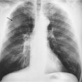
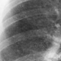
Although the plain chest X-ray is one of the most useful imaging techniques, it is limited by the fact that it is a two-dimensional image and small or subtle abnormalities can be overlooked. In other circumstances the chest X-ray will identify an abnormality but will give limited information as to its extent or detailed appearance. Remember also that, although particularly useful for detecting lung abnormalities, the chest X-ray is a very poor way of imaging the mediastinum.
Types of CT scan
High-resolution CT scanning (HRCT)
Intravenous contrast would not be used with HRCT, since contrast would need to be given before each individual image was taken – something that is clearly impossible.
Interpreting the images
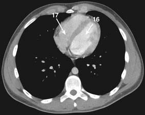
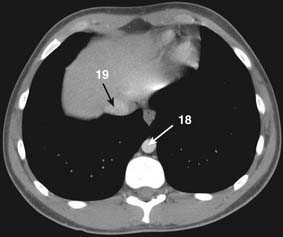
Interpretation of the CT scan requires significant expertise and should only be done by a radiologist. As well as having the experience to interpret the image slices, radiologists have access to software that will allow them to display the images in other forms, for example in the sagittal and coronal planes as well as cross-sectional and three-dimensional images. The CT scans we have included in this book have been chosen because they illustrate situations in which, working on the wards, emergency department or outpatients, you are likely to order scans and a basic knowledge of how they are interpreted will be of use. The next section will give you a basic knowledge of the anatomy of the scan that should help you visualize abnormalities reported on by the radiologist.
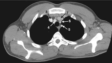
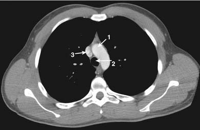
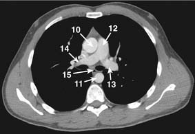
Finding your way around the CT scan

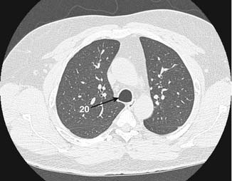
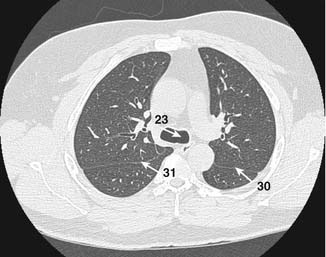
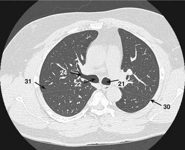
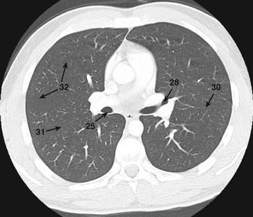
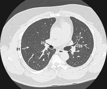
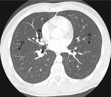
Now look at the lung windows. Start from the top image. You will need to know how to identify the lobes of the lung, so that you can localize any pathology.
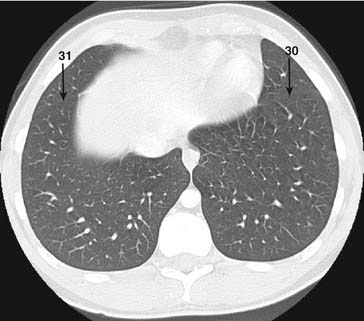
See images on p. 35–38. Identify the major fissures on the left (30) and right (31) and the minor fissure (32) on the right. On the left the major fissure separates the upper and lower lobes. On the right the major fissure separates the lower lobe from the rest of the lung and the minor fissure separates the upper and middle lobes. The minor fissure can be difficult to spot.
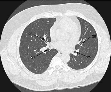
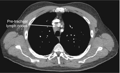
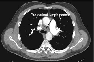
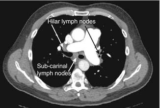
Now that you have identified the normal structures, look for enlarged lymph nodes. You need to distinguish lymph nodes from blood vessels.