The Complement System

Published on 22/03/2015 by admin

Filed under Pediatrics

Last modified 22/04/2025

Print this page

rate 1 star rate 2 star rate 3 star rate 4 star rate 5 star
Your rating: none, Average: 0 (0 votes)

This article have been viewed 1401 times

Chapter 127 The Complement System

Complement was originally defined as the nonspecific, heat-labile complementary principle required with specific antibody to lyse bacteria. The 1st 4 components were numbered in the order of their discovery and are termed the classical pathway. Unfortunately, the components fix to the immune complex in a different order, C1423. Beyond this confusing start, complement is a logical, exquisitely balanced, and highly influential system that is fundamental to the clinical expression of host defense and inflammation.

The complement system, an essential component of innate immunity, is broadly conceptualized as the classical, lectin, and alternative pathways, which interact and depend on each other for their full activity; the membrane attack complex (C5b6789), formed from activity of any pathway; the 8 serum and 4 membrane regulatory proteins that are defined to date; and 8 fully defined cell membrane receptors that bind complement components or fragments (Table 127-1). The 27 circulating components and regulators together compose about 15% of the globulin fraction of serum. The normal concentrations of serum complement components vary by age (Chapter 708); newborn infants have mild to moderate deficiencies of all components.

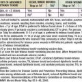
Table 127-1 CONSTITUENTS OF THE COMPLEMENT SYSTEM

SERUM COMPONENTS

Classical Pathway

C1q

C1r

C1s

C4

C2

C3

Alternative Pathway

Factor B

Factor D

Lectin Pathway

Mannose-binding lectin (MBL), ficolins 1, 2, 3 (M, L, H, respectively)

MBL-associated serine proteases (MASPs) 1, 2

Membrane Attack Complex

C5

C6

C7

C8

C9

Regulatory Protein, Enhancing

Properdin

Regulatory Proteins, Downregulating

C1 inhibitor (C1 INH)

C4-binding protein (C4-bp)

Factor H

Factor I

S protein (vitronectin)

Clusterin

Carboxypeptidase N (anaphylatoxin inactivator)

MEMBRANE REGULATORY PROTEINS

CR1 (CD35)

Membrane cofactor protein (MCP; CD46)

Decay-accelerating factor (DAF, CD55)

CD59 (membrane inhibitor of reactive lysis; protectin)

MEMBRANE RECEPTORS

CR1 (CD35)

CR2 (CD21)

CR3 (CD11b/CD18)

CR4 (CD11c/CD18)

C3a receptor

C5a receptor

C1q receptors

Complement receptor of the Ig superfamily (CRIg)

CR, complement receptor; Ig, immunoglobulin.

After C1423, complement nomenclature is logical and consists of only a few rules. Fragments of components resulting from cleavage by other components acting as enzymes are assigned lowercase letters (a, b, c, d, e); with the exception of C2 fragments, the smaller piece that is released into surrounding fluids is assigned the lowercase letter a, and the major part of the molecule, bound to other components or to some part of the immune complex, is assigned letter b—for example, C3a and C3b. Components of the alternative pathway, B and D, have been assigned uppercase letters, as have the control proteins I and H, which downregulate both pathways. C3, and especially its major fragment C3b, is a component of both the classical and alternative pathways.

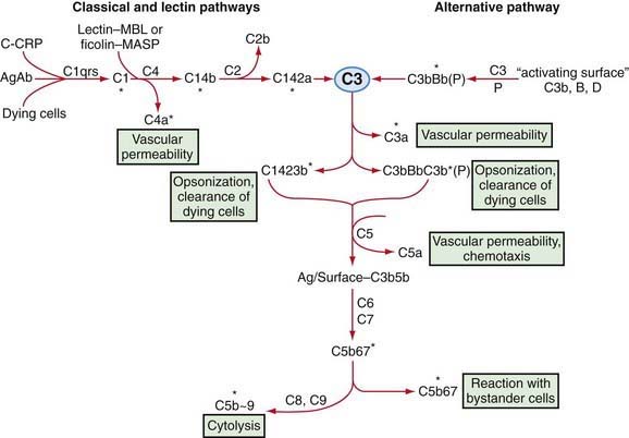
Complement is a system of interacting proteins. The biologic functions of the system depend on the interactions of individual components, which occur in sequential, cascade fashion. Activation of each component, except the 1st, depends on activation of the prior component or components in the sequence. Interaction occurs along three pathways (Fig. 127-1): the classical pathway, in the order antigen–antibody–C142356789; the lectin (carbohydrate-binding) pathway, in the order microbial carbohydrate–lectin (mannose-binding lectin [MBL] or ficolin)–MBL-associated serine protease–C42356789; and the alternative pathway, in the order activator–C3bBD–C356789. Antibody accelerates the rate of activation of the alternative pathway, but activation can occur on appropriate surfaces in the absence of antibody. The classical and the alternative pathways interact with each other through the ability of both to activate C3.

Activation of the early-acting components of complement (C1423) results in the generation of a series of active enzymes, C1, C42, and C423, on the surface of the immune complex or underlying cell. These enzymes cleave and activate the next component in the sequence. In contrast, the interaction among C5b, C6, C7, C8, and C9 is nonenzymatic and depends on changes in molecular configuration.

Classical and Lectin Pathways

The classical pathway sequence begins with fixation of C1, by way of C1q, to the Fc, non–antigen-binding part of the antibody molecule after antigen-antibody interaction. The C1 tricomplex changes configuration and the C1s subcomponent becomes an active enzyme, C1 esterase. Certain bacteria, Mycoplasma, RNA viruses, and the lipid A component of bacterial endotoxin can activate C1q directly and trigger the full complement cascade.

As part of the innate immune response, broadly reactive “natural” antibodies and C-reactive protein (CRP), which reacts with carbohydrate from microorganisms and with dying cells, can substitute for specific antibody in the fixation of C1q and initiate reaction of the entire sequence. Endogenous agents, including uric acid crystals, amyloid deposits, DNA, and components of damaged cells such as apoptotic blebs and mitochondrial membranes, can activate C1q directly. But in this case, the ligand-C1q complex interacts strongly with the inhibitors C4-binding protein and factor H, allowing some C3-mediated opsonization and phagocytosis but limiting the full inflammatory response typically triggered by microbes. C1q synthesized in the brain and retina enables the complement-dependent pruning of synapses that is essential for normal nervous system development.

There are 4 recognition molecules in the lectin pathway: MBL and ficolins 1, 2, and 3 (also termed ficolins M, L, and H, respectively). MBL is the prototype of the collectin family of carbohydrate-binding proteins (lectins) that are believed to play an important part in innate, nonspecific immunity; its structure is homologous to that of C1q. These lectins, in association with MBL-associated serine proteases 1 and 2 (MASPs 1/2), can bind to mannose, lipoteichoic acid, and other carbohydrates on the surface of bacteria, fungi, parasites, and viruses. MASPs then function there like C1s to cleave C4 and C2 and activate the complement cascade. The peptide C4a has weak anaphylatoxin activity and reacts with mast cells to release the chemical mediators of immediate hypersensitivity, including histamine. C3a and C5a, released later in the sequence, are potent anaphylatoxins, and C5a is also an important chemotactic factor. Fixation of C4b to the complex permits it to adhere to neutrophils, macrophages, B cells, dendritic cells, and erythrocytes.

Cleavage of C3 and generation of C3b is the next step in the sequence. The serum concentration of C3 is the highest of any component, and its activation is the most crucial step in terms of biologic activity. Cleavage of C3 can be achieved through the C3 convertase of the classical pathway, C142, or of the alternative pathway, C3bBb. Once C3b is fixed to a complex or dead or dying host cell, it can bind to cells with receptors for C3b (complement receptor 1 [CR1]), including B lymphocytes, erythrocytes, and phagocytic cells (neutrophils, monocytes, and macrophages), leading in the last case to phagocytosis. Efficient phagocytosis of most microorganisms in vitro, especially by neutrophils, requires binding of C3 to the microbe. The severe pyogenic infections that commonly occur in C3-deficient patients indicate that phagocytosis in vivo is also inefficient without C3. The biologic activity of C3b is controlled by cleavage by factor I to iC3b, which promotes phagocytosis on binding to the iC3b receptor (CR3) on phagocytes. Further degradation of iC3b by factor I and proteases yields C3dg, then C3d; C3d binds to CR2 on B lymphocytes and thereby serves as a co-stimulator of antigen-induced B-cell activation.

Alternative Pathway

The alternative pathway can be activated by C3b generated through classical pathway activity or proteases from neutrophils or the clotting system. It can also be activated by a form of C3 created by low-grade, spontaneous reaction of native C3 with a molecule of water, a “tickover” that occurs constantly in plasma. Once formed, C3b or the hydrolyzed C3 can bind to any nearby cell or to factor B. Factor B attached to C3b in the plasma or on a surface can be cleaved to Bb by the protease factor D. The complex C3bBb becomes an efficient C3 convertase, which generates more C3b through an amplification loop. Properdin can bind to C3bBb, increasing stability of the enzyme and protecting it from inactivation by factors I and H, which modulate the loop and the pathway.

Certain “activating surfaces” promote alternative pathway activation if C3b is fixed to them, including bacterial teichoic acid or endotoxin, virally infected cells, antigen-IgA complexes, and cardiopulmonary bypass and renal dialysis membranes. These surfaces act by protecting the C3bBb enzyme from the control otherwise exercised by factors I and H. Rabbit red blood cell membrane is such a surface, which serves as the basis for an assay of serum alternative pathway activity. Endotoxin may alter normally “nonactivating” cell surfaces in vivo so that C3bBb is relatively protected from inactivation, which may partially explain the activation of complement in patients with gram-negative bacteremia. Sialic acid on the surface of microorganisms or cells prevents formation of an effective alternative pathway C3 convertase by promoting activity of factors I and H. Nevertheless, significant activation of C3 can occur through the alternative pathway, and the resultant biologic activities are qualitatively the same as those achieved through activation by C142 (see Fig. 127-1).

Control Mechanisms

Without control mechanisms acting at multiple points, there would be no effective complement system, and unbridled consumption of components would generate severe, potentially lethal damage to the host. At the 1st step, C1 inhibitor (C1 INH) inhibits C1r and C1s enzymatic activity and, thus, the cleavage of C4 and C2. C1 INH also inhibits MASP-2, factors XIa and XIIa of the clotting system, and kallikrein of the contact system. Activated C2 has a short half-life, and this relative instability limits the effective life of C42 and C423. The alternative pathway enzyme that activates C3, C3bBb, also has a short half-life, though it can be prolonged by the binding of properdin (P) to the enzyme complex. P can also bind directly to microbes and promote assembly of the alternative pathway C3 convertase.

Serum contains the enzyme carboxypeptidase N, which cleaves the N-terminus arginine from C4a, C3a, and C5a, thereby limiting their biologic activity. Factor I inactivates C4b and C3b; factor H accelerates inactivation of C3b by factor I; and an analogous factor, C4-binding protein (C4-bp), accelerates C4b cleavage by factor I, thus limiting assembly of the C3 convertase. Three protein constituents of cell membranes, CR1, membrane cofactor protein (MCP), and decay-accelerating factor (DAF), promote the disruption of C3 and C5 convertases assembled on those membranes. Another cell membrane-associated protein, CD59, can bind C8 or both C8 and C9 and thereby interfere with insertion of the membrane attack complex (C5b6789). The serum proteins S protein and clusterin can inhibit attachment of the C5b67 complex to cell membranes, bind C8 or C9 in a full membrane attack complex, or otherwise interfere with the formation or insertion of this complex. S protein also promotes macrophage uptake of dying neutrophils. The genes for the regulatory proteins factor H, C4-bp, MCP, DAF, CR1, and CR2 are clustered on chromosome 1.

Participation in Host Defense

Neutralization of virus by antibody can be enhanced with C1 and C4 and further enhanced by the additional fixation of C3b through the classical or alternative pathway. Complement may, therefore, be particularly important in the early phases of a viral infection when antibody is limited. Antibody and the full complement sequence can also eliminate infectivity of at least some viruses by the production of typical complement “holes,” as seen by electron microscopy. Fixation of C1q can opsonize (promote phagocytosis) through binding to the C1q receptor.

C4a, C3a, and C5a can bind to mast cells and thereby trigger release of histamine and other mediators, leading to vasodilatation and the swelling and redness of inflammation. C5a can enhance macrophage phagocytosis of C3b-opsonized particles and induce macrophages to release the cytokines tumor necrosis factor and interleukin 1. C5a is a major chemotactic factor for neutrophils, monocytes, and eosinophils, which can efficiently phagocytize microorganisms opsonized with C3b or cleaved C3b (iC3b). Further inactivation of cell-bound C3b by cleavage to C3d and C3dg removes its opsonizing activity, but it can still bind to B cells. Fixation of C3b to a target cell can enhance its lysis by natural killer cells or macrophages.

Insoluble immune complexes can be solubilized if they bind C3b, apparently because C3b disrupts the orderly antigen-antibody lattice. Binding C3b to a complex also allows it to adhere to C3 receptors (CR1) on red blood cells, which then transport the complexes to hepatic and splenic macrophages for removal. This phenomenon may at least partially explain the immune complex disease found in patients who lack C1, C4, C2, or C3.

The complement system serves to link the innate and adaptive immune systems. C4b or C3b coupled to immune complexes promotes their binding to antigen-presenting macrophages, dendritic cells, and B cells. Coupling of antigen to C3d allows binding to CR2 on B cells, which reduces the amount of antigen needed to trigger an antibody response by a factor of up to 10,000.

Neutralization of endotoxin in vitro and protection from its lethal effects in experimental animals require C1 INH and later-acting components of complement, at least through C6. Finally, activation of the entire complement sequence can result in lysis of virus-infected cells, tumor cells, and most types of microorganisms. Bactericidal activity of complement has not appeared to be important to host defense, except for the occurrence of Neisseria infections in patients lacking later-acting components of complement (Chapter 128).