The breast

Published on 10/03/2015 by admin

Filed under Obstetrics & Gynecology

Last modified 22/04/2025

Print this page

rate 1 star rate 2 star rate 3 star rate 4 star rate 5 star
Your rating: none, Average: 0 (0 votes)

This article have been viewed 1350 times

Chapter 40 The breast

The adult breast is of various sizes and is divided into 15–25 lobes, separated from each other by fibrous septa which radiate from the nipple. Each lobe has its own duct system, which terminates in a dilated area beneath the nipple and then opens on to the surface of the nipple as a punctate orifice. Each lobe is divided into lobules, each of which contains 10–100 acini surrounded by fatty tissue, lymphatics and blood vessels.

During the menstrual cycle, the female breast undergoes cyclical changes induced by oestradiol and progesterone. Oestradiol induces growth of the acini and, combined with progesterone in the luteal phase, causes duct development, increased vascular congestion, and fluid transudation into the breast tissues. The result is that in the late luteal phase the breasts are fuller, heavier, and may be painful.

BREAST DISEASES

In some women the normal cyclical enlargement of the breasts is exaggerated, with the result that the duct systems increase in size and the breasts become tender and nodular. The change may affect one segment of each breast – usually the upper, outer segment – but may involve all segments.

The condition is termed benign breast disease, which has replaced the previous diagnostic terms of mazoplasia, fibroadenosis and chronic mastitis. Benign breast disease may be localized or diffuse. Its aetiology is not known.

Diffuse breast disease

This form is found most often in women aged 30–50. The symptoms vary from mild discomfort to severe tenderness and pain. They are worse in the luteal phase of the menstrual cycle, but may persist throughout. Palpation of the breasts reveals coarse nodular areas, as if bundles of string were in the breast.

There appears to be a considerable psychological element in the cause of breast tenderness and pain. Many of the women have or have had chronic pelvic pain and premenstrual syndrome (PMS). For this reason, it is often helpful to explore the woman’s lifestyle and to talk about relationship problems. As in the management of PMS, it often helps if the woman keeps a daily diary of her symptoms, in terms of type, severity and duration, before any treatment is offered. This is because treatment is not very effective, although some women find relief by wearing a well-fitting bra day and night. There is no clear evidence that a low-fat diet or reducing caffeine intake has any effect and randomized controlled trials have failed to demonstrate any benefit from vitamin B1 or evening primrose oil. If the tenderness and pain is severe and the above treatments have been tried, the woman may choose to try a hormonal approach. The three agents that have been shown to reduce chronic breast pain are danazol 200 mg daily, tamoxifen 10 mg daily and bromocriptine 2.5–5 mg daily. Of these the one with the least side-effects is the antioestrogen tamoxifen so that it is currently the drug of choice. In one-third of patients neither breast pain nor discomfort is relieved by any of the available treatments. If the pain is severe and debilitating, then mastectomy as a last resort can be considered.

Patients should be given a full, clear explanation of the possibility that treatment will not be successful, and the opportunity to talk with their doctor.

A recent study has found that a woman who has benign breast disease has a slightly increased risk of developing breast cancer in the premenopause. It would be wise to encourage such women to have mammograms regularly from the age of 40.

BREAST CANCER

Breast cancer is the second most common cancer in women, and affects 1 woman in 8 in the USA, usually after the age of 50. Those women at greater risk of developing breast cancer are summarized in Box 40.1. Early detection is the only way to control the disease, as by the time the cancer can be palpated easily, spread is likely to have occurred.

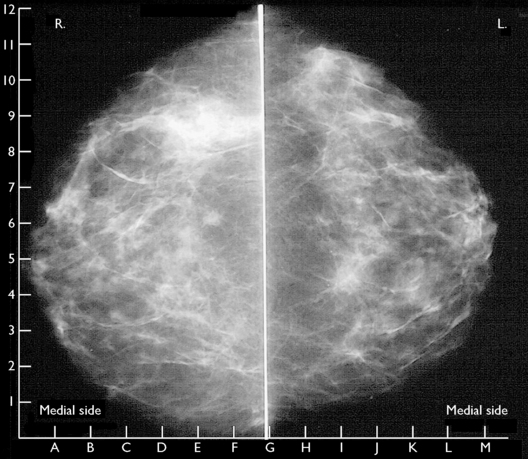
For this reason, programmes to persuade women to learn and practise breast self-examination have been developed in many countries (see p. 2). In addition, health authorities recommend that women over 35 have an annual breast examination by a doctor. This should be supplemented by mammography between the ages of 40 and 45, then annually from the age of 50 (Fig. 40.1). Two views should be taken, as this increases the detection of breast cancer and reduces recall rates. About 95% of women who have mammographic and clinical screening will have no evidence of breast cancer, 5% will require further investigation, and 1% will need biopsy to establish or exclude breast cancer. In women at high risk (Box 40.1), MRI has been shown to have a high sensitivity for detection of malignancy particularly in women younger than 40 years.

The aetiology of breast cancer has not been elucidated. A genetic factor exists, as breast cancer tends to be found in families. Three genes, BRCA1, BRCA2 and a mutation of TP53, carry a high risk of breast cancer in younger women, although these account for only a small proportion of cases. For women with BRCA1 mutation the life-time risk of developing breast cancer is between 55–85%, and 16–60% for ovarian cancer. For BRCA2 the risks are similar for breast cancer but less, 11–27%, for ovarian cancer. Childbearing before the age of 30 seems to be protective.

Treatment

Early breast cancer is best treated by breast-conserving surgery and radiation therapy to the axilla. The effectiveness of adjuvant treatment using chemotherapy or tamoxifen is being studied. A meta-analysis of 145 000 women with early breast cancer has shown that those who received additional treatment following surgery had higher 5-, 10- and 15-year survival rates. For women under the age of 50 adjuvant chemotherapy for 6 months, tamoxifen for 2 years or ovarian ablation had approximately equal efficacy in prolonging survival. The treatments reduced the annual breast cancer death rate by 30%. For women over 50, tamoxifen or chemotherapy increased the survival at 10 years by 12%. Tamoxifen had fewer adverse effects.

More advanced breast carcinoma is treated by modified radical mastectomy or radiotherapy. Survival is increased in postmenopausal women under 70 who have positive lymph nodes and are oestrogen receptor positive, if they are given chemotherapy (including doxorubicin, paclitaxel and cyclophosphamide) initially, followed by tamoxifen for 2–5 years. If the woman is over 70 chemotherapy may be omitted. Women who have positive nodes but are receptor negative should be prescribed chemotherapy, whereas women who have negative nodes but are receptor positive should take tamoxifen for 2–5 years.

These treatments eliminate oestrogen secretion, with the result that the patient is likely to suffer severe menopausal symptoms (hot flushes, dry vagina, bone loss), which may interfere with her social and sexual life. The use of hormone replacement therapy remains controversial. For this reason, each patient should be given sufficient information to enable her to make an informed choice.

For women with the BRCA1–2 mutations prophylactic mastectomy may be considered although this does not totally remove the risk of developing breast cancer. Bilateral oophorectomy in the premenopausal woman reduces the risk of breast cancer by 50% and ovarian cancer by 80–95%. Selective oestrogen receptor modulators (SERMS) including tamoxifen and raloxifene decreased the risk by 50% but all have significant side-effects which need to be taken into consideration when counselling these women.