The Acute Abdomen

Published on 06/06/2015 by admin

Filed under Pediatrics

Last modified 22/04/2025

Print this page

rate 1 star rate 2 star rate 3 star rate 4 star rate 5 star
Your rating: none, Average: 0 (0 votes)

This article have been viewed 4641 times

5 The Acute Abdomen

Abdominal pain is a chief complaint frequently encountered in the pediatric office, urgent care, and emergency department settings. Although typically minor and self-limited, acute abdominal pain may also signify a medical or surgical process requiring immediate treatment. The clinician’s role is to identify patients who have serious or potentially life-threatening conditions, such as acute appendicitis, bowel obstruction, or peritonitis. The most difficult challenge lies in making a timely diagnosis so treatment can be initiated and potential morbidity prevented.

In this chapter, a clinical guideline is presented for the evaluation and management of children with acute abdominal pain. Appendicitis is the most common surgical emergency in children and adolescents and deserves special mention. Key features of its pathophysiology, clinical presentation, evaluation, and management are highlighted throughout this chapter.

Etiology and Pathogenesis

Abdominal pain falls into three clinical categories: visceral, parietal (somatic), and referred pain. A general understanding of these is helpful in determining the cause of abdominal pain.

Visceral pain is poorly localized and is often described as dull and aching. It is caused by stretching, distension, or ischemia of the viscera. Parietal (somatic) pain is well localized, discrete, and often described as sharp and intense in character. Pain is stimulated by stretching, inflammation, or ischemia of the parietal peritoneum. The pattern of pain in appendicitis has features of both visceral and parietal pain. Initially, the pain is visceral in nature: vague, poorly localized, and periumbilical. As the peritoneum becomes inflamed over the ensuing 12 to 48 hours, the pain migrates to and localizes in the right lower quadrant (RLQ).

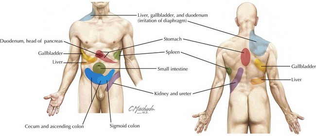
Referred pain is often perceived at sites distant from the affected organ and may be described either as sharp and localized or as a vague aching sensation (Figure 5-1). Examples include irritation of the parietal pleura of the lung perceived as abdominal wall pain and inflammation of the gallbladder perceived as scapular pain.

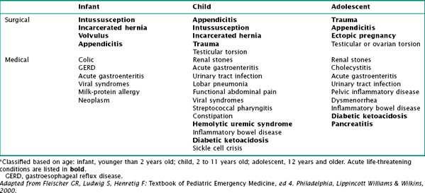
It is helpful to classify the cause of acute abdominal pain by age (Table 5-1). There are many potential causes of abdominal pain, including infectious, anatomic, traumatic, inflammatory, functional, and oncologic.

Clinical Presentation

Differential Diagnosis

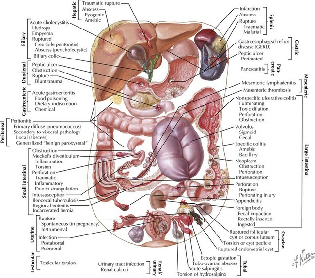
The differential diagnosis for abdominal pain in children is very broad, and it can be approached in different ways (Figure 5-2). Certain conditions occur more commonly at specific ages and therefore it may be useful to classify causes of acute abdominal pain based on age. Pain may also be classified as surgical or medical (Table 5-1).

Across all ages, gastroenteritis and appendicitis are the most common medical and surgical causes of acute abdominal pain, respectively. Malrotation with midgut volvulus is the single most devastating abdominal surgical emergency of childhood (see Chapter 109).

Life-threatening causes of abdominal pain include those related to trauma, intestinal obstruction, and peritoneal irritation. Examples of processes that present with intestinal obstruction are intussusception, midgut volvulus, and extrinsic obstruction caused by adhesions from prior surgery. Peritoneal irritation may be secondary to inflammation or perforated viscus; examples include necrotizing enterocolitis in a neonate, acute appendicitis, and ruptured ectopic pregnancy.

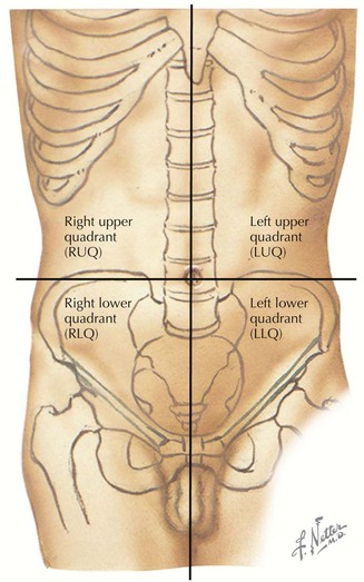
Abdominal pain can also be classified based on the location of the pain. A classic approach is by dividing the abdomen into four quadrants (Figure 5-3). This can help the practitioner direct the workup to rule in or out the most common diagnoses based on the location of symptoms. For example, hepatic and gallbladder disease usually present with right upper quadrant pain (see Chapter 115). Appendicitis classically presents with migration of pain to the RLQ. Gastritis or peptic ulcer disease may present with left upper quadrant pain (see Chapter 108).

Pain Character

Acute abdominal pain caused by a medical or surgical emergency typically increases in intensity over time, may awaken the child at night, and likely interferes with activity. In addition to the age of patient and the location of the pain, other important features of the history include the onset, frequency and duration, pattern, associated symptoms, and pertinent medical history.

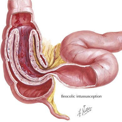
Infants and young children can seldom localize their pain, and parents often describe an inconsolable child who lies with his or her legs drawn up to the chest. Asking the parents if they think the child is in pain can be helpful to distinguish pain from fussiness or irritability. Pain that is intermittent, with paroxysms of cramping inconsolable pain alternating with return to normal state, is characteristic of intussusception (Figure 5-4). Peritoneal irritation is suggested by pain that is worse with movements that change the tension of the abdominal wall, such as a bumpy car ride or walking. Pain that improves after vomiting or a bowel movement reflects a small bowel or large bowel cause, respectively.

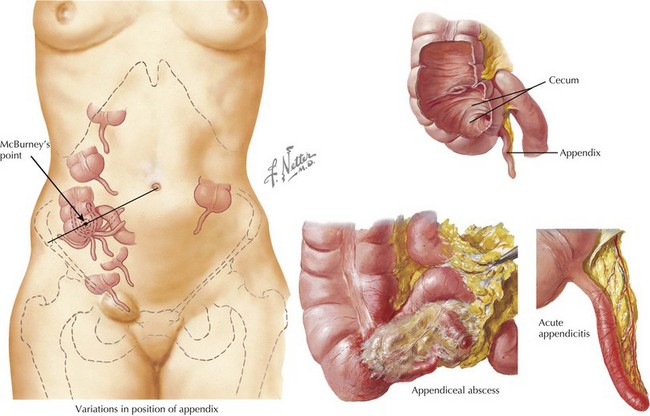
The pain of acute appendicitis is progressive in character without intermittent relief. The initial pain of early evolving appendicitis is classically periumbilical. As the inflammatory process progresses to affect the surrounding abdominal wall structures, the pain shifts to the RLQ (Figure 5-5). This progression is very helpful if obtained on history. Patients may describe a sudden temporary decrease in pain, which often coincides with perforation and temporary relief of intraluminal pressure.

Associated Symptoms

Certain infectious processes are associated with abdominal pain: fever, headache, and sore throat suggest streptococcal pharyngitis (see Chapter 35); dysuria and vomiting may be attributable to a urinary tract infection (see Chapter 93); tachypnea and cough point to a lower lobar pneumonia (see Chapter 91). An associated rash may suggest a vasculitis such as Henoch-Schönlein purpura as the cause of the abdominal pain (see Chapter 28). Whereas nonbloody diarrhea suggests gastroenteritis, bloody diarrhea could also signal hemolytic uremic syndrome, inflammatory bowel disease, or bacterial enteritis. Abdominal pain that is accompanied by vomiting but without diarrhea should prompt a more careful evaluation for potentially life-threatening conditions such as intussusception, midgut volvulus, adhesive small bowel obstruction, or pancreatitis.

Appendicitis is classically accompanied by anorexia before the onset of pain, although this is not always the case. Fever is variable, and vomiting usually occurs after the pain has migrated to the RLQ. Diarrhea is occasionally seen with appendicitis, often caused by pelvic irritation after perforation. The diagnosis of appendicitis can be difficult in children because classic symptoms are often not present, especially in infants and very young children.

Pertinent Medical History

Certain chronic medical conditions are associated with abdominal complications. Patients with sickle cell disease are predisposed to cholecystitis, splenic sequestration, and abdominal vaso-occlusive crises (see Chapter 53). Patients with diabetic ketoacidosis often present with accompanying abdominal pain (see Chapter 4). Patients with Hirschsprung’s disease are at risk for enterocolitis and toxic megacolon, and those with nephrotic syndrome may develop primary bacterial peritonitis. Patients with a history of abdominal surgery are predisposed to adhesions, which may cause obstruction.

Physical Examination

Clinicians can gain considerable information from the patient’s general appearance even as observed from afar. A child in severe pain may prefer to be curled up in a position that shortens the rectus muscles and protects the abdomen. Patients with peritonitis often appear acutely ill and prefer to lie still. Intussusception should always be considered in a young child with altered mental status. Clinicians should pay attention to vital signs, specifically for the presence of fever, tachycardia, or hypotension. Signs of poor perfusion may be seen with peritonitis or hypovolemia.

The abdominal examination is optimally performed when the child is quiet, calm, and cooperative. Clinician should consider examining young patients in the parent’s lap, if possible. Start in the area away from reported pain. Evaluate for distension and the presence or absence of bowel sounds. Palpate while paying close attention to presence of masses or focal tenderness. A mass may be indicative of neoplasm, intussusception, or stool. Reproducible focal tenderness points to an intraabdominal inflammatory process. Patients with peritoneal irritation present with percussion pain, involuntary guarding, or rebound tenderness. Rebound is elicited by deep palpation followed by a sudden release. Pay attention to the child’s face when attempting these maneuvers because they may not always be able to express localization. The rectal examination is sometimes useful in helping to narrow the differential diagnosis: note the presence of hard stool in the rectal vault (constipation), blood (intussusception, inflammatory bowel disease, infection, milk-protein allergy), or an inflamed retrocecal appendix.

Signs on examination that suggest an acute surgical cause include marked abdominal distension, severe tenderness, rigidity, involuntary guarding, and rebound tenderness. Classic presentations of acute surgical conditions include a neonate with bilious vomiting (midgut volvulus), colicky abdominal pain (intussusception), and RLQ pain (appendicitis). Surgical subspecialists should be alerted early when a surgical cause is suspected. A delay in the diagnosis of appendicitis is associated with rupture and associated complications, especially in young children younger the age of 2 years. A delay in diagnosis increases risk of perforation and postoperative complications to as high as 39%.

Overall, the cause is less likely to be serious in an otherwise healthy child who has a nonfocal examination, is without obvious discomfort to deep palpation, and is without constitutional or extraabdominal findings.

Evaluation and Management

The clinician’s first priority is to stabilize patients who are critically ill, focusing on identifying and addressing abnormalities in airway, breathing, and circulation. Next, the clinician must identify potential acutely life-threatening processes requiring emergent surgical intervention (Table 5-1). Typically, these are distinguished based on the patient’s history and physical examination, as discussed previously. Laboratory testing and imaging studies may be useful adjuncts in identifying the diagnosis.

Imaging Studies

Imaging studies should be performed when the history and physical examination reveal focal findings or suggest concerning diagnoses.

Abdominal plain films are useful in evaluating for obstruction (air-fluid levels, distended bowel), mass, or perforation (free air). An optimal radiographic study requires supine and upright views to evaluate the bowel gas pattern and movement of intestinal loops. Although the results are most often normal, radiographic signs of appendicitis include a sentinel loop (localized bowel obstruction), an appendicolith, or obliteration of the psoas shadow.

Any child with suspected midgut volvulus (i.e., report of bilious vomiting in an infant) needs an emergent contrast study to clarify the rotational status of his or her small intestine. Failure to diagnose and surgically correct midgut volvulus in a timely manner can lead to infarction of the entire small intestine. Infarction is a preventable catastrophe that may result in long-term total parenteral nutrition dependence and potentially death.

Ultrasonography (US) is the imaging modality of choice for evaluation of the biliary tree and suspected urolithiasis and in the diagnosis of ovarian or testicular pathology, such as torsion or tubo-ovarian abscess. Additionally, US aids in diagnosis of intussusception, pyloric stenosis and appendicitis. Advantages are that it is safe, noninvasive, and does not use radiation; however, the ability to obtain results often depends on the skill of the technician. The sensitivity and specificity of US for diagnosing appendicitis are greater than 90%. Signs of appendicitis on US include a noncompressible tubular structure in RLQ, wall thickness larger than 2 mm, overall diameter larger than 6 mm, free fluid in the RLQ, thickening of the mesentery, and localized tenderness with compression.

Because of the risks of radiation, especially in children, computed tomography (CT) should be reserved to rule in or out significant pathology. CT with contrast is most useful when considering a wide variety of diagnoses, such as intraabdominal mass, pancreatitis, intraabdominal abscess, and appendicitis. Noncontrast CT is indicated in the evaluation of urolithiasis when US is inconclusive. The sensitivity and specificity of CT for diagnosing appendicitis are greater than 90% and 85% to 90%, respectively. The signs of appendicitis on CT include wall thickness larger than 2 mm, appendicolith, abscess or phlegmon, free fluid greater than expected for age, thickening of the mesentery, and fat stranding.