[level-membership-for-anesthesiology-category]
The 17 Segment Model
John C. Sciarra
(Update note: In the ancient history of the first edition, there were 16 segments. Apparently we have evolved a 17th segment!)
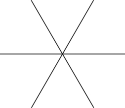
First off, draw a line. Just a straight line. As seen below this is a straight line.

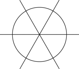
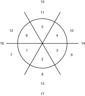
Grip your pencil tightly because I am going to ask you to do something completely different. Draw a circle on top of your star.
%
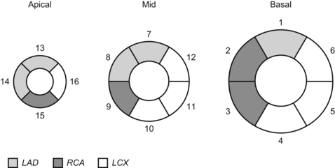
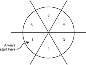
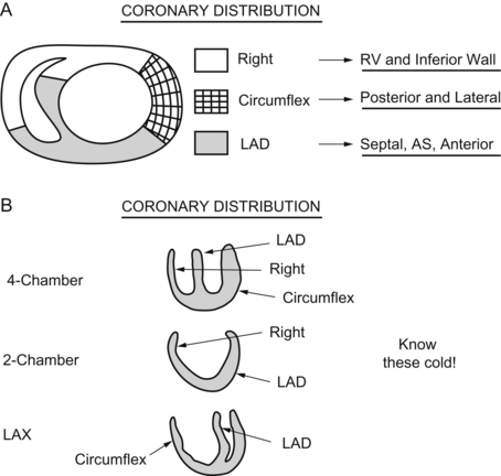
This is the image you might see in most cardiology textbooks—if you ever dared to crack one open.
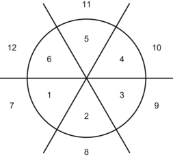
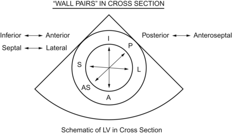
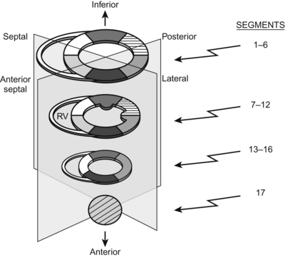
Alright, let’s get back to the world of anesthesia. This next image is the one that has the positional planes lined up as the heart sits in the chest and as the probe is at the back of the heart. Our orientation is the top of the page is the back of the heart—the inferior portion—and the front of the heart is at the bottom of the page or TEE image—anterior. This should all be crystal clear in the cool exploded 3-D image I made below, which I drew from an actual exploded heart.
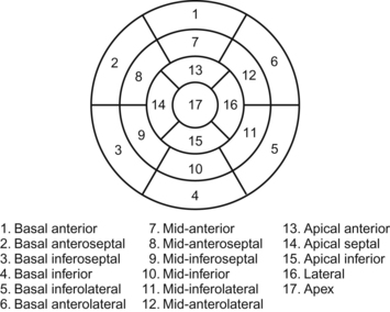
To review the coronary anatomy. Which really shouldn’t be necessary—but what the heck:
LM → LAD → Diags (the “D” in LAD leads into the “D” in diagonals).
Circ → OMs (circumferential looks like the circumference of the “O” in Obtuse Marginal).
(I hope those little memory hints help.)
%
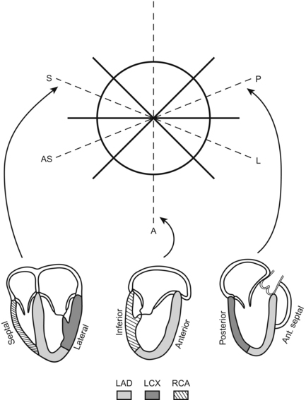
So there you have it. The star makes it all come together. I recommend trying to draw the 17 segment star in a few days to see if you remembered it. Then in the future, during say, some exam, you see a question involving the coronary arteries, wall segments, or ECG changes, just draw the star and figure it out. Nothing will stop your star power!
Questions
1. Ischemia in the RCA would give wall motion abnormalities in segment 2 or 5?
2. Segment 14 is hyperkinetic. You tell the surgeon which graft is down?
3. The patient from the beginning of the chapter gets better, gets a gun and goes all gangster on his shooter for revenge. He aims for the LAD. Does he hit segment 2 or 12?
Answers
[/level-membership-for-anesthesiology-category][not-level-membership-for-anesthesiology-category]
The 17 Segment Model
John C. Sciarra
(Update note: In the ancient history of the first edition, there were 16 segments. Apparently we have evolved a 17th segment!)
First off, draw a line. Just a straight line. As seen below this is a straight line.
Grip your pencil tightly because I am going to ask you to do something completely different. Draw a circle on top of your star.
[/not-level-membership-for-anesthesiology-category]
