19 Tendons
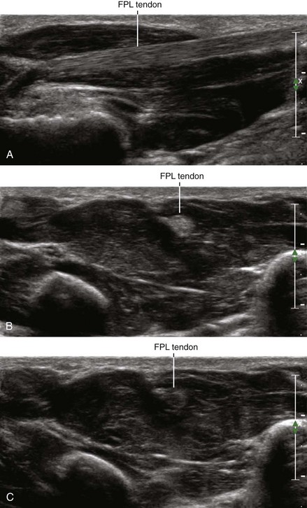
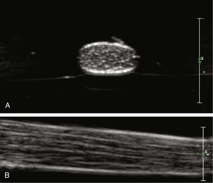
Tendons are the strong structures that connect muscle to bone. Their fibrillar echotexture (fiber-like, appearing as the fine hairs of a violin bow) results from parallel collagen bundles. Because of this ordered architecture, tendons are highly anisotropic, meaning that the received echoes are highly dependent on the angle of insonation.1–3
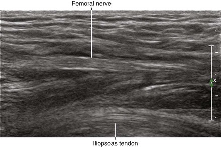
Tendons and nerves are both imaged during regional block procedures, and therefore some commentary regarding their discriminatory features is appropriate. Although the two structures can appear similar, tendons and nerves are primarily distinguished by tracing their course. Because tendons only form at the ends of muscle, changes in cross-sectional area along the course are substantial. For nerves, the cross-sectional area is relatively constant along the nerve path. In addition, the amplitudes of the received echoes from tendons are more sensitive to transducer inclination than nerves (tendons are more anisotropic than nerves). At high frequencies of insonation (≥10 MHz), the fibrillar echotexture of tendons can be distinguished from the fascicular echotexture of nerves (Table 19-1).4
Table 19-1 Characteristics of Nerves and Tendons that Can Be Identified with Ultrasound Imaging
| Characteristic | Nerve | Tendon |
|---|---|---|
| Echotexture | Fascicular | Fibrillar |
| Elemental composition | Fascicles (coarse, thick, wavy, less numerous) | Fibrils (fine, thin, straight, more numerous) |
| Internal architecture | Plexiform (combine and divide) | Parallel fibrils |
| Cross-sectional area | Constant along nerve path | Forms at the ends of muscle |
| Overall shape | Round, oval, or triangular | Round, oval, or triangular; C or S |
| Shape can change along path | Shape does not change along path | |
| Branching | Yes | None |
| Anisotropy | Moderately sensitive | Highly sensitive |
| Adjacent vessels | Often | Infrequent |
| Border | Not as distinct | Distinct paratenon |
| Compressibility | More compressible | Less compressible |
Tendons can have central or eccentric location within muscle depending on whether the muscle is bipennate or unipennate, respectively. Multipennate muscles have more than one tendon. Normal tendons are avascular with no flow detectable either on color-flow or power Doppler examinations.5 Direct injections into tendons have been associated with tendon rupture.6
Clinical Pearls
• The position of musculotendinous junctions is variable, making it difficult to identify tendons based on anatomic position alone.
• Tendons consist of a network of thin parallel and longitudinally oriented specular echoes that resemble fibrils. The septa consist of loose connective tissue with elastic fibers, vessels, and thin muscle fibers.
• Nerves typically have fewer and thicker echogenic lines than do tendons.
1 Fornage BD. The hypoechoic normal tendon: a pitfall. J Ultrasound Med. 1987;6:19–22.
2 Crass JR, van de Vegte GL, Harkavy LA. Tendon echogenicity: ex vivo study. Radiology. 1988;167:499–501.
3 Connolly DJ, Berman L, McNally EG. The use of beam angulation to overcome anisotropy when viewing human tendon with high-frequency linear array ultrasound. Br J Radiol. 2001;74:183–185.
4 Silvestri E, Martinoli C, Derchi LE, et al. Echotexture of peripheral nerves: correlation between US and histologic findings and criteria to differentiate tendons. Radiology. 1995;197:291–296.
5 O’Connor PJ, Grainger AJ, Morgan SR, et al. Ultrasound assessment of tendons in asymptomatic volunteers: a study of reproducibility. Eur Radiol. 2004;14:1968–1973.
6 Ford LT, DeBender J. Tendon rupture after local steroid injection. South Med J. 1979;72:827–830.