Temporomandibular Disorders
Effects of Occlusion, Orthodontic Treatment, and Orthognathic Surgery
• Incidence of Temporomandibular Disorders in the General Population and the Basic Treatment Approach
• Review of Occlusal Factors and Association with Temporomandibular Disorders
• Occlusal Conditions More Frequently Associated with Temporomandibular Disorders
• Orthodontic Treatment and Its Effects on Temporomandibular Disorders
• Association of Jaw Deformities, Orthognathic Surgery, and Temporomandibular Disorders
• Brief Overview of Okeson’s Thinking About Temporomandibular Disorders and the Effects of Occlusion
• Personal Thoughts About the Role of Open Joint Procedures for Temporomandibular Disorders
Incidence of Temporomandibular Disorders in the General Population and the Basic Treatment Approach
The prevalence of temporomandibular disorders (TMDs) in the general population as reported in the literature varies widely. It is estimated that an average of 32% of the population reports at least one symptom of TMD, whereas an average of 55% demonstrates at least one clinical sign.* Schiffman and colleagues completed a cross-sectional epidemiologic study that indicated that, at any given time, between 40% and 75% of individuals who were not currently seeking treatment had at least one sign of TMD, and about one third of individuals reported at least one symptom of TMD.296 Mongini and colleagues documented a prevalence of TMD symptoms in children and teenagers of 12% and 20%, respectively.210 Wanman and colleagues showed an incidence of joint noise in young adults in their late teens to be as high as 17.5% over a 2-year period.345 TMDs remain a frequent cause of visits to primary care physicians, otolaryngologists, internists, and pediatricians. Despite the high incidence of TMDs, their natural history suggests that, in up to 40% of patients, symptoms resolve spontaneously.296
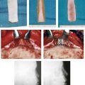
TMD includes various signs and symptoms of the temporomandibular joint (TMJ), the masticatory muscles, and related structures (Figs. 9-1 through 9-8). These may include a spectrum of referred head and neck pain; joint noise (e.g., popping, clicking, crepitus); reduced or altered mandibular range of movement as a result of muscle spasm or disc displacement; condylar head erosion; and direct pain on palpation of either the TMJ or the masticatory muscles. The signs and symptoms associated with TMDs can also lead to misdiagnosis.16,112,171,216,342
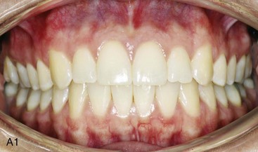
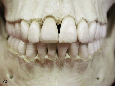
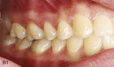
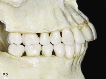
Figure 9-1 A, Intraoral frontal view of dentition juxtaposed with the underlying alveolar bone. B, Intraoral lateral view of dentition juxtaposed with the underlying alveolar bone. From Okeson JP: Management of temporomandibular disorders and occlusion, St. Louis, 2012, Elsevier, p 3, Figures 1-1, A and B, 1-3, and 1-4.
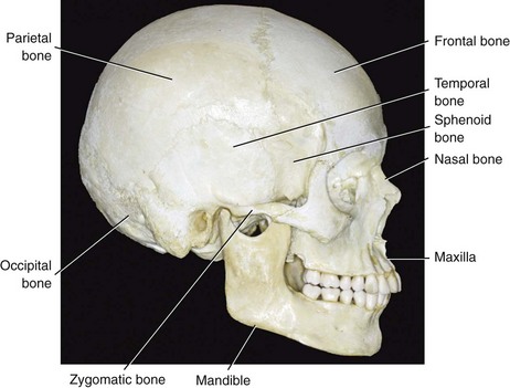
Figure 9-2 Lateral view of the craniofacial skeleton. From Okeson JP: Management of temporomandibular disorders and occlusion, St. Louis, 2012, Elsevier, p 4, Figure 1-5.
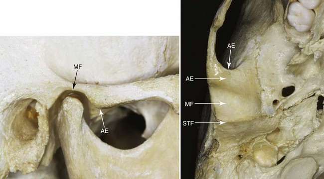
Figure 9-3 Bony structures (components) of the temporomandibular joint. MF, Mandibular fossa; AE, articular eminence; STF, squamotympanic fissure. From Okeson JP: Management of temporomandibular disorders and occlusion, St. Louis, 2012, Elsevier, p 4, Figure 1-12.
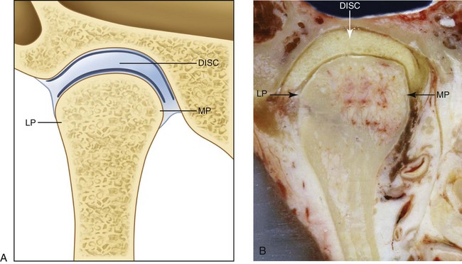
Figure 9-4 Anterior cross-sectional view of the articular disc, the fossa, and the condyle. A, Illustration. B, Cadaver specimen. The disc adapts to the morphology of the fossa and the condyle. LP, Lateral pole; MP, medial pole. From Okeson JP: Management of temporomandibular disorders and occlusion, St. Louis, 2012, Elsevier, p 8, Figure 1-14.
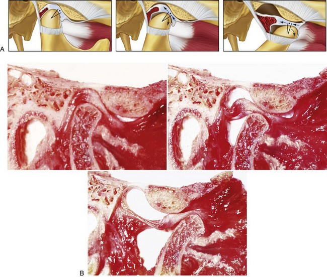
Figure 9-5 A, Lateral view illustrations and B, cadaver specimen views of normal movement of the condyle and disc during vertical mouth opening. As the disc moves out of the fossa, it rotates posteriorly on the condyle. First, rotational movement occurs (predominantly in the lower joint space). After that, translation takes place (predominantly in the superior joint space) as mouth opening continues. A from Okeson JP: Management of temporomandibular disorders and occlusion, St. Louis, 2012, Elsevier, p 17, Figure 1-30. B courtesy Terry Tanaka, MD, San Diego, Calif.
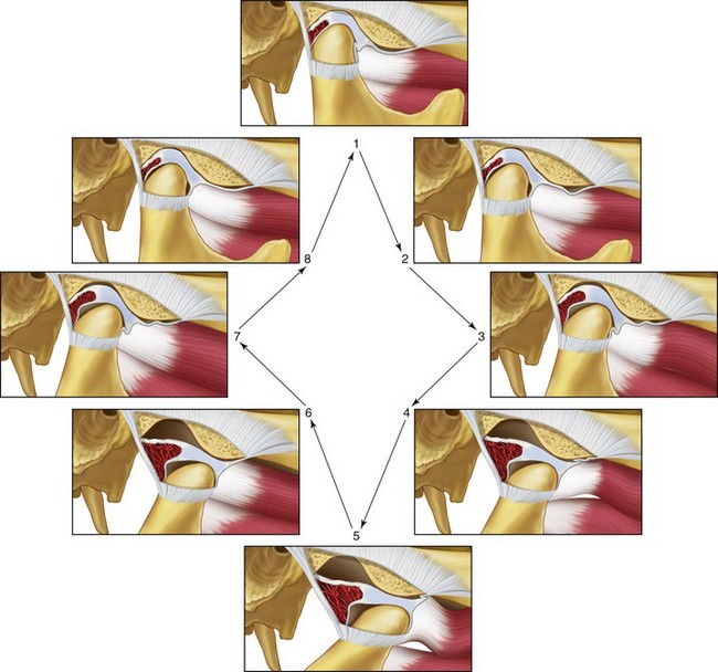
Figure 9-6 Illustrations of the normal functional movement of the condyle and disc during the full range of opening and closing. The disc is rotated posteriorly on the condyle as the condyle is translated anteriorly out of the fossa. The closing movement is the exact opposite of the opening movement. From Okeson JP: Management of temporomandibular disorders and occlusion, St. Louis, 2012, Elsevier, p 19, Figure 1-31.
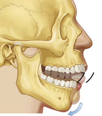
Figure 9-7 Illustration of rotational movement of the mandible. This movement occurs with the condyles in the terminal hinge position. This pretranslation opening will occur until the anterior teeth are approximately 20 to 25 mm apart. From Okeson JP: Management of temporomandibular disorders and occlusion, St. Louis, 2012, Elsevier, p 64, Figure 4-7.
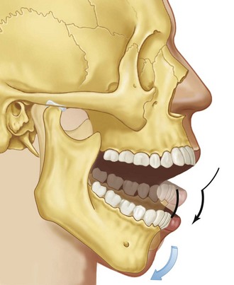
Figure 9-8 The second stage of movement during mouth opening is illustrated. Note that the condyle is translated down the articular eminence as the mouth rotates open to its maximum limit. From Okeson JP: Management of temporomandibular disorders and occlusion, St. Louis, 2012, Elsevier, p 64, Figure 4-8.
A wide variety of often contradictory “conservative” treatment modalities (i.e., non-invasive or potentially reversible) have been recommended to alleviate the symptoms of TMD and to prevent recurrence.* Enthusiasm by clinicians for the direct surgical alteration of the structures that comprise the TMJ to treat TMD have waxed and waned over the past half century.60,71,141,143,200,222,241,344 Occlusal factors (i.e., degrees of malocclusion) are often claimed to be associated with TMD. Although it is sometimes stated that orthodontic treatment increases the prevalence of TMD, it is also claimed that orthodontic maneuvers (for at least some types of malocclusion) may reduce the signs and symptoms of TMD. Studies document that the correction of a baseline jaw deformity with malocclusion through orthodontics and orthognathic surgery improves the presenting TMD in the majority of the treated patients, whereas a minority get worse.6,29,31,67,102,131,152,232,236,347,349,356,359 This is described in greater detail later in this chapter.
Mandibular Range of Motion
The expected normal mandibular range of motion and inter-incisor mouth opening is discussed periodically in the literature.9,70,93,115,146,179,197,234,235,249,308,309,311–313,315,316,318 Even a 6-year-old child can often open the mouth to a maximum 40 mm or more. Measurements of maximum vertical mouth opening with and without discomfort are considered useful parameters of TMJ health. A restricted mouth opening is considered by many to be a distance of less than 40 mm. Only 1.2% of young adults open their mouths less than 40 mm. However, 15% of the healthy elderly population open their mouths less than 40 mm. Any lateral or protrusive movement of less than 8 mm is considered restricted, although the significance of this measurement is unclear.
Review of Occlusal Factors and Association with Temporomandibular Disorders
Clinicians from a spectrum of medical and dental specialties often consider malocclusion to be the major etiologic factor of TMD.† An assumed strong association between TMD and the malpositioning of the teeth serves as one reason why the diagnosis and treatment of TMD is generally considered within the purview of dentistry. Many of the therapeutic options for the treatment of TMD are based on the idea of “normalizing” or “neutralizing” the occlusion.135,355 Common treatment approaches to do so include the reversible use of an occlusal appliance or definitive techniques such as occlusal adjustment (equilibration), restorative dentistry, orthodontics, and orthognathic surgery. Concurrently, it is also true that either the onset of TMD or the worsening of current symptoms is frequently blamed on any one of the previously mentioned treatment regimens.
The dental literature contains clinical studies and reports that claim there are causal relationships between occlusal factors and the symptoms and signs associated with TMD. Egermark-Eriksson and colleagues examined 402 children and reported an association between signs and symptoms of TMD, specific malocclusions (i.e., Class II, Class III, anterior crossbite, and anterior open bite), and occlusal supra-contacts.87 Brandt studied 1342 children and found a correlation between TMD and degrees of overbite (deep and open) and overjet.33 Nilner examined 749 teenagers and young adults and reported an association with centric relation (CR)–centric occlusion (CO) discrepancies (i.e., slides), balancing-side contact slides, and symptoms and signs of TMD.220 Confusion in the literature arises as independent research studies have drawn contrary conclusions. DeBoever and colleagues examined 135 individuals with signs and symptoms of TMD and found no specific associations with malocclusion.59 Gunn and colleagues similarly found no association between TMD and occlusion in 151 migrant children that they studied.121 Dworkin and colleagues examined 592 individuals within a specific health maintenance organization and found no association between TMD signs and symptoms and specific occlusions.80
In their review article, Seligman and Pullinger pointed out that the majority of existing clinical research (up to that point in time) did not support a clear relationship between occlusion and TMD symptoms.302 They further clarified that the majority of published research relied on symptom-based data rather than discrete disease classifications. Seligman pointed out that symptoms like joint sounds (e.g., popping, clicking, crepitus) and pain (e.g., in the TMJ region and the masticatory system) lack specificity.303 In addition, most published studies did not differentiate between TMD diagnostic categories (e.g., disc displacement, myalgia, TMJ osteoarthritis). Most published studies also suffered from a lack of or an inadequate control group. Seligman and colleagues attempted to avoid these shortcomings by completing a multifactorial analysis of the risks and relative odds of an individual having a TMD as a function of 1 of 11 common occlusal features.259,304 They looked for correlations between each of these 11 specific occlusal factors (Box 9-1) and 1 of 6 TMJ diagnostic categories (Box 9-2), including asymptomatic normal controls without TMD.259
• Overjets ranging from –1 to +6 mm
• Overbites ranging from –2 to +10 mm
• Midline discrepancies of up to 5 mm
• Anteroposterior molar relationships from –6 to +6 mm
Pullinger and colleagues concluded that occlusion cannot be considered the most important factor in the definition of TMD but that specific occlusal factors do make biologic contributions to TMD and thus cannot be ignored.254–261 In most individuals, they observed that the human biologic system will adapt to a variety of malocclusions in an attempt to achieve TMJ stability. The authors surmised that some occlusal features will place greater adaptive demands on the individual’s biologic system than others. Although most individuals are able to compensate for their occlusal disharmony without problems, adaptation for others may lead to a greater risk for TMD. On the basis of this data, the authors stressed the importance of five occlusal conditions, which in isolation or combination are more likely to reach the threshold that exceeds adaptation for an individual and therefore results in TMJ dysfunction. The high-risk malocclusions are discussed later in this chapter.
Occlusal Conditions More Frequently Associated with Temporomandibular Disorders
Anterior Open Bite
Research by Pullinger and colleagues showed a strong relationship between osteoarthritis of the condylar head with a loss of condylar height and an anterior open-bite malocclusion.254–261 Interestingly, anterior open bite was not commonly associated with disc displacement disorders with or without reduction. Myalgia (i.e., masticatory muscle discomfort) was also a frequent symptom among patients with an anterior open bite. It is not stated in what percentage of individuals the anterior open-bite malocclusion occurred only secondarily as a result of osteoarthritis followed by a loss of condylar height with clockwise rotation of the mandible. Therefore, it is unclear what percentage of the examined individuals with anterior open bites had these finding as part of a developmental jaw deformity.
Centric Relation–Centric Occlusion Slides
Limited occlusal slides (i.e., 1 mm) were common in all patient groups, including the normal controls. CR–CO slides of more than 2 mm were only found in the patients with TMDs. Pullinger found that larger slides were frequently associated with osteoarthritis of the condylar head. He questioned the advisability of the clinicians’ automatic attempt to “relieve” the occlusal slides in all individuals—whether by irreversible (e.g., occlusal equilibration, orthodontics, surgery, restorative dentistry) or reversible (e.g., occlusal splints) means—because the observed slide may be either adaptive or inconsequential.260
 Note:
Note: Unilateral Maxillary (Lingual) Crossbite
The occlusal feature of unilateral maxillary crossbite occurs in about 10% of the adult population, and it is frequently found in association with TMJ disc displacement with or without disc reduction. Approximately 25% of the patients with non-reducing disc displacements were found to have unilateral maxillary lingual crossbites. This indicated to Pullinger and colleagues that disc displacement may be a biologic adaptive response of the individual to a unilateral maxillary lingual crossbite.260 The investigators theorized that these individuals may later be prone to osteoarthritis. They suggested that the orthodontic treatment of a unilateral crossbite discovered during childhood should be considered as a preventative measure to reduce the adaptive demands on the masticatory system. They did not come to the same conclusion for the correction of a longstanding unilateral posterior crossbite in an adult, because skeletal adaptation would have already occurred. Unfortunately, the investigators did not clarify if the crossbites in their study patients were the result of developmental jaw deformities (e.g., asymmetrical mandibular excess) or simply due to the malpositioning of the teeth. They did not specifically comment on whether orthognathic surgery should be carried out in conjunction with orthodontics (if needed) in young adults to correct the presenting malocclusion as a way to prevent the progression of TMD.
Missing Posterior Teeth (≥5)
The investigators found that, when five or more posterior teeth were missing, the incidence of TMD increased. This includes TMD categories of both osteoarthritis and disc displacement with or without osteoarthritis. Pullinger and colleagues further claimed that, when two or four of the “posterior teeth” (bicuspids) were removed as part of an orthodontic treatment protocol, there was a negligible effect on TMD.260 Other studies carried out by Kayser156 and Witter354 have also documented acceptable masticatory function after bicuspid extractions and orthodontic treatment without increased signs and symptoms of TMD.
 Note:
Note: The multifactorial analysis of occlusal factors and TMD disease categories carried out by Pullinger and colleagues showed that, except for the few clearly defined occlusal features described previously, there was a low probability of a malocclusion being associated with TMD. Seligman and colleagues estimated that the overall contribution of occlusal factors for the defining of patients with TMD was only in the range of 10% to 20%.302 The corollary is that 80% to 90% of the causative factors cannot be explained by patients’ presenting malocclusions.
In the Pullinger and Seligman study, it must be assumed that many of the significant overjets observed in the adult subjects were secondary deformities after condylar resorption (i.e., condylysis).260 The authors did not present evidence to suggest that the presence of significant overbite or overjet plays a role in the pathophysiology of non-arthritic disorders of the TMJ, and they did not show that it was a causative factor in the observed osteoarthritis. They do believe that a combination of unilateral retruded centric position in the presence of a clinically apparent retruded centric position–inter-contact position slide may encourage TMJ disc displacement but that unilateral retruded centric position per se is not associated with TMD. They found that lost molar support (i.e., 4 or 5 posterior teeth) may be associated with osteoarthrosis. They concluded that a dental midline discrepancy is not a predictor of TMD development during adulthood. Anterior open bite is frequent in the presence of condylysis or osteoarthritic changes in the TMJ, but it is probably the result of (rather than the cause of) the osteoarthritic changes. Interestingly, when open bites are observed in children, they are rarely associated with TMJ symptoms. In the pediatric population, the majority of observed open bites represent a developmental jaw deformity rather than a result of osteoarthritis. Nevertheless, it is recommended that, when either a child or an adult is found to have an anterior open bite, radiographic analysis of the condylar head should be undertaken to check for osteoarthritic changes.
Orthodontic Treatment and Its Effects on Temporomandibular Disorders
Review of the Literature
The relationships among malocclusion, orthodontic treatment to improve the occlusion, and the effects on TMD is discussed frequently in the literature.* A spectrum of orthodontic treatment philosophies has been championed by individual practitioners on the basis of presumed effects on TMD. The longitudinal nature of orthodontic treatment, which is often carried out over several years, means that a changing TMD history would also be expected in a control group that is not simultaneously undergoing orthodontic treatment. Therefore, the occurrence of joint noise (e.g., popping, clicking) or myalgia (i.e., masticatory muscle discomfort) during orthodontic treatment may either be the result of the treatment or consistent with the natural history of the untreated individual.
Sadowsky and colleagues reported on 75 adult study patients who had undergone orthodontic treatment with full appliances during adolescence at least 10 years earlier.290 The treated group was compared with a group of 75 similar adults with untreated malocclusions. The authors also went on to increase both their control and subject groups and to report these additional findings several years later.291 In the two sequenced studies, respectively, 15% to 21% of the treated subjects presented with one or more signs of TMD (e.g., joint noise), and 29% to 42% had at least one or more symptoms of TMD. There were no statistically significant differences between the treated and untreated (control) groups.
Larsson and colleagues evaluated 23 individuals who had undergone orthodontic treatment during adolescence at least 10 years earlier.172 Mild TMD was recorded in 8 out of 23 (35%) of the patients, whereas only 1 (4%) had severe TMD. The authors compared this with published epidemiologic controls and stated that there was no difference in the incidence of TMD in the treated group versus the control group.
Dahl and colleagues examined 51 subjects 5 years after the completion of orthodontic treatment and compared them with an untreated control group.54 The authors found no statistical differences in TMD between the two groups. The number of subjects in both groups who had at least one mild TMD symptom was relatively high (70% in the treated group versus 90% in the untreated group). Mild symptoms (e.g., joint noise), masticatory muscle fatigue, and stiffness in the lower jaw were frequently observed in both groups.
A study by Smith and Freer examined 87 individuals who were treated with full orthodontic bracketing during adolescence.320 The treatment group (N = 87) was compared with an untreated control group (N = 28). Four years after treatment ended, TMD symptoms were found in 21% of the treated group and 14% of the control group; this was not found to be statistically significant. The one symptom that was statistically more common in the treatment group was a “soft click” (64% of the treated group versus 36% of the untreated group).
Rendell and colleagues examined 462 individuals (90% adolescents and 10% adults) who were undergoing orthodontic treatment in a graduate orthodontic dental school clinic.267 With the use of a modified Helkimo Index, only 11 of the patients presented with TMD signs or symptoms before treatment. Of the other 451 patients, during the 18-month study, none developed TMD signs or symptoms. For those with preexisting TMD signs or symptoms (N = 11), there was no consistent change in the level of pain or dysfunction reported during treatment.
Kremenak and colleagues reported pretreatment and posttreatment TMD examination data from 109 patients who were treated with full orthodontics.166,167 The Helkimo Index scores were also compared. No statistically significant differences were noted between the subjects’ pretreatment and posttreatment scores. Ninety percent of the patients had Helkimo Index scores that either remained the same or improved, and 10% of the treated patients had scores that worsened. The authors concluded that the orthodontic treatment provided to their patients was not an important etiologic factor for the development of TMD.
Hirata and colleagues examined 102 individuals before and after orthodontic treatment for signs and symptoms of TMD.128 They compared their treatment group with 41 untreated controls who were matched for age. The incidence of TMD for the treatment and control groups was not statistically significantly different.
 Note:
Note: Orthodontic Treatment Undertaken Specifically to Relieve Temporomandibular Disorders
In everyday dental practice, the establishment of a preferred intra-arch morphologic relationship of the teeth and then the achievement of a satisfactory inter-arch relationship of the teeth in one jaw to those in the other jaw (i.e., occlusion) serves as the basis of treatment objectives. According to a review of the literature, the establishment of a “normal” occlusion through orthodontic mechanics or via other dental means carried out for the purpose of eliminating or preventing the worsening of TMD does not seem warranted. Pullinger and Seligman have stated five potential exceptions to this, which were described previously in this chapter.255–261 McNamara and colleagues also stated that prudent basic dental treatment objectives will generally include the establishment of the six keys to ideal occlusion as advocated by Andrews, when feasible; the establishment of a limited CR–CO slide (i.e., <2 mm); the limiting of an anterior open bite; the alleviation of significant overjet (i.e., >6 to 7 mm); the limiting of unilateral maxillary lingual crossbites in children; and the establishment of a stable posterior occlusion.
Orthodontic Treatment Undertaken Specifically to Prevent Temporomandibular Disorders
The literature also contains studies that suggest that specific orthodontic maneuvers are effective for the prevention of TMD. The studies by Magnusson and colleagues187–192 and the study by Egermark and colleagues89 suggest preventative TMD advantages related to orthodontic occlusal corrections. At 5 and 10 years, these investigators reevaluated a group of 402 children and adolescents who had originally undergone a full course of orthodontic treatment. The Helkimo Index was used to measure clinical signs of TMD in the now older age group (i.e., ≥25 years old). Interestingly, the Helkimo Index outcome was lower in those who had undergone orthodontic treatment as compared with the control group. The studies by Dahl and colleagues and Sadowsky and colleagues also demonstrated a trend (although not a statistically significant one) toward the decreased prevalence of TMD signs and symptoms in orthodontically treated patients.53,290,291 These studies would require further confirmation before accepting them as credible indications of a cause-and-effect relationship.
Summary of Effects of Orthodontic Treatment on Temporomandibular Disorders
McNamara, Seligman, and Okeson summarize their understanding of the relationship between orthodontic treatment and TMDs as follows203:
1. Signs and symptoms of TMD do occur in healthy individuals.
2. Signs and symptoms of TMD increase with age, particularly during adolescence.
3. Orthodontic treatment performed during adolescence generally does not increase or decrease the odds of developing TMD later in life.
4. The extraction of teeth as part of an orthodontic treatment plan does not increase the risk of TMD.
5. There is no elevated risk for TMD associated with any particular type of conventional orthodontic mechanics.
6. Although a stable occlusion is a reasonable orthodontic treatment goal, the inability to achieve a specific gnathologic ideal occlusion does not result in TMD signs or symptoms.
7. No specific method of TMD prevention has been demonstrated to be effective.
8. When more severe TMD signs and symptoms are present, simple forms of treatment can alleviate them in most patients.
 Note:
Note: McNamara, Seligman, and Okeson suggest that until reliable criteria are developed for the treatment of TMD, the dental and medical professions should be encouraged to manage presenting TMJ symptoms with reversible therapies and to only consider permanent alterations in the occlusion (for the specific purpose of treating or preventing TMD) in patients with very unique circumstances. This is in agreement with the National Institure of Health conference guidelines which states that reversible therapies should be used in the primary treatment of TMD219
A summary of evidence-based systematic reviews of TMDs was carried out by Rinchuse and McMinn.283 Their systematic review resulted in the following conclusions:
1. Occlusion, which was once considered the primary and sole cause of TMD, now at best is recognized as having a secondary role in causing it.
2. Conventional orthodontic treatment that improves the occlusion does not cause TMD.
3. The use of occlusal adjustments in orthodontic patients has no evidence-based support for the treatment of TMD.
4. There is evidence-based support for the use of occlusal splints and biofeedback for the treatment of TMD.
Association of Jaw Deformities, Orthognathic Surgery, and Temporomandibular Disorders
Review of the Literature
The association of a jaw deformity with malocclusion and the effects of orthodontic and surgical correction on TMD signs and symptoms have been examined in clinical studies for the past several decades.* The quality of the research and therefore the value of the conclusions vary. There are just three longitudinal prospective controlled clinical trial studies that have been carried out to assess the effects of orthognathic surgery on TMD: the studies by Onizawa and colleagues,232 Panula and colleagues,236 and Dervis and colleagues.67
The study by Onizawa and colleagues sought to evaluate changes in the symptoms of TMD after orthognathic surgery.232 TMJ symptoms in preoperative orthognathic patients (N = 30) were compared with those of healthy control volunteers (N = 30). Changes in TMD symptoms were evaluated in both groups at 3- and 6-month postoperative intervals. The authors found no significant differences in the incidence of TMJ sounds, deviation of mouth opening, or tenderness on direct palpation of the TMJ and masticatory muscles between patients who were scheduled to undergo orthognathic surgery and control volunteers. The treatment group did not report TMJ symptoms significantly more often than the control subjects. At both the 3- and 6-month postoperative intervals, half or more of the treatment group showed no change in TMJ sounds, deviation of mouth opening, or tenderness on palpation of the TMJ or masticatory muscles. At each evaluation time frame, some individuals in both the control group and the study group exhibited improvement in TMJ symptoms, whereas others showed a change for the worse. Findings of postoperative TMJ sounds were observed to be associated with a reduction of mandibular mobility (i.e., mouth opening). The authors concluded that TMD symptoms did not always show improvement after surgical correction and that, for some patients, the symptoms changed for the worse.
Panula and colleagues carried out research to study the effects of orthognathic surgery on TMJ dysfunction.236 They completed a prospective follow-up study to examine the influence of orthognathic procedures for the correction of dentofacial deformities on signs and symptoms of TMJ dysfunction. Sixty consecutive patients with dentofacial deformities were examined preoperatively and then twice after orthognathic surgery. The Helkimo Anamnestic and Dysfunction Indices were also given as a questionnaire, and the results were used as part of the determination. The prevalence of headache was also assessed. The average follow-up was 4 years after the initial preoperative examination. A group (N = 20) with a similar type and grade of dentofacial deformity who elected not to undergo surgical correction or any other occlusal therapy served as the control group. The majority of the treatment group (73%) was found to have signs and symptoms of TMJ dysfunction or TMD during the preoperative examination. At the final postsurgery examination (i.e., 4 years after surgery), the prevalence of TMD had been reduced to 60%. There was also a dramatic improvement in the prevalence of headache, which was initially experienced by 63% of patients and reduced to 25% of patients at the final visit. The authors concluded that signs and symptoms of TMD, including pain (headache) levels, could be reduced with orthognathic treatment. The risk of an orthognathic patient experiencing new TMD was extremely low. No association could be shown between TMD and any specific type or magnitude of dentofacial deformity.
A study was conducted by Dervis and colleagues with the objective of looking at the long-term prevalence of TMD in individuals with a dentofacial deformity who then underwent orthognathic surgery as compared with a control group.67 Signs and symptoms of TMD in both the dentofacial deformity patients and the control group were evaluated before surgery, 1 week after release of intermaxillary fixation, and 1 and 2 years after surgery with the use of the Helkimo Anamnestic and Dysfunction Indices. At both the initial preoperative examination and the final examination (i.e., 2 years after surgery), signs and symptoms of TMD in the patients who were undergoing orthognathic surgery were compared with those of healthy control subjects. The lengths of condylar pathways during opening and with lateral and protrusive movements were also evaluated with the use of axiography. A statistically significant reduction of TMD symptoms and signs 2 years after surgery as compared with before surgery was found. Interestingly, at the initial examination, the patients with dentofacial deformities who were scheduled to undergo orthognathic surgery did not report TMD signs and symptoms with a greater frequency than other healthy control subjects. At the final examination (i.e., 2 years after surgery), greater improvements in TMD symptoms in the patients who had undergone orthognathic surgery were found as compared with healthy control subjects. The authors also found a statistically significant decrease in masticatory muscle tenderness after surgery. The authors concluded that the “functional status” of the TMJ can be improved with orthognathic surgery. There was no clear association between TMD symptoms and a specific type or pattern of dentofacial deformity.
When comparing and contrasting the three published longitudinal prospective controlled studies that were conducted to determine the effects of orthognathic procedures on TMD symptoms and signs (i.e., Onizawa and colleagues,232 Panula and colleagues,236 and Dervis and colleagues67), several observations can be made:
1. Patients with dentofacial deformities who were scheduled to undergo orthognathic surgery were not found to have significant differences in their TMD signs and symptoms as compared with patients in the control groups.
2. After orthognathic surgery and orthodontics, signs and symptoms related to TMD were frequently improved.
3. Despite the trend for diminished signs and symptoms of TMD after orthognathic surgery and orthodontics, individuals did not always show improvement; for some, the symptoms changed for the worse.
4. After orthognathic surgery and orthodontic treatment, a decrease in masticatory muscle tenderness was frequent. A decrease in joint tenderness on palpation was also a common occurrence.
5. In one of the three studies reviewed, a decrease in maximum mouth opening was observed, but this was not found to be of clinical importance.
Karabouta and colleagues reviewed 280 individuals with different patterns of mandibular deformities (e.g., prognathism, retrognathia, open bite, asymmetry) who underwent orthognathic surgery that included sagittal split ramus osteotomies of the mandible. Before surgery, the patients were found to present subjective or objective TMJ dysfunction symptoms with an incidence of 41%.154 After surgery, the incidence of such symptoms in the same patients was reduced to 11%. Individuals with no preoperative TMJ dysfunction symptoms developed such symptoms with an incidence of 4% after surgery.
White and colleagues set out to review the prevalence of TMD in patients with dentofacial deformities who were undergoing orthognathic surgery.349 Preoperatively, 49% of their study group presented with TMD. After orthognathic surgery, in the symptomatic patients, 89% had improved temporomandibular function, 3% were unchanged, and 8% developed increased symptoms. Of the preoperative asymptomatic patients, 8% developed TMD after surgery. In the study group, TMD was significantly greater in patients with Class II skeletal deformities as compared with those with Class III tendencies. Overall, temporomandibular function generally improved in both treatment groups (i.e., Class II and Class III) after successful surgery.
Hori and colleagues reported three individual (non-consecutive) cases in which there was a worsening of preexisting TMJ dysfunction after sagittal split ramus osteotomy of the mandible and 6 weeks of intermaxillary fixation.131 All three of their patients initially presented specifically seeking treatment of malocclusion for the relief of TMD. All three patients had active TMD before and after their orthognathic procedures.
Aghabeigi and colleagues studied the effect of orthognathic surgery on the TMJ of patients who presented with an anterior open bite.6 This was a retrospective survey study of patients (N = 83) who presented with an anterior open bite who then underwent orthognathic surgery. The preoperative prevalence of head and neck pain was 32%, TMJ dysfunction was found in 40% of patients, and limited mouth opening was found in 7%. The overall prevalence of TMJ signs and symptoms was not found to be significantly different after orthognathic surgery. In the study group, an abnormal psychological profile was the most significant factor associated with the presence and persistence of TMJ pain. The authors concluded that orthognathic surgery does not significantly influence TMD in patients with anterior open bites. Female patients—particularly those with abnormal psychological profile—were at higher risk for persistent postoperative TMJ pain.
Westermark and colleagues studied a consecutive series of individuals to understand the effects of orthognathic surgery on the signs and symptoms of TMD.347 The authors sent a survey questionnaire to a consecutive series of patients (N = 1516) who initially presented with dentofacial deformities and who then underwent orthognathic surgeries that were performed by a group of experienced surgeons at a single institution. The survey was given at least 2 years after surgery. The questions asked in the survey related to joint pain, chewing pain, joint noise, grinding, daytime headache, and morning headache. Preoperatively, 43% of the patients reported subjective symptoms of TMD; postoperatively, 28% of the patients did. This difference suggested an overall beneficial effect of orthognathic surgery on TMD signs and symptoms. Patients with Class II skeletal deformities did not improve as much as with those with Class III skeletal prognathism.
Yamada and colleagues studied the incidence of condylar morphologic change, disc displacement, and signs and symptoms of TMD in individuals set to undergo orthognathic surgery.359 The study showed preoperative condylar morphologic change (unilateral or bilateral) in 36% of the subjects and in 24% of each joint. Preoperative disc displacement (unilateral or bilateral) was seen in 41% of the subjects and in 30% of each joint. The percentage of condylar morphologic change and disc displacement was higher for individuals with anterior open-bite skeletal Class II growth patterns and lower for patients with skeletal Class III deformities.
Wolford and colleagues studied changes in TMD after orthognathic surgery in a series of 25 patients who were scheduled to undergo bimaxillary surgery.356 They used magnetic resonance imaging and clinical verification of TMJ articular disc displacement. This was a retrospective evaluation with an average follow up of 2.2 years. Presurgically, 16% of the patients had only TMJ pain, 64% had only TMJ sounds, and 20% had both TMJ pain and sounds. Postoperatively, 24% of the patients had only TMJ pain, 16% had only TMJ sounds, and 60% had both TMJ pain and sounds. Thirty-six percent of the patients had TMJ pain preoperatively; this jumped to 84% after surgery. In addition, the authors claimed that 6 out of 25 of their patients (24%) developed condylar resorption postoperatively, thereby explaining the high incidence of observed Class II open-bite malocclusion. The authors demonstrated that patients with preexisting TMJ dysfunction who undergo bimaxillary osteotomies via the techniques that they used have a high probability of the significant worsening of TMJ dysfunction after surgery. This reported high incidence of TMD and condylar resorption after orthognathic surgery is contrary to all other studies reported in the literature.
Borstlap and colleagues reported about a consecutive series of individuals who were scheduled to undergo sagittal split ramus osteotomies for both Class II mandibular retrognathism and Class III mandibular prognathism.31 Patients were reviewed preoperatively, immediately postoperatively, and at 3, 6, and 24 months postoperatively. Any signs and symptoms of TMD were documented, including clicking, preauricular pain, maximum mouth opening, protrusive movements, and lateral excursions. The authors documented that 56% of the patients with preexisting TMD had relief of their signs and symptoms. Interestingly, 22% of individuals without preexisting TMD went on to develop clinical signs or symptoms after surgery.
Bock and colleagues studied the importance of temporomandibular function as a factor in the achievement of patient satisfaction after orthognathic surgery.29 The authors’ orthognathic surgery patients (N = 102) were examined at an average of 47 months after surgery. The average age of their patients was 24 years. A patient satisfaction questionnaire and other modalities were reviewed, including the Helkimo Index, clinical findings, nerve function parameters, and the incidence of intraoperative and postoperative complications. The results of their study showed that 91% of patients were satisfied or very satisfied with their overall orthognathic results. Answers to the questionnaires revealed that only 79% of patients would undergo treatment again (i.e., 21% of the patients would not). Statistically significant differences were found in the Helkimo Index and in the pain reported both in the TMJs and with jaw movements between the satisfied and dissatisfied postoperative patients. The authors surmised that the 21% of patients who would not undergo treatment again were more likely to have a greater degree of TMD and limitations with regard to mandibular range of movement. Statistically significant differences were found between those who were satisfied and those who were dissatisfied with surgery on the basis of their Helkimo Indices. Those with pain on palpation in the TMJ region and those with pain experienced during motion of the mandible were more likely to be overall dissatisfied with the treatment. The persistence of postoperative symptoms of TMD, including pain, was the single most important correlation with low patient satisfaction.
Frey and colleagues studied the effects of mandibular advancement procedures and counterclockwise rotation on signs and symptoms of TMD.102 The study patients were followed for a minimum of 2 years after orthognathic surgery. A series of patients with Angle Class II malocclusions (N = 127) who were undergoing sagittal split ramus osteotomies with advancement and counterclockwise rotation of the mandible were studied to evaluate signs and symptoms of TMD at specific intervals after surgery. The results of the study indicate that counterclockwise rotation of the mandible was associated with more masticatory muscle tenderness early after surgery, especially in those who were undergoing more than 7 mm of advancement. The combination of extensive mandibular advancement with counterclockwise rotation was also associated with increased TMD symptoms early after surgery. Interestingly, all TMJ symptoms declined by 2 years after surgery. The authors conclude that surgical advancement and counterclockwise rotation of the mandible (i.e., sagittal split ramus osteotomy) is associated with a slight increase in masticatory muscle discomfort during initial recovery. All symptomatology tended to decline over time, which suggests that the amount of advancement and the amount of mandibular counterclockwise rotation should not be considered as risk factors for the long-term development of TMD in patients without preexisting symptoms.
Jung and colleagues retrospectively evaluated the effects of bilateral intraoral vertical ramus osteotomies with setback for the management of Class III skeletal patterns on symptoms that are associated with TMD.152 Patients were evaluated preoperatively and at intervals (i.e., 1, 3, 6, 12, 18, and 24 months) after surgery for specific parameters, including mouth opening, clicking, and TMJ pain. The authors reported that 94% of those with preoperative joint noise achieved resolution. After surgery, 8% of individuals who were free of noise before surgery developed joint noise afterward. On average, mouth opening was 50 mm before surgery and 49 mm 6 months after surgery. Joints that were free of pain before surgery remained so. Those individuals with preoperative TMJ pain remained free of pain at 18 months. The authors concluded that bilateral intraoral vertical ramus osteotomy with setback as a method of managing Class III skeletal deformities was also effective for the relief of undesirable TMJ symptoms (e.g., joint noise, pain) without negatively affecting mouth opening.
Additional thoughts Concerning Effects of Orthodontics and Orthognathic Surgery on Temporomandibular Disorders
It may not be correct to conclude that orthodontic treatment and orthognathic surgery carried out for the correction of a presenting jaw deformity with malocclusion are the responsible factors for any observed reduction or resolution of TMD signs and symptoms.18 During the decades that have passed since McCarty and Farrar first defined the diagnoses of internal derangement of the TMJ and then described a disc repositioning procedure to treat it, quality research to evaluate their recommended treatment has been scant.200 The experience of orthopedic surgeons with regard to the treatment of derangements of the knee should remind us of the need for clinical practice to be based on sound supporting evidence. Moseley and colleagues carried out a controlled clinical trial of arthroscopic surgery (i.e., lysis and lavage or arthroplasty) for osteoarthritis of the knee that should give us pause when considering irreversible TMD treatment.212 Clinician opinions regarding the effectiveness of lysis and lavage or arthroplasty to manage knee pathology in anticipation of improved function and relief of pain have varied but are currently without consensus. Therefore, a randomized clinical trial that included a placebo group was designed by Moseley and colleagues to address this controversy. To blind the placebo group, a sham surgical procedure was designed as part of the study. The placebo subjects were given a general or regional anesthetic, and incisions were made; however, the arthroscope was not inserted into the joint, and no therapeutic procedure was carried out. The results of the study were that neither treatment group (i.e., lysis and lavage or arthroplasty) offered improved clinical outcomes as compared with the placebo group for osteoarthritis. As a consequence of this evidence-based study, many orthopedic surgeons have changed treatment recommendations for their patients. In any case, Medicare (i.e., the U.S. government medical insurance plan for the elderly) made a decision that it would no longer cover arthroscopy for osteoarthritis of the knee. Restorative dentists, orthodontists, and orthognathic surgeons should remain circumspect when advising patients regarding irreversible treatment specifically for the purpose of relieving TMD.
Brief Overview of Okeson’s Thinking About Temporomandibular Disorders and the Effects of Occlusion
Okeson believes that, during the normal use of the masticatory system, either local or systemic events can occur that may influence function.165,227–231 Each individual has a varied ability to tolerate these events without any adverse head and neck effects. It is likely that an individual’s tolerance can be influenced by these local or systemic factors. When an event exceeds the individual’s physiologic tolerance, the system begins to reveal changes. Each component of the masticatory system can tolerate a certain amount of change. When functional change exceeds a critical level, alteration of the tissues begins. The tissue breakdown site is generally seen in the structure with the least tolerance. Therefore, the breakdown site varies from individual to individual. This is influenced by factors such as the individual’s unique maxillofacial morphology, previous head and neck trauma, and local tissue conditions. The weakest link in the masticatory system will break first (i.e. masticatory muscles, TMJs, supporting structures of the teeth, the teeth themselves). Typically, the weakest structures in the system are the masticatory muscles; this is evidenced by muscle tenderness and pain during mandibular movements.
Personal Thoughts About the Role of Open Joint Procedures for Temporomandibular Disorders
A critical review of the literature suggests that outcomes for open TMJ procedures are often unpredictable and may result in further TMD.73–77 The success rate for “redo” open joint surgery is even less favorable. Among the criteria for an open joint procedure are the following: that the clinician must be confident that the source of the pain or dysfunction is arising from within the joint; that the appropriate use of conservative measures has failed; that the current symptoms are intolerable to a patient who is psychosocially stable; and that a significant long-term success rate for the selected open joint procedure in the specific patient can be assured.
References
1. Academy of Denture Prosthetics. Glossary of prosthodontic terms (appendix). J Prosthet Dent. 1956; 692:5–34.
2. Ackerman, M. The myth of Janus: orthodontic progress faces orthodontic history. Am J Orthod Dentofacial Orthop. 2003; 123:594–596.
3. Ackerman, M. Evidence-based orthodontics for the 21st century. J Am Dent Assoc. 2004; 135:162–167.
4. Adams, S, Zander, HA. Functional tooth contacts in lateral and in centric relation. J Am Dent Assoc. 1964; 69:465–473.
5. Agerberg, A. Occlusal and temporomandibular joint relations: a comparative study. Cranio. 1987; 5:233–238.
6. Aghabeigi, B, Hiranaka, D, Keith, DA, et al. Effect of orthognathic surgery on the temporomandibular joint in patients with anterior open bite. Int J Adult Orthodon Orthognath Surg. 2001; 16:153.
7. Ahlborg, G, Finne, K. Changes in temporomandibular joint pain dysfunction after surgical correction of dentofacial anomalies. Int J Oral Maxillofac Surg. 1986; 15:707–714.
8. Ahlgren, J, Posselt, U. Need of functional analysis and selective grinding in orthodontics: a clinical and electromyographic study. Acta Odontol Scand. 1963; 21:187–217.
9. Ahlgren, J. Mechanism of mastication. Acta Odontol Scand. 1966; 24(Suppl 44):1–109.
10. Al-Ani, Z, Gray, RJ, Davies, SJ, et al. Stabilization splint therapy for the treatment of temporomandibular myofascial pain: a systematic review. J Dent Educ. 2005; 69:1242–1250.
11. Alexander, PC. Analysis of the cuspid protected occlusion. J Prosthet Dent. 1963; 13:309–317.
12. Alexander, SR, Moore, RN, DuBois, LM. Mandibular condyle position: comparison of articulator mountings and magnetic resonance imaging. Am J Orthod Dentofacial Orthop. 1993; 104:230–239.
13. American Academy of Pediatric Dentistry, University of Texas Health Science Center at San Antonio Dental School. Treatment of temporomandibular disorders in children: summary statements and recommendations. J Am Dent Assoc. 1990; 120:265–269.
14. Andrews, LF. The six keys to normal occlusion. Am J Orthod. 1972; 62:296–309.
15. Angle, EH. Malocclusion of teeth, ed 7. Philadelphia: SS White Dental Mfg Co; 1907.
16. Ash, MM. Current concepts in the etiology, diagnosis and treatment of TMJ and muscle dysfunction. J Oral Rehabil. 1986; 13:1.
17. Ash, MM, Ramjford, S. Occlusion, ed 4. Philadelphia: Saunders; 1996.
18. Assael, LA. Temporomandibular disorders in surgical practice: does science support treatment decisions? J Oral Maxillofac Surg. 2009; 67:935–936.
19. Aubrey, RB. Occlusal objectives in orthodontic treatment. Am J Orthod. 1978; 74:162–175.
20. Auerbach, SM, Laskin, DM, Frantsve, LM, Orr, T. Depression, pain, exposure to stressful life events, and long-term outcomes in temporomandibular disorder patients. J Oral Maxillofac Surg. 2001; 59:628–634.
21. Barghi, N, Agular, T, Martinez, C, et al. Prevalence of types of temporomandibular joint clicking in subjects missing posterior teeth. J Prosthet Dent. 1987; 57:617–620.
22. Bean, LR, Thomas, CA. Significance of condylar positions in patients with temporomandibular disorders. J Am Dent Assoc. 1987; 114(1):76–77.
23. Begg, PR. Stone Age man’s dentition. Am J Orthod. 1954; 40:298–312.
24. Bell, WE. Clinical diagnosis of the pain-dysfunction syndrome. J Am Dent Assoc. 1969; 79:154.
25. Bell, WH, Gonyea, W, Finn, RA, et al. Muscular rehabilitation after orthognathic surgery. Oral Surg Oral Med Oral Pathol. 1983; 56:229.
26. Belser, UC, Hannam, AG. The influence of altered working-side occlusal guidance on masticatory muscles and related jaw movements. J Prosthet Dent. 1985; 53:406–413.
27. Berry, DC, Watkinson, AC. Mandibular dysfunction and incisor relationship. Br Dent J. 1978; 144:74–77.
28. Berry, DC, Singh, BP. Daily variation in occlusal contacts. J Prosthet Dent. 1983; 50:386–391.
29. Bock, JJ, Fuhrmann, RAW. The importance of temporomandibular function for patient satisfaction following orthognathic surgery. J Orofac Orthop. 2007; 68:299–307.
30. Boero, RP. The physiology of splint therapy: a literature review. Angle Orthod. 1989; 59:165–180.
31. Borstlap, WA, Stoelinga, PJW, Hoppenreijs, TJM, van’t Hof, MA. Stabilisation of sagittal split advancement osteotomies with miniplates: a prospective, multicentre study with two-year follow-up Part I. Clinical parameters. Int J Oral Maxillofac Surg. 2004; 33:433–441.
32. Boyd, SB, Karas, ND, Sinn, DP. Recovery of mandibular mobility following orthognathic surgery. J Oral Maxillofac Surg. 1991; 49:924.
33. Brandt, D. Temporomandibular disorders and their association with morphologic malocclusion in children. In: Carlson DS, McNamara JA, Ribbens KA, eds. Developmental aspects of temporomandibular joint disorders. Monograph 16, Craniofacial Growth Series. Ann Arbor, Mich: Center for Human Growth and Development, The University of Michigan; 1985:296–297.
34. Brodie, AG. Differential diagnosis of joint conditions in orthodontia. Angle Orthod. 1934; 4:160–170.
35. Brodie, AG. The temporomandibular joint. Ill Dent J. 1939; 8:2–12.
36. Buckley, MJ, Tulloch, JFC, White, RP, et al. Complications of orthognathic surgery: a comparison between wire and rigid fixation. Int J Adult Orthodon Orthognath Surg. 1989; 4:69.
37. Budtz-Jorgensen, E, Luan, W, Holm-Pedersen, P, Fejerskov, O. Mandibular dysfunction related to dental, occlusal and prosthetic conditions in a selected elderly population. Gerodontics. 1985; 1:28–33.
38. Bush, FM. Malocclusion, masticatory muscle, and temporomandibular joint tenderness. J Dent Res. 1985; 64:129–133.
39. Carlsson, GE, Egermark, I, Magnusson, T. Predictors of bruxism, other oral parafunctions, and tooth wear over a 20-year follow-up period. J Orofac Pain. 2003; 17:50–57.
40. Carlsson, GE, Magnusson, T, Egermark, I. Prediction of demand for treatment of temporomandibular disorders based on a 20-year follow-up study. J Oral Rehabil. 2004; 31:511–517.
41. Celenza, FV. The centric position: replacement and character. J Prosthet Dent. 1973; 30:591–598.
42. Chiappone, RC. A gnathologic approach to orthodontic finishing. J Clin Orthod. 1975; 9:405–417.
43. Christensen, GJ. Is occlusion becoming more confusing? A plea for simplicity. J Am Dent Assoc. 2004; 135:767–770.
44. Clark, JR, Evans, RD. Functional occlusion: I. A review. J Orthod. 2001; 28:76–81.
45. Cohen, WE. A study of occlusal interferences in orthodontically treated occlusions and untreated normal occlusions. Am J Orthod. 1965; 51:647.
46. Cordray, FE. A crisis in orthodontists? It’s time to look within. Am J Orthod Dentofacial Orthop. 1992; 101:472–476.
47. Cordray, FE. Centric relation treatment and articulator mountings in orthodontics. Angle Orthod. 1996; 66(2):153–158.
48. Cordray, FE. Most condylar discrepancies not detectable clinically. Am J Orthod Dentofacial Orthop. 2001; 120:15A.
49. Cordray, FE. Three-dimensional analysis of models articulated in the seated condylar position from a deprogrammed asymptomatic population: a prospective study. Part 1. Am J Orthod Dentofacial Orthop. 2006; 129:619–630.
50. Costen, JB. Neuralgia and ear symptoms associated with disturbed function of the temporomandibular joint. JAMA. 1936; 107:252–258.
51. Crawford, SD. Condylar axis position, as determined by the occlusion and measured by the CPI instrument, and signs and symptoms of temporomandibular dysfunction. Angle Orthod. 1999; 69:103–115.
52. Creekmore, DC, Cetlin, NM, Ricketts, RM, et al. JCO roundtable: diagnosis and treatment planning. J Clin Orthod. 1992; 26:585–606.
53. Crider, A, Glaros, AG, Gevirtz, RN. Efficacy of biofeedback-based treatments for temporomandibular disorders. Appl Psychophysiol Biofeedback. 2005; 30:333–345.
54. Dahl, BL, Krogstad, BO, Ogaard, B, Eckersberg, T. Signs and symptoms of craniomandibular disorders in two groups of 19-year-old individuals, one treated orthodontically and the other not. Acta Scand. 1988; 46:89–93.
55. Dahlberg, G, Petersson, A, Westesson, P-L, Eriksson, L. Disk displacement and temporomandibular joint symptoms in orthognathic surgery patients. Oral Surg Oral Med Oral Pathol. 1995; 79:273–277.
56. Dawson, PE. Optimum TMJ condyle position in clinical practice. Int J Periodontics Restorative Dent. 1985; 3:11–31.
57. Dawson, PE. Evaluation, diagnosis and treatment of occlusal problems, ed 2. St. Louis: C. V. Mosby; 1989.
58. Dawson, PE. Evidence-based versus experience-based views on occlusion and TMD. Am J Orthod Dentofacial Orthop. 2005; 128:150–151.
59. De Boever, JA, Adriaens, PA. Occlusal relationship in patients with pain-dysfunction symptoms in the temporomandibular joint. J Oral Rehabil. 1983; 10:1–7.
60. De Bont, LG, Stegenga, B. Pathology of temporomandibular joint internal derangement and osteoarthrosis. Int J Oral Maxillofac Surg. 1993; 22:71.
61. De Clercq, CA, Neyt, LF, Mommaerts, MY, et al. Condylar resorption in orthognathic surgery: A retrospective study. Int J Adult Orthodon Orthognath Surg. 1994; 9:233.
62. De Clercq, CA, Abeloos, J, Mommaerts, MY. Temporomandibular joint symptoms in an orthognathic surgery population. J Craniomaxillofac Surg. 1995; 23:195–199.
63. De Laat, A, van Steenberghe, D. Occlusal relationships and temporomandibular joint dysfunction: Part I. Epidemiologic findings. J Prosthet Dent. 1985; 54:835–842.
64. De Laat, A, van Steenberghe, D, Lesaffe, E. Occlusal relationship and temporomandibular joint dysfunction: Part II. Correlation between occlusal and articular parameters and symptoms of TMJ dysfunction by means of stepwise logistic regression. J Prosthet Dent. 1986; 55:116–121.
65. De Laat, A, Stappaerts, K, Papy, S. Counseling and physical therapy as treatment for myofascial pain of the masticatory system. J Orofac Pain. 2003; 17:42–49.
66. Dechow, PC, Carlson, DS. Occlusal force after mandibular advancement in adult rhesus monkey. J Oral Maxillofac Surg. 1986; 44:887.
67. Dervis, E, Tuncer, E. Long-term evaluations of temporomandibular disorders in patients undergoing orthognathic surgery compared with a control group. Oral Surg Oral Med Oral Pathol Oral Radiol Endod. 2002; 94:554–560.
68. Dibbets, JMH, van deer Weele, L. Orthodontic treatment in relation to symptoms attributed to dysfunction of the temporomandibular joint: a 10-year report of the University of Groningen study. Am J Orthod Dentofacial Orthop. 1987; 91:193–199.
69. Dibbets, JMH, van deer Weele, L. Orthodontic treatment modalities and TMJ dysfunction. In: Michigan T, ed. Craniofacial growth theory and orthodontic concept. Ann Arbor, Mich: Center for Human Growth and Development, The University of Michigan; 1990:153–167.
70. Dijkstra, P, Kropmans, T, Stegenga, B. The association between generalized joint hypermobility and temporomandibular joint disorders: a systematic review. J Dent Res. 2002; 81:158–163.
71. Dingman, RO. Diagnosis and treatment of lesions of the TMJ. Am J Orthod Oral Surg. 1940; 26:374–390.
72. Dionne, RA. Pharmacologic treatments for temporomandibular disorders. Oral Surg Oral Med Oral Pathol Oral Radiol Endod. 1997; 83:134–142.
73. Dolwick, MF, Katzberg, RW, Helms, CA. Internal derangement of the temporomandibular joint: fact or fiction? J Prosthet Dent. 1983; 49:415–418.
74. Dolwick, MF, Nitzan, DW. TMJ disk surgery: 8-year follow-up evaluation. Fortschr Kiefer Gesichtschir. 1990; 35:162–167.
75. Dolwick, MF, Dimitroulis, G. Is there a role for temporomandibular joint surgery? Br J Oral Maxillofac Surg. 1994; 32:307–313.
76. Dolwick, MF, Dimitroulis, G. A re-evaluation of the importance of disc position in temporomandibular disorders. Aust Dent J. 1996; 41:184–187.
77. Dolwick, MF. Temporomandibular joint surgery for internal derangement. Dent Clin North Am. 2007; 51:195–208.
78. Droukas, B, Lindee, C, Carlsson, GE. Occlusion and mandibular dysfunction: a clinical study of patients referred for functional disturbances of the masticatory system. J Prosthet Dent. 1985; 53:402–406.
79. Du, S, Rinchuse, DJ, Zullo, T, Rinchuse, DJ. Reliability of three occlusal classification systems. Am J Orthod Dentofacial Orthop. 1998; 113:463–470.
80. Dworkin, SF, Huggins, KH, LeResche, L, et al. Epidemiology of signs and symptoms of temporomandibular disorders: clinical signs in cases and controls. J Am Dent Assoc. 1990; 120:273–281.
81. Dworkin, SF, LeResce, L. Research diagnostic criteria for temporomandibular disorders: review, criteria, examinations and specifications, critique. J Craniomandib Disord. 1992; 6:301–355.
82. Dworkin, SF. The case for incorporating biobehavioral treatment into TMD management. J Am Dent Assoc. 1996; 127:1607–1610.
83. Dworkin, SF, Sherman, J, Mancl, L, et al. Reliability, validity, and clinical utility of the research diagnostic criteria for Temporomandibular Disorders Axis II Scales: depression, non-specific physical symptoms, and graded chronic pain. J Orofac Pain. 2002; 16:207–220.
84. Egermark, I, Carlsson, GE, Magnusson, T. A prospective long-term study of signs and symptoms of temporomandibular disorders in patients who received orthodontic treatment in childhood. Angle Orthod. 2005; 75:645–650.
85. Egermark-Eriksson, I, Ingervall, B. Anomalies of occlusion predisposing to occlusal interferences in children. Angle Orthod. 1982; 52:293–299.
86. Egermark-Eriksson, I. Mandibular dysfunction in children and in individuals with dual bite. Swed Dent J. 1982; 6:9–20.
87. Egermark-Eriksson, I. The dependence of mandibular dysfunction in children on functional and morphologic malocclusion. Am J Orthod. 1983; 83:187–194.
88. Egermark-Eriksson, I, Carlsson, GE, Magnusson, T, Thilander, B. A longitudinal study on malocclusion in relation to signs and symptoms of cranio-mandibular disorders in children and adolescents. Eur J Orthod. 1990; 12:399–407.
89. Egermark, I, Thilander, B. Craniomandibular disorders with special reference to orthodontic treatment: an evaluation from childhood to adulthood. Am J Orthod Dentofacial Orthop. 1992; 101:28–34.
90. Egermark, I, Blomqvist, JE, Cromvik, U, Isaksson, S. Temporomandibular dysfunction in patients treated with orthodontics in combination with orthognathic surgery. Eur J Orthod. 2000; 22:537–544.
91. Ekberg, E, Nilner, M. Treatment outcome of appliance therapy in temporomandibular disorder patients with myofascial pain after 6 and 12 months. Acta Odontol Scand. 2004; 62:343–349.
92. Ellis, PE, Benson, PE. Does articulating study casts make a difference to treatment planning? J Orthod. 2003; 30:45–49.
93. Farrar, WB. Characteristics of the condylar path in internal derangements of the TMJ. J Prosthet Dent. 1978; 39:319.
94. Farrar, WB, McCarty, WL, Jr. The TMJ dilemma. J Ala Dent Assoc. 1979; 63:19–26.
95. Farrar, WB. Craniomandibular practice: the state of the art; definition and diagnosis. J Cranio Pract. 1982-1983; 1:4–12.
96. Feinerman, DM, Piecuch, JF. Long-term effects of orthognatic surgery on the temporomandibular joint: comparison of rigid and nonrigid fixation methods. Int J Oral Maxillofac Surg. 1995; 24:268–272.
97. Ferrando, M, Andreu, Y, Galdón, MJ, et al. Psychological variables and temporomandibular disorders: distress, coping, and personality. Oral Surg Oral Med Oral Pathol Oral Radiol Endod. 2004; 98:153–160.
98. Flynn, B, Brown, DT, Lapp, TH, et al. A comparative study of temporomandibular symptoms following mandibular advancement by bilateral sagittal split osteotomies: rigid versus nonrigid fixation. Oral Surg Oral Med Oral Pathol. 1990; 70:372.
99. Forssel, H, Kirveskari, T, Kangasniemi, P. Response to occlusal treatment in headache patients previously treated to mock occlusal adjustment. Acta Odontol Scand. 1987; 45:77–80.
100. Forssell, H, Kalso, E, Koskela, P, et al. Occlusal treatments in temporomandibular disorders: A qualitative systematic review of randomized controlled trials. Pain. 1999; 83:549–560.
101. Franks, AST. The dental arch of patients presenting with temporomandibular joint dysfunction. Br J Oral Surg. 1967; 5:157–166.
102. Frey, DR, Hatch, JP, Van Sickels, JE, et al. Effects of surgical mandibular advancement and rotation on signs and symptoms of temporomandibular disorder: a 2-year follow-up study. Am J Orthod Dentofacial Orthop. 2008; 33:490. e1–490. e8.
103. Fujimura, K, Segami, N, Sato, J, et al. Comparison of the clinical outcomes of patients having sounds in the temporomandibular joint with skeletal mandibular deformities treated by vertico-sagittal ramus osteotomy or vertical ramus osteotomy. Oral Surg Oral Med Oral Pathol Oral Radiol Endod. 2005; 99:24.
104. Gazit, E, Liebermann, MA. Occlusal contacts following orthodontic treatment. Angle Orthod. 1985; 55:316–320.
105. Gerstner, GE, Marchi, F, Haerian, D. Relationship between antero-posterior maxillomandibular morphology and masticatory jaw movement patterns. Am J Orthod Dentofacial Orthop. 1999; 115:256–266.
106. Gesch, D, Bernhardt, O, Kocher, T, et al. Association of malocclusion and functional occlusion with signs of temporomandibular disorders in adults: results of the population-based study of health in Pomerania. Angle Orthod. 2004; 74:512–520.
107. Gianelly, AA. Orthodontics, condylar position and TMJ status. Am J Orthod Dentofacial Orthop. 1989; 95:521–523.
108. Gianelly, AA, Anderson, CK, Boffa, J. Longitudinal evaluation of condylar position in extraction and nonextraction treatment. Am J Orthod Dentofacial Orthop. 1991; 100:416–420.
109. Gianelly, AA, Cozzanic, M, Boffa, J. Condylar position and maxillary first premolar extraction. Am J Orthod Dentofacial Orthop. 1991; 99:473–476.
110. Girardot, RA, Jr. Comparison of condylar position in hyperdivergent and hypodivergent facial skeletal types. Angle Orthod. 2001; 71:240–246.
111. Glindbueg, RW, Laskin, DM, Blaustein, DI. The effects of immobilization on the primate temporomandibular joint: a histochemical study. J Oral Maxillofac Surg. 1982; 40:3.
112. Gobetti, JP, Turp, JC. Fibrosarcoma misdiagnosed as a temporomandibular disorder: a cautionary tale. Oral Surg Oral Med Oral Pathol Oral Radiol Endod. 1998; 4:404–409.
113. Goodman, P, Greene, CS, Laskin, DM. Response of patients with myofascial pain-dysfunction syndrome to mock equilibration. J Am Dent Assoc. 1976; 92:955–959.
114. Graham, MM, Buxbaum, J, Staling, LM. A study of occlusal relationships and the incidence of myofacial pain. J Prosthet Dent. 1982; 47:549–555.
115. Green, JR, Moore, CA, Ruark, JL, et al. Development of chewing in children from 12 to 48 months: a longitudinal study of EMG patterns. J Neurophysiol. 1997; 77:2704–2716.
116. Greene, CS, Lerman, MD, Sutcher, HD, Laskin, DM. The TMJ pain-dysfunction syndrome: heterogenity of the patient population. J Am Dent Assoc. 1969; 79:1169.
117. Greene, CS, Mohl, ND, McNeil, C, et al. Temporomandibular disorders and science. J Prosth Dent. 1998; 80:214–215.
118. Greene, CS. The etiology of temporomandibular disorders: implications for treatment. J Orofac Pain. 2001; 15:93–105.
119. Greene, CS, Obrez, A. Mandibular repositioning in the treatment of temporomandibular disorder: critical analysis. Alpha Omegan. 2003; 96:40–46.
120. Griffiths, RH. Guidelines for the examination, diagnosis and management of temporomandibular disorders. J Am Dent Assoc. 1983; 106:75–77.
121. Gunn, SM, Woolfolk, MW, Faja, BW. Malocclusion and TMJ symptoms in migrant children. J Craniomandib Disord. 1988; 2:196–200.
122. Guralnick, W, Kaban, LB, Merrill, RG. Temporomandibular-joint afflictions. N Engl J Med. 1978; 299:123–129.
123. Hackney, FL, Van Sickels, JE, Nummikoski, PV. Condylar displacement and temporomandibular joint dysfunction following bilateral sagittal split osteotomy and rigid fixation. J Oral Maxillofac Surg. 1989; 47:223.
124. Hart, DL, Lundquist, DO, Davis, HC. The effect of vertical dimension on muscular strength. J Orthop Sports Phys Ther. 1981; 3:57–61.
125. Heikinheimo, K, Salmi, K, Myllarniemi, S, Kirveskari, P. A longitudinal study of occlusal disorders at the ages 12 and 15. Eur J Orthod. 1990; 12:190–197.
126. Helkimo, M. Studies on function and dysfunction of the masticatory system: II. Index for anamnestic and clinical dysfunction and occlusal state. Swed Dent J. 1974; 67:101–121.
127. Henrikson, T, Nilner, M. Temporomandibular disorders and the need for stomatognathic treatment in orthodontically treated and untreated girls. Eur J Orthod. 2000; 22:283–292.
128. Hirata, RH, Heft, MW, Hernandez, B, King, GJ. Longitudinal study of signs of temporomandibular disorders (TMD) in orthodontically created and untreated groups. Am J Orthod Dentofacial Orthop. 1992; 101:35–40.
129. Hirsch, C, John, MT, Drangsholt, MT, Mancl, LA. Relationship between overbite/overjet and clicking or crepitus of the temporomandibular joint. J Orofac Pain. 2005; 19:218–225.
130. Hoffman, PJ, Silverman, SI, Garfinkel, L. Comparison of condylar position in centric relation and in centric occlusion in dentulous subjects. J Prosthet Dent. 1973; 30:582–588.
131. Hori, M, Okaue, M, Hasegawa, M, et al. Worsening of pre-existing TMJ dysfunction following sagittal split osteotomy: a study of three cases. J Oral Sci. 1999; 41:133.
132. Huang, G. Occlusal adjustment for treating and preventing temporomandibular disorders. Am J Orthod Dentofacial Orthod. 2004; 126:138–139.
133. Huber, NU, Hall, EH. A comparison of the signs of temporomandibular joint dysfunction and occlusal discrepancies in a symptom-free population of men and women. Oral Surg Oral Med Oral Pathol. 1990; 70:180–183.
134. Hwang, HS, Behrents, RG. The effect of orthodontic treatment on centric discrepancy. J Cranio Pract. 1996; 14:132–137.
135. Ingervall, B. Recording of retruded positions of mandible in children: a comparison between registrations in general anaesthesia and with children awake. Odontol Rev. 1968; 19(4):413–421.
136. Ingervall, B. Tooth contacts of the functional and non-functional side in children and young adults. Arch Oral Biol. 1972; 17:191–200.
137. Ingervall, B. Functionally optimal occlusion: the goal of orthodontic treatment. Am J Orthod. 1976; 70:81–90.
138. Ingervall, B. Orthodontic treatment in adults with temporomandibular dysfunction symptoms. Am J Orthod. 1978; 73:551–559.
139. Ireland, VE. The problem of the clicking jaw. Proc R Soc Med. 1951; 44:363–374.
140. Isaacson, D. A biologic concept of occlusion. J Prevent Dent. 1976; 3:12–16.
141. Israel, HA, Diamond, B, Saed-Nejad, F, Ratcliffe, A. The relationship between parafunctional masticatory activity and arthroscopically diagnosed temporomandibular joint pathology. J Oral Maxillofac Surg. 1999; 57:1034–1039.
142. Israel, HA, Syrop, SB. The important role of motion in the rehabilitation of patients with mandibular hypomobility: a review of the literature. Cranio. 1997; 15:74–83.
143. Israel, HA, Diamond, B, Saed-Nejad, F, Ratcliffe, A. Osteoarthritis and synovitis as major pathoses of the temporomandibular joint: comparison of clinical diagnosis with arthroscopic morphology. J Oral Maxillofac Surg. 1998; 56:1023–1027.
144. Iwase, M, Sugimori, M, Kurachi, Y, et al. Changes in bite force and occlusal contacts in patients treated for mandibular prognathism by orthognathic surgery. J Oral Maxillofac Surg. 1998; 56:850.
145. Iwase, M, Ohashi, M, Tachibana, H, et al. Bite force, occlusal contact area and masticatory efficiency before and after orthognathic surgical correction of mandibular prognathism. Int J Oral Maxillofac Surg. 2006; 35:1102.
146. Jankelson, B, Hoffman, GM, Hendron, JA, Jr. The physiology of the stomatognathic system. J Am Dent Assoc. 1952; 46:375–386.
147. Johansson, A, Unell, L, Carlsson, GE, et al. Gender difference in symptoms related to temporomandibular disorders in a population of 50-year-old subjects. J Orofac Pain. 2003; 17:29–35.
148. Johansson, A, Unell, L, Carlsson, GE, et al. Risk factors associated with symptoms of temporomandibular disorders in a population of 50- and 60-year-old subjects. J Oral Rehabil. 2006; 33:473–481.
149. John, MT, Hirsch, C, Drangsholt, MT, et al. Overbite and overjet are not related to self-report of temporomandibular disorder symptoms. J Dent Res. 2002; 81:164–169.
150. Johnston, LE. Gnathologic assessment of centric slides in postretention orthodontic patients. J Prosthet Dent. 1988; 60:712–715.
151. Juhl, GI, Jensen, TS, Norholt, SE, et al. Incidence and symptoms of temporomandibular disorders following third molar surgery: a case controlled study. J Oral Rehabil. 2009; 36:199.
152. Jung, H-D, Jung, Y-S, Park, H-S. The chronologic prevalence of temporomandibular joint disorders associated with bilateral intraoral vertical ramus osteotomy. J Oral Maxillofac Surg. 2009; 67:797–803.
153. Kaplan, RL. Concepts of occlusion: gnathology as a basis for a concept of occlusion. Dent Clin North Am. 1963; 7:577–590.
154. Karabouta, I, Martis, C. The TMJ dysfunction syndrome before and after sagittal split osteotomy of the rami. J Maxillofac Surg. 1985; 13:185–188.
155. Katzberg, RW, Westesson, P, Tallents, RH, Drake, CM. Orthodontics and temporomandibular joint internal derangement. Am J Orthod Dentofacial Orthop. 1996; 109:515–520.
156. Kayser, AF. Shortened dental arches and oral function. J Oral Rehabil. 1981; 8:457–462.
157. Kerstens, HCJ, Tuinzing, DB, Van der Kwast, WAM. Temporomandibular joint symptoms in orthognathic surgery. J Craniomaxillofac Surg. 1989; 17:215–218.
158. Kerstens, HCJ, Tuinzing, DB, Golding, RP, et al. Condylar atrophy and osteoarthrosis after bimaxillary surgery. Oral Surg Oral Med Oral Pathol. 1990; 69:274.
159. Kim, M, Graber, T, Viana, M. Orthodontics and temporomandibular disorders: a meta-analysis. Am J Orthod Dentofacial Orthop. 2002; 121:438–446.
160. Kim, YG, Oh, SH. Effect of mandibular setback surgery on occlusal force. J Oral Maxillofac Surg. 1997; 55:121.
161. Kircos, LT, Ortendahl, DA, Arakawa, M. Magnetic resonance imaging of the TMJ disc in asymptomatic volunteers. J Oral Maxillofac Surg. 1987; 45:852–854.
162. Kirveskari, P, Alanen, P, Jamsa, T. Association between craniomandibular disorders and occlusal interferences. J Prosthet Dent. 1989; 62:66–69.
163. Kirveskari, P, Le Bell, Y, Salonen, M, et al. Effect of elimination of occlusal interference on signs and symptoms of craniomandibular disorder in young adults. J Oral Rehabil. 1989; 16:21–26.
164. Klar, NA, Kulbersh, R, Freeland, T, Kaczynski, R. Maximum intercuspation-centric relation disharmony in 200 consecutively finished cases in a gnathologically oriented practice. Semin Orthod. 2003; 9:109–116.
165. Klasser, GD, Okeson, JP. The clinical usefulness of surface electromyography in the diagnosis and treatment of temporomandibular disorders. J Am Dent Assoc. 2006; 137:763–771.
166. Kremenak, CR, Kinser, DD, Harman, HA, et al. Orthodontic risk factors for Temporomandibular disorders (TMD) I: Premolar extractions. Am J Orthod Dentofacial Orthop. 1992; 101:13–20.
167. Kremenak, CR, Kinser, DD, Melcher, TJ, et al. Orthodontics as a risk factor for temporomandibular disorders (TMD) II. Am J Orthod Dentofacial Orthop. 1992; 101:21–27.
168. Kulbersh, R, Kaczynski, R, Freeland, T. Orthodontics and gnathology. Semin Orthod. 2003; 9:93–95.
169. Kulbersh, RDM, Navarro, M, Kaczynski, R. Condylar distraction effects of standard edgewise therapy versus gnathologically based edgewise therapy. Semin Orthod. 2003; 9:117–127.
170. Kydd, WL. Psychosomatic aspects of temporomandibular joint dysfunction. J Am Dent Assoc. 1959; 59:31–44.
171. Lader, E. Lyme disease misdiagnosed as TMJ syndrome. A case report. N Y State Dent J. 1989; 55(9):50–52.
172. Larsson, E, Ronnerman, A. Mandibular dysfunction symptoms in orthodontically treated patients ten years after completion of treatment. Eur J Orthod. 1981; 3:89–94.
173. Laskin, DM. Etiology of the pain-dysfunction syndrome. J Am Dent Assoc. 1969; 79:147–153.
174. Laskin, DM, Ryan, WA, Greene, CS. Incidence of the temporomandibular symptoms in patients with major skeletal malocclusions: a survey of oral and maxillofacial surgery training programs. Oral Surg. 1986; 61:537–541.
175. Laskin, DM. Temporomandibular disorders: a term past its time? J Am Dent Assoc. 2008; 139:124–128.
176. LeResche, L, Truelove, EL, Dworkin, SF. Temporomandibular disorders: a survey of dentists’ knowledge and beliefs. J Am Dent Assoc. 1993; 124:90–105.
177. Levitt, SR, McKinney, MW. Validating the TMJ scale in a national sample of 10,000 patients: demographic and epidemiologic characteristics. J Orofac Pain. 1994; 8:25–35.
178. Lieberman, MA, Gazit, E, Fuchs, C, Lilos, P. Mandibular dysfunction in 10-18 year old school children as related to morphological occlusion. J Oral Rehabil. 1985; 12:209–214.
179. Lindauer, SJ, Sabol, G, Isaacson, RJ, Davidovitch, M. Condylar movement and mandibular rotation during jaw opening. Am J Orthod Dentofacial Orthop. 1995; 107:573–577.
180. Link, JJ, Nickerson, JW. Temporomandibular joint internal derangements in an orthognathic surgery population. Int J Adult Orthodon Orthognath Surg. 1992; 7:161–169.
181. Lobbezoo, F, van Denderen, RJ, Verheij, JG, Naeije, M. Reports of SSRI-associated bruxism in the family physician’s office. J Orofac Pain. 2001; 15:340–346.
182. Lopez-Gavito, G, Wallen, TR, Little, RM, Joondeph, DR. Anterior open-bite malocclusion: a longitudinal ten-year postretention evaluation of orthodontically treated patients. Am J Orthod. 1985; 87:175–186.
183. Lundh, H, Westesson, PL, Kopp, S. A three-year follow-up of patients with reciprocal temporomandibular joint clicking. Oral Surg Oral Med Oral Pathol. 1983; 63:530–535.
184. Luther, F. Orthodontics and the temporomandibular joint: where are we now? Part 1. Orthodontics and temporomandibular disorders. Angle Orthod. 1998; 68:295–304.
185. Luther, F. Orthodontics and the temporomandibular joint: where are we now? Part 2. Functional occlusion, malocclusion, and TMD. Angle Orthod. 1998; 68:305–318.
186. MacMillan, HW. Unilateral vs bilateral balanced occlusion. J Am Dent Assoc. 1930; 17:1207–1220.
187. Magnusson, T, Ahlborg, G, Finne, K, et al. Changes in temporomandibular joint pain-dysfunction after surgical correction of dentofacial deformities. Int J Oral Maxillofac Surg. 1984; 15:707.
188. Magnusson, T, Enbom, L. Signs and symptoms of mandibular dysfunction after introduction of experimental balancing-side interferences. Acta Odontol Scand. 1984; 42:129–135.
189. Magnusson, T, Ahlborg, G, Svartz, K. Function of the masticatory system in 20 patients with mandibular hypo- or hyperplasia after correction by a sagittal split osteotomy. Int J Oral Maxillofac Surg. 1990; 19:289.
190. Magnusson, T, Egermark, I, Carlsson, GE. A longitudinal epidemiologic study of signs and symptoms of temporomandibular disorders from 15 to 35 years of age. J Orofac Pain. 2000; 14:310–319.
191. Magnusson, T, Egermark, I, Carlsson, GE. Treatment received, treatment demand, and treatment need for temporomandibular disorders in 35-year-old subjects. Cranio. 2002; 20:11–17.
192. Magnusson, T, Egermarki, I, Carlsson, GE. A prospective investigation over two decades on signs and symptoms of temporomandibular disorders and associated variables. A final summary. Acta Odontol Scand. 2005; 63:99–109.
193. Malki, GA, Zawawi, KH, Melis, M, Hughes, CV. Prevalence of bruxism in children receiving treatment for attention deficit hyperactivity disorder: a pilot study. J Clin Pediatr Dent. 2004; 29:63–67.
194. Management of temporomandibular disorders. National Institute of Health Technology Assessment Conference Statement. J Am Dent Assoc. 1996; 106(1):75–77.
195. Manfredini, D, di Poggio, AB, Romagnoli, M, et al. Mood spectrum in patients with different painful temporomandibular disorders. Cranio. 2004; 22:234–240.
196. Mann, AW, Pankey, LC. Concepts of occlusion: the P. M. philosophy of occlusal rehabilitation. Dent Clin North Am. 1963; 7(3):621–636.
197. Masserman, T. A concept of jaw function with a related clinical application. J Prosthet Dent. 1963; 13:130–140.
198. McAdam, DB. Tooth loading and cuspal guidance in canine and group function occlusions. J Prosthet Dent. 1976; 35:283–290.
199. McCain, JP, Sanders, B, Koslin, MG, et al. Temporomandibular joint arthroscopy: a 6-year multicenter retrospective study of 4,831 joints. J Oral Maxillofac Surg. 1992; 50:926–930.
200. McCarty, W. Diagnosis and treatment of internal derangements. In: Solberg WK, Clark GT, eds. Temporomandibular joint problems: biologic diagnoses and treatment. Chicago: Quintessence Publishing, 1979.
201. McLaughlin, RP. Malocclusion and the temporomandibular joint—a historical perspective. Angle Orthod. 1988; 85:185–191.
202. McLean, DW. Physiologic vs pathologic occlusion. J Am Dent Assoc. 1938; 25:1583–1594.
203. McNamara, JA, Jr., Seligman, DA, Okeson, JP. Occlusion, orthodontic treatment, and temporomandibular disorders: a review. J Orofac Pain. 1995; 9:73–90.
204. McNeely, ML, Armijo Olivo, S, Magee, DJ. A systematic review of the effectiveness of physical therapy interventions for temporomandibular disorders. Phys Ther. 2006; 86:710–725.
205. McNeil, C, Mohl, ND, Rugh, JD, Tanaka, TT. Temporomandibular disorders: diagnosis, management, education, and research. J Am Dent Assoc. 1990; 120:253–260.
206. Medlicott, MS, Harris, SR. A systematic review of the effectiveness of exercise, manual therapy, electrotherapy, relaxation training, and biofeedback in the management of temporomandibular disorder. Phys Ther. 2006; 86:955–973.
207. Meng, HP, Dibbets, JMH, van de Weele, L, Boering, C. Symptoms of temporomandibular joint dysfunction and predisposing factors. J Prosthet Dent. 1987; 57:215–222.
208. Mohlin, B, Kopp, S. A clinical study on the relationship between malocclusions, occlusal interferences and mandibular pain and dysfunction. Swed Dent J. 1978; 2:105–112.
209. Mohlin, B, Ingervall, B, Thilander, B. Relationship between malocclusion and mandibular dysfunction in Swedish men. Eur J Orthod. 1980; 2:229–237.
210. Mongini, F. Anatomic and clinical evaluation of the relationship between the temporomandibular joint and occlusion. J Prosthet Dent. 1977; 38:539.
211. Monje, F, Delgaad, E, Navarro, MJ, et al. Changes in temporomandibular joint after mandibular subcondylar osteotomy: an experimental study in rats. J Oral Maxillofac Surg. 1993; 51:1221.
212. Moseley, JB, O’Malley, K, Petersen, NJ, et al. A controlled trial of arthroscopic surgery for osteoarthritis of the knee. N Engl J Med. 2002; 347:81–88.
213. Moulton, RE. Psychiatric considerations in maxillofacial pain. J Am Dent Assoc. 1955; 51:408.
214. Moyers, RE. An electromyographic analysis of certain muscles in temporomandibular movement. Am J Orthod. 1950; 36:481–515.
215. Moyers, RE. The development of occlusion and temporomandibular joint disorders. In: Carlson DS, McNamara JA, Ribbens KA, eds. Developmental aspects of temporomandibular joint disorders. Monograph 16, Craniofacial Growth Series. Ann Arbor, Mich: Center for Human Growth and Development, The University of Michigan; 1985:115–120.
216. Muñoz, M, Goizueta, C, Gil-Díez, JL, Díaz, FJ. Osteocartilaginous exostosis of the mandibular condyle misdiagnosed as temporomandibular joint dysfunction. Oral Surg Oral Med Oral Pathol Oral Radiol Endod. 1998; 5:494–495.
217. Nesbitt, BA, Moyers, RE, Tenhave, T. Adult temporomandibular joint disorder symptomatology and its association with childhood occlusal relations. In: Carlson DS, McNamara JA, Ribbens KA, eds. Developmental aspects of temporomandibular joint disorders. Monograph 16, Craniofacial Growth Series. Ann Arbor, Mich: Center for Human Growth and Development, The University of Michigan, 1985.
218. NIH Technology Assessment Conference Statement: management of temporomandibular disorders. J Am Dent Assoc. 1996; 127:1595–1606.
219. NIH Technology Assessment Panel on integration of behavioral and relaxation approaches into the treatment of chronic pain and insomnia: integration of behavioral and relaxation approaches into the treatment of chronic pain and insomnia. JAMA. 1996; 276:313–318.
220. Nilner, M. Prevalence of functional disturbances and disorders of the stomatognathic system in 15-18 year olds. Swed Dent J. 1981; 5:189–197.
221. Nilsson, IM. Reliability, validity, incidence and impact of temporomandibular pain disorders in adolescents. Swed Dent J Suppl. 2007; 7–86.
222. Nitzan, DW, Dolwick, MF, Heft, MW. Arthroscopic lavage and lysis of the temporomandibular joint: a change in perspective. J Oral Maxillofac Surg. 1990; 48:798–802.
223. Nomura, K, Vitti, M, Oliveira, AS, et al. Use of the Fonseca’s questionnaire to assess the prevalence and severity of temporomandibular disorders in Brazilian dental undergraduates. Braz Dent J. 2007; 18:163–167.
224. O’Reilly, MT, Rinchuse, DJ, Close, J. Class II elastics and extractions and temporomandibular disorders: a longitudinal prospective study. Am J Orthod Dentofacial Orthop. 1993; 103:459–463.
225. O’Ryan, F, Epker, BN. Surgical orthodontics and the temporomandibular joint: II. Mandibular advancement via modified sagittal split ramus osteotomies. Am J Orthod. 1983; 83:418.
226. O’Ryan, F, Epker, BN. Temporomandibular joint function and morphology: observations on the spectra of normalcy. Oral Surg. 1984; 58:272–279.
227. Okeson JP, ed. Orofacial pain: guidelines for assessment, diagnoses and management. Chicago: Quintessence, 1996.
228. Okeson, JP. Current diagnostic classification schema and assessment of patients with temporomandibular disorders. Oral Surg Oral Med Oral Pathol Oral Radiol Endod. 1997; 83:61–64.
229. Okeson, JP. Management of temporomandibular disorders and occlusion, ed 7. St. Louis: Elsevier; 2012.
230. Okeson, JP. Bell’s orofacial pains: the clinical management of orofacial pain, ed 6. Chicago: Quintessence Publishing; 2004.
231. Okeson, JP. The international classification of headache disorders, 2nd ed. Cephalalgia. 2004; 24(Suppl 1):24–160.
232. Onizawa, K, Schmelzen, R, Vogt, S. Alteration of temporomandibular joint symptoms after orthognathic surgery comparison with healthy volunteers. J Oral Maxillofac Surg. 1995; 53:117.
233. Owens, S, Buschang, PH, Throckmorton, GS, et al. Masticatory performance and areas of occlusal contact and near contact in subjects with normal occlusion and malocclusion. Am J Orthod Dentofacial Orthop. 2002; 121:602–609.
234. Pameijer, JH, Glickman, I, Roeber, FW. Intraoral occlusal telemetry: Part III. Tooth contacts in chewing, swallowing and bruxism. J Periodontol. 1969; 40:253–258.
235. Pameijer, JA, Brion, W, Glickman, I, Roeber, FW. Intraoral occlusal telemetry: Part V. Effect of occlusal adjustment upon tooth contact during chewing and swallowing. J Prosthet Dent. 1970; 24:492–497.
236. Panula, K, Somppi, M, Finne, K, Oikarinen, K. Effects of orthognathic surgery on temporomandibular joint dysfunction: a controlled prospective 4-year follow-up study. Int J Oral Maxillofac Surg. 2000; 29:183–187.
237. Paquette, DE, Beattie, JR, Johnston, LE, Jr. A long-term comparison of nonextraction and premolar extraction edgewise therapy in “borderline” Class II patients. Am J Orthod Dentofacial Orthop. 1992; 102:1–14.
238. Parker, MW, Holmes, EK, Terezhalmy, GT. Personality characteristics of patients with temporomandibular disorders: diagnostic and therapeutic implications. J Orofac Pain. 1993; 7:337–344.
239. Parker, WS. Centric relation and centric occlusion: an orthodontic responsibility. Am J Orthod. 1978; 74:481–500.
240. Peck, S. Philosophy and evidence-based orthodontics. Angle Orthod. 1997; 67:403.
241. Perrott, DH, Alborzi, A, Kaban, LB, Helms, CA. A prospective evaluation of the effectiveness of temporomandibular joint arthroscopy. J Oral Maxillofac Surg. 1990; 48:1029–1032.
242. Perry, HT, Lammie, GA, Main, J, Teuscher, GW. Occlusion in a stress situation. J Am Dent Assoc. 1960; 60:626–633.
243. Perry, HT. Relation of occlusion to temporomandibular joint dysfunction. J Am Dent Assoc. 1969; 79:137–141.
244. Perry, HT. Adolescent temporomandibular joint dysfunction. Am J Orthod. 1973; 63:517–525.
245. Perry, HT. Mandibular function: an orthodontic responsibility. Am J Orthod. 1975; 67:316.
246. Perry, HT. Temporomandibular joint and occlusion. Angle Orthod. 1976; 46:284–293.
247. Peterssen, A, Willmar-Hogeman, K. Radiographic changes of the temporomandibular joint after oblique sliding osteotomy of the mandibular rami. Int J Oral Maxillofac Surg. 1989; 18:27.
248. Plata, M, Barghi, N, Rey, R. Clinical evaluation of induced occlusal disharmonies. J Dent Res. 1982; 61:203–208.
249. Posselt, U. Studies in the mobility of the human mandible. Acta Odontol Scand. 1952; 10(Suppl 10):1–160.
250. Posselt, U. The temporomandibular joint syndrome and occlusion. J Prosthet Dent. 1971; 25:432–438.
251. Poveda Roda, R, Bagan, JV, Diaz Fernandez, JM, et al. Review of temporomandibular joint pathology: Part I. Classification, epidemiology and risk factors. Med Oral Patol Oral Cir Bucal. 2007; 12:E292–E298.
252. Prentiss, HJ. Preliminary report upon the temporomandibular articulation in the human type. Dent Cosmos. 1918; 60:505.
253. Proffit, WR. Who said that? Am J Orthod. 1985; 87:166.
254. Pullinger, AG, Solberg, WK, Hollender, L, Petersson, A. Relationship of mandibular condylar position to dental occlusal factors in an asymptomatic population. Am J Orthod Dentofacial Orthop. 1987; 91:200–206.
255. Pullinger, AG, Seligman, DA, Solberg, WK. Temporomandibular disorders: Part II. Occlusal factors associated with temporomandibular joint tenderness and dysfunction. J Prosthet Dent. 1988; 59:363–367.
256. Pullinger, AG, Seligman, DA. Overbite and overjet characteristics of refined diagnostic groups of temporomandibular disorder patients. Am J Orthod Dentofacial Orthop. 1991; 100:401–415.
257. Pullinger, AG, Seligman, DA. Trauma history in diagnostic groups of temporomandibular disorders. Oral Surg Oral Med Oral Pathol. 1991; 71:529–534.
258. Pullinger, AG, Seligman, DA. The degree to which attrition characterizes differentiated patient groups of temporomandibular disorders. J Orofac Pain. 1993; 7:196–208.
259. Pullinger, AG, Seligman, DA, Gornbein, JA. A multiple logistic regression analysis of the risk and relative odds of temporomandibular disorders as a function of common occlusal features. J Dent Res. 1993; 72:968–979.
260. Pullinger, AG, Seligman, DA. Quantification and validation of predictive values of occlusal variables in temporomandibular disorders using a multifactorial analysis. J Prosthet Dent. 2000; 83:66–75.
261. Pullinger, AG, Seligman, DA. Multifactorial analysis of differences in temporomandibular joint hard tissue anatomic relationships between disk displacement with and without reduction in women. J Prosthet Dent. 2001; 86:407–419.
262. Ramfjord, SP. Dysfunction temporomandibular joint and muscle pain. J Prosthet Dent. 1961; 11:353–374.
263. Ramfjord, SP, Ash, MM. Occlusion, ed 3. Philadelphia: Saunders; 1983.
264. Ramfjord, SP. Current concepts on the development of TMJ dysfunction. In: Carlson DS, McNamara JA, Ribbens KA, eds. Developmental aspects of temporomandibular joint disorders. Monograph 16, Craniofacial Growth Series. Ann Arbor, Mich: Center for Human Growth and Development, The University of Michigan; 1985:81–97.
265. Randow, K, Carlsson, K, Edlund, J, Oberry, T. The effect of an occlusal interference on the masticatory system: an experimental investigation. Odontol Rev. 1976; 27:245–256.
266. Raphael, KG, Klausner, JJ, Nayak, S, Marbach, JJ. Complementary and alternative therapy use by patients with myofascial temporomandibular disorders. J Orofac Pain. 2003; 17:36–41.
267. Rendell, JK, Norton, LA, Gay, T. Orthodontic treatment and temporomandibular disorders. Am J Orthod Dentofacial Orthop. 1992; 101:84–87.
268. Reynders, RM. Orthodontics and temporomandibular disorders: a review of the literature (1966-1988). Am J Orthod Dentofacial Orthop. 1990; 97:463–471.
269. Reynolds, JM. The organization of occlusion for natural teeth. J Prosthet Dent. 1971; 26:56–67.
270. Ricketts, RM. Clinical implications of the temporomandibular joint. J Am Dent Assoc. 1966; 52:416–439.
271. Rinchuse, DJ, Sassouni, V. An evaluation of eccentric occlusal contacts in orthodontically treated subjects. Am J Orthod Dentofacial Orthop. 1982; 82(3):251–256.
272. Rinchuse, DJ, Rinchuse, DJ. The impact of the American Dental Association’s guidelines for the examination, diagnosis, and management of temporomandibular disorders on orthodontic practice. Am J Orthod. 1983; 83:518–522.
273. Rinchuse, DJ, Sassouni, V. An evaluation of functional occlusal interferences in orthodontically treated and untreated subjects. Angle Orthod. 1983; 53(2):122–130.
274. Rinchuse, DJ. Does orthodontics cause TMJ disorders? Orthod Rev. 1987; 1:11.
275. Rinchuse, DJ. Counterpoint: preventing adverse effects on the temporomandibular joint through orthodontic treatment. Am J Orthod Dentofacial Orthop. 1987; 91:500–506.
276. Rinchuse, DJ, Rinchuse, DJ. Ambiguities of Angle’s classification. Angle Orthod. 1989; 59:295–298.
277. Rinchuse, DJ, Abraham, J, Medwid, L, Mortimer, R. TMJ sounds: are they a common finding or are they indicative of pathosis/dysfunction? Am J Orthod Dentofacial Orthop. 1990; 98:512–545.
278. Rinchuse, DJ. CR-CO discrepancy. Angle Orthod. 1995; 65:4–6.
279. Rinchuse, DJ, Rinchuse, DJ. Orthodontics justified as a profession. Am J Orthod Dentofacial Orthop. 2002; 121:93–96.
280. Rinchuse, DJ, Kandasamy, S. Evidence-based versus experience-based views on occlusion and TMD. Am J Orthod Dentofacial Orthop. 2005; 127:249–254.
281. Rinchuse, DJ, Sweitzer, EM, Rinchuse, DJ, Rinchuse, DL. Understanding science and evidence-based decision making in orthodontics. Am J Orthod Dentofacial Orthop. 2005; 127:618–624.
282. Rinchuse, DJ, Kandasamy, S. Articulators in orthodontics: an evidence-based perspective. Am J Orthod Dentofacial Orthop. 2006; 129:299–308.
283. Rinchuse, DJ, McMinn, JT. Summary of evidence-based systematic reviews of canine protected occlusion. Am J Orthod Dentofacial Orthop. 2006; 130:715–720.
284. Rinchuse, DJ, Kandasamy, S. Centric relation: a historical and contemporary orthodontic perspective. J Am Dent Assoc. 2006; 137:494–501.
285. Rinchuse, DJ, Kandasamy, S, Sciote, J. A contemporary and evidence-based view of canine protected occlusion. Am J Orthod Dentofacial Orthop. 2007; 132:90–102.
286. Riolo, ML, Brandt, D, Tenhave, T. Associations between occlusal characteristics and signs and symptoms of TMJ dysfunction in children and young adults. Am J Orthod Dentofacial Orthop. 1987; 92:467–477.
287. Roth, RH. Temporomandibular pain-dysfunction and occlusal relationships. Angle Orthod. 1973; 43:136–154.
288. Roth, RH. Functional occlusal for the orthodontist, Part I. J Clin Orthod. 1981; 15(1):32–51.
289. Roth, RH. Functional occlusion for the orthodontist, Part II. J Clin Orthod. 1981; 25:100–123.
290. Sadowsky, C, BeGole, EA. Long-term status of temporomandibular joint function and functional occlusion after orthodontic treatment. Am J Orthod. 1980; 78:201–212.
291. Sadowsky, C, Polson, AM. Long-term status of temporomandibular disorders and functional occlusion after orthodontic treatment: results of two long-term studies. Am J Orthod. 1984; 86:386–390.
292. Sanroman, JF, Gonzalez, JM, Del Hoyo, A, et al. Morphometric and morphological changes in the temporomandibular joint after orthognathic surgery: a magnetic resonance imaging and computed tomography prospective study. J Craniomaxillofac Surg. 1997; 25:139.
293. Schellhas, KP, Piper, MA, Omlie, MR. Facial skeleton remodeling due to temporomandibular joint degeneration: an imaging study of 100 patients. Am J Neuroradiol. 1990; 11:541–551.
294. Schellhas, KP, Piper, MA, Bessette, RW, et al. Mandibular retrusion, temporomandibular joint derangement, and orthognathic surgery planning. Plast Reconstr Surg. 1992; 90:218.
295. Schellhas, KP, Pollei, SR, Wilkes, CH. Pediatric internal derangements of the temporomandibular joint: effect on facial development. Am J Orthod Dentofac Orthop. 1993; 104:51–59.
296. Schiffman, E, Fricton, JR. Epidemiology of TMJ and craniofacial pain. In: Fricton JR, Hathaway KM, eds. TMJ and craniofacial pain: diagnosis and management. St. Louis: Ishiyaku Euroamerica, 1988.
297. Schütz, TC, Andersen, ML, Tufik, S. Effects of COX-2 inhibitor in temporomandibular joint acute inflammation. J Dent Res. 2007; 86:475–479.
298. Schuyler, C. Fundamental principles in the correction of occlusal disharmony, natural and artificial. JADA. 1935; 22:1193–1202.
299. Schuyler, C. Factors contributing to traumatic occlusion. J Prosthet Dent. 1961; 11:708–716.
300. Schwartz, LL. A TMJ pain-dysfunction syndrome. J Chron Dis. 1956; 3:284–293.
301. Schwartz, LL, Cobin, HP. Symptoms association with the temporomandibular joint. Oral Surg. 1957; 10:339–344.
302. Seligman, DA, Pullinger, AG. The role of intercuspal occlusal relationships in temporomandibular disorders: a review. J Craniomandib Disord. 1991; 5:96–106.
303. Seligman, DA, Pullinger, AG. The degree to which dental attrition in modern society is a function of age and of canine contact. J Orofac Pain. 1995; 9:266–275.
304. Seligman, DA, Pullinger, AG. A multiple stepwise logistic regression analysis of trauma history and 16 other history and dental cofactors in females with temporomandibular disorders. J Orofac Pain. 1996; 10:351–361.
305. Seligman, DA. Emphasis on TMD. J Am Dent Assoc. 1997; 128:140–142.
306. Seligman, DA, Pullinger, AG. Analysis of occlusal variables, dental attrition, and age for distinguishing healthy controls from female patients with intracapsular temporomandibular disorders. J Prosthet Dent. 2000; 83:76–82.
307. Seligman, D, Pullinger, A. Reply. J Prosthet Dent. 2000; 84:114–115.
308. Shanahan, TE, Alexander, L. Mandibular and articular movements. J Prosthet Dent. 1962; 12:82–86.
309. Shanahan, TE, Alexander, L. Mandibular and articular movements. Part IV. Concepts of lateral movements and condyle paths. J Prosthet Dent. 1963; 14:279–289.
310. Shaw, DM. Form and function in teeth and a rational unifying principle applied to interpretation. Int J Orthod. 1924; 10:703–718.
311. Sheppard, IM, Jacobson, HG, Zaino, C, Poppel, MH. Dynamics of occlusion. J Am Dent Assoc. 1959; 58(3):77–84.
312. Sheppard, IM, Sheppard, SM. Maximal incisal opening: a diagnostic index? J Dent Med. 1965; 20:13–15.
313. Sheppard, IM, Sheppard, SM. Range of condylar movement during mandibular opening. J Prosthet Dent. 1965; 15:263–271.
314. Shupe, RJ, Mohammed, SE, Christensen, LV, et al. Effects of occlusal guidance on jaw muscle activity. J Prosthet Dent. 1984; 51:811–818.
315. Sicher, H. Temporomandibular articulation in mandibular overclosure. J Am Dent Assoc. 1948; 36:131–139.
316. Sicher, H. Positions and movements of the mandible. J Am Dent Assoc. 1954; 48:620–625.
317. Sicher, H. Structural and functional basis for disorders of the temporomandibular joint articulation. J Oral Surg. 1955; 13:275–279.
318. Sicher, H, DuBrul, EL. Oral anatomy. St. Louis: Mosby; 1970.
319. Silverman, MM. Comparative accuracy of the gnathological and neuromuscular concepts. J Am Dent Assoc. 1978; 96:559–565.
320. Smith, A, Freer, TJ. Post-orthodontic occlusal function. Austral Dent J. 1989; 34:301–309.
321. Solberg, WK, Bibb, CA, Nordstrom, BB, Hanson, TL. Malocclusion associated with temporomandibular joint changes in young adults at autopsy. Am J Orthod. 1986; 89:326–330.
322. Sostmann, M, Meyer, J, Berten, JL. TMJ function following orthognathic surgery. Dtsch Stomatol. 1991; 41:487–489.
323. Storey, AT. Orthodontic treatment and temporomandibular function: the etiology of TM disorders. In: Carlson DS, ed. Craniofacial growth theory and orthodontic treatment. Monograph 23, Craniofacial Growth Series. Ann Arbor, Mich: Center for Human Growth and Development, The University of Michigan; 1999:106–107.
324. Stowell, AW, Gatchel, RJ, Wildenstein, L. Cost-effectiveness of treatments for temporomandibular disorders: biopsychosocial intervention versus treatment as usual. J Am Dent Assoc. 2007; 138:202–208.
325. Sved, A. Equilibration as a post-retention measure. Dent Clin North Am. 1960; 815–820.
326. The American Academy of Orofacial Pain. Temporomandibular disorders, guidelines for classification, assessment, and management. Carol Stream, Ill: Quintessence Publishing Co Inc; 1993.
327. Thilander, B, Rubio, G, Pena, L, de Mayorga, C. Prevalence of temporomandibular dysfunction and its association with malocclusion in children and adolescents: an epidemiologic study related to specified stages of dental development. Angle Orthod. 2002; 72:146–154.
328. Thompson, JR. Concepts regarding function of the stomatognathic system. J Am Dent Assoc. 1954; 48:626–637.
329. Thompson, JR. Abnormal function of the stomatognathic system and its orthodontic implications. Am J Orthod. 1962; 48:758–765.
330. Thompson, JR. Differentiation of functional and structural dental malocclusion and its implication to treatment. Angle Orthod. 1972; 42:252–262.
331. Thompson, JR. Abnormal function of the temporomandibular joints and related musculature: orthodontic implications, Part I. Angle Orthod. 1986; 56(2):143–163.
332. Thompson, JR. Abnormal function of the temporomandibular joints and related musculature: orthodontic implications, Part II. Angle Orthod. 1986; 56(3):181–195.
333. Throckmorton, GS, Buschang, PH, Hayasaki, H, Pinto, AS. Changes in the masticatory cycle following treatment of posterior unilateral crossbite in children. Am J Orthod Dentofacial Orthop. 2001; 120:521–529.
334. Timm, TA, Herremans, EL, Ash, M. Occlusion and orthodontics. Am J Orthod. 1976; 70:138–145.
335. Timmis, DP, Aragon, SB, Van Sickels, JE. Masticatory dysfunction with rigid and nonrigid osteosynthesis of sagittal split osteotomies. Oral Surg Oral Med Oral Pathol. 1986; 62:119–123.
336. Truelove, E, Huggins, KH, Mancl, L, Dworkin, SF. The efficacy of traditional, low-cost and nonsplint therapies for temporomandibular disorder: a randomized controlled trial. J Am Dent Assoc. 2006; 137:1099–1107.
337. Tsukiyama, Y, Baba, K, Clark, GT. An evidence-based assessment of occlusal adjustment as a treatment for temporomandibular disorders. J Prosthet Dent. 2001; 86:57–66.
338. Turk, DC. Psychosocial and behavioral assessment of patients with temporomandibular disorders: diagnostic and treatment implications. Oral Surg Oral Med Oral Pathol Oral Radiol Endod. 1997; 83:65–71.
339. Turner, JA, Dworkin, SF, Mancl, L, et al. The roles of beliefs, catastrophizing, and coping in the functioning of patients with temporomandibular disorders. Pain. 2001; 92:41–51.
340. Upton, G, Scott, R, Hayward, J. Major maxillomandibular malrelations and temporomandibular joint pain-dysfunction. J Prosthet Dent. 1984; 51:686.
341. Vanderos, A. The relationship between craniomandibular dysfunction and malocclusion in white children with unilateral cleft lip and palate. Cranio. 1989; 7:200–204.
342. Vesper, M, Röder, K, Siegert, J, et al. Acute temporomandibular joint arthritis after Lyme borreliosis. Mund Kiefer Gesichtschir. 2001; 4:258–260.
343. Wabeke, KB, Hansson, TL, Hoogstraten, J, van der Kuy, P. Temporomandibular joint clicking: a literature overview. J Craniomandib Disord. 1989; 3:163–173.
344. Wang, B, Yang, C, Cai, XY, et al. Malocclusion as a common occurrence in temporomandibular joint arthroscopic disc repositioning: outcomes at 49 days after surgery. J Oral Maxillofac Surg. 2011; 69:1587–1593.
345. Wänman, A. Longitudinal course of symptoms of craniomandibular disorders in men and woman: a 10-year followup study of an epidemiologic sample. Acta Odontol Scand. 1996; 54:337–342.
346. Weiland, FJ. The role of occlusal discrepancies in the long-term stability of the mandibular arch. Eur J Orthod. 1994; 16:521–529.
347. Westermark, A, Shayeghi, F, Thor, A. Temporomandibular dysfunction in 1516 patients before and after orthognathic surgery. Int J Adult Orthodon Orthognath Surg. 2001; 16:145–151.
348. Westesson, P-L, Eriksson, L, Kurita, K. Reliability of a negative clinical temporomandibular joint examination: prevalence of disk displacement in asymptomatic temporomandibular joints. Oral Surg Oral Med Oral Path. 1989; 68:551–554.
349. White, CS, Dolwick, MF. Prevalence and variance of temporomandibular dysfunction in orthognathic surgery patients. Int J Adult Orthodon Orthognath Surg. 1992; 7:7–14.
350. Will, LA, Joondeph, DR, Hohl, TH, et al. Condylar position following mandibular advancement: its relationship to relapse. J Oral Maxillofac Surg. 1984; 42:578.
351. Williamson, EH. Occlusion: understanding or misunderstanding. Angle Orthod. 1976; 46:86–93.
352. Williamson, EH. Temporomandibular dysfunction in pretreatment adolescent patients. Am J Orthod. 1977; 72:429–433.
353. Wisth, PJ. Mandibular function and dysfunction in patients with mandibular prognathism. Am J Orthod. 1984; 85:193–198.
354. Witter, DJ, Elteren, PV, Kayser, AF, Van Rossum, GM. Oral comfort in shortened dental arches. J Oral Rehab. 1990; 17:137–143.
355. Woda, A, Vigneron, P, Kay, D. Non-functional and functional occlusal contacts: a review of the literature. J Prosthet Dent. 1979; 42:335–341.
356. Wolford, LM, Reiche-Fischel, O, Mehra, P. Changes in TMJ dysfunction after orthognathic surgery. J Oral Maxillofac Surg. 2002; 61:655.
357. Wood, DP. The CR-CO discrepancy: author’s response. Angle Orthod. 1995; 65:6–8.
358. Wyatt, WE. Preventing adverse effects on the temporomandibular joint through orthodontic treatment. Am J Orthod Dentofacial Orthop. 1987; 91:493–499.
359. Yamada, K, Hanada, K, Hayashi, T, et al. Condylar bony change, disk displacement, and signs and symptoms of TMJ disorders in orthognathic surgery patients. Oral Surg Oral Med Oral Pathol Oral Radiol Endod. 2001; 91:603.
360. Zarb, GA, Speck, JE. The treatment of temporomandibular joint dysfunction: a retrospective study. J Prosthet Dent. 1977; 38:420.
*References 22, 24, 39, 40, 50, 80, 81, 83, 94, 95, 116–118, 120, 122, 126, 139, 147, 148, 151, 161, 170, 173, 175–177, 181, 183, 190–195, 199, 207, 214, 218, 220–221, 223, 244, 251, 252, 262, 264, 270, 277, 300, 301, 319, 326, 343, 345, 348
*References 10, 13, 20, 30, 53, 65, 72, 82, 91, 97, 113, 119, 142, 204–206, 213, 219, 238, 266, 297, 324, 328, 336, 338, 339, 360
†References 1, 4, 5, 8, 11, 12, 14, 15, 17, 21, 23, 26–28, 37, 38, 41, 43, 44, 51, 56–58, 63, 64, 78, 79, 85–88, 92, 99–101, 106, 114, 125, 129, 130, 132, 133, 136, 140, 149, 153, 162, 163, 178, 186, 188, 196, 198, 201, 202, 208–210, 215, 217, 233, 239, 242, 243, 246, 248, 250, 254–261, 263, 265, 269, 278, 280, 283–287, 298, 299, 302–307, 310, 314, 317, 321, 327, 333, 337, 341, 346, 351, 357
*References 2, 3, 19, 34, 35, 42, 45–49, 52, 68, 69, 84, 89, 90, 104, 107–109, 127, 134, 137, 138, 150, 155, 159, 164, 168, 169, 182, 184, 185, 203, 224, 237, 240, 245, 253, 268, 271–276, 279, 281, 282, 288–291, 323, 325, 329–332, 334, 352, 358
*References 6, 7, 25, 32, 33, 36, 55, 61, 62, 66, 67, 96, 98, 103, 105, 110, 111, 123, 124, 131, 144, 145, 154, 157, 158, 160, 174, 180, 187, 189, 211, 225, 226, 232, 236, 247, 292–295, 322, 335, 340, 347, 349, 350, 353, 356, 359