147
Telangiectasias
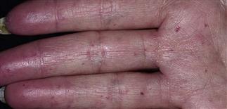
Multiple punctuate telangiectasias on the palms and fingers can be seen in connective tissue disorders such as CREST syndrome.
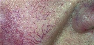
Close-up view shows dilated arterioles coalescing to form a telangiectasia.
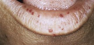
Numerous telangiectasias involving the lips and tongue can be a sign of hereditary hemorrhagic telangiectasia or CREST syndrome.
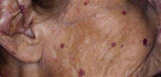
Multiple telangiectasias appear on the sun exposed cheeks in predisposed individual. Patients with rosacea may also have erythema and pustules.
DESCRIPTION
Common asymptomatic dilatations of capillaries, venules, and arterioles within the subpapillary plexus.
HISTORY
Occur in various clinical settings.
Primary telangiectasia
• Hereditary hemorrhagic telangiectasia (Osler–Rendu–Weber syndrome). Autosomal dominant. Telangiectasias on mucosae, skin, and internal organs. Earliest sign: recurrent epistaxis in childhood. Characteristic telangiectasias occur in early adulthood and are prominent on tongue, palate, nasal mucosa, palms, soles, nail beds. Normal lifespan. Increased risk of life-threatening hemorrhage. • Hereditary benign telangiectasia. Autosomal dominant. Widespread telangiectasias. No mucosal or internal organ involvement. No associated bleeding diathesis. • Ataxia telangiectasia (Louis–Bar syndrome). Autosomal recessive condition with progressive cerebellar ataxia, telangiectasias, immune dysfunction. The earliest sign, ataxia, is evident when the child begins to walk, and usually by the age of 3 years. Telangiectasias appear later on conjunctivae, face, neck, upper trunk by age 5. Café-au-lait macules, skin ulcerations, poikiloderma, premature gray hair, dry skin, sclerodermatous skin changes, eczema, and hirsutism may also occur. Caused by defective DNA repair of chromosomal breakages. • Generalized essential telangiectasia. Women affected more often than men. Telangiectasias first appear on legs and then gradually, progressively, and symmetrically extend to involve trunk and arms. • Unilateral nevoid telangiectasia. Unilateral dermatomal distribution of fine telangiectasias. Most common dermatomes are the trigeminal and the third and fourth cervical nerves. May be congenital or acquired. Congenital: males more than females. Acquired: females more than males. Can begin during puberty and pregnancy, and resolve in adulthood and after delivery, suggesting causative role for estrogen.
Secondary telangiectasias
• Basal cell carcinoma, rosacea, collagen vascular disorders, corticosteroid atrophy, CREST, scleroderma, chronic graft-versus-host disease. • Secondary telangiectasias occur in the setting of altered dermal connective tissue as a result of injury or chronic inflammation. Damage may be from ultraviolet radiation (actinic damage), ionizing radiation, or treatment with topical or intralesional corticosteroids.
PHYSICAL FINDINGS
• Non-palpable pink to red dilated dermal vessel with a diameter of 1 mm or less; easily blanched with diascopy. • Lesions may appear as discrete vessels or clustered as telangiectatic ‘mats.’
TREATMENT
Telangiectasia may be ablated with laser surgery or pinpoint electrocautery. Individual lesions may require several treatments.


