Techniques and Devices for Lead Extraction

Published on 21/06/2015 by admin

Filed under Cardiovascular

Last modified 22/04/2025

Print this page

rate 1 star rate 2 star rate 3 star rate 4 star rate 5 star
Your rating: none, Average: 5 (1 votes)

This article have been viewed 2812 times

26 Techniques and Devices for Lead Extraction

Lead extraction is increasingly required and is directly related to the increased numbers of cardiac implantable electronic devices (CIEDs). The number of extractions has increased because of expanded indications, such as the need to upgrade to newer technology in patients with occluded veins or lead-device safety alerts, and improved access to extraction providers. The components of the CIED, the leads and pulse generators, have a limited functional life. As the population and the devices age, components of the system require removal for a variety of reasons, including infection, lead malfunction, venous stenosis/occlusion, and safety alerts.

This chapter discusses the indications, techniques, and devices for lead extraction in detail, using definitions and information from the 2009 Heart Rhythm Society (HRS) Expert Consensus on Facilities, Training, Indications, and Patient Management for transvenous lead extraction.1

image Definitions

Implant lead removal technically means removal of a CIED lead using any technique. However, leads with short implantation duration can usually be removed with simple traction, and this is defined as a “lead removal procedure.”

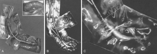
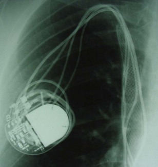
However, with prolongation of the duration of implantation, segments of chronically implanted leads are encased in encapsulating fibrous tissue and may be bound to the vein and/or heart wall, bound to another lead, or both (Figs. 26-1 to 26-4). Removal of chronically implanted leads from these binding sites becomes very difficult and potentially hazardous with traction alone. The tensile strength of encapsulating fibrous tissue is greater than that of the surrounding tissue, and thus leads cannot easily be removed without risking a tear or avulsion of the vein or heart wall. In such cases the removal, separation, and freeing of leads from encapsulating fibrous tissue are defined as lead extraction. As a rule, if one or more of the leads implanted in the patient that require removal are more than 1 year old or require extraction tools for removal, the procedure is labeled as a “lead extraction procedure,” and if not, then a “lead removal without extraction.”

Extraction techniques include traction, countertraction, counterpressure, and tissue disruption, cutting locally with an instrument, laser, or electrosurgery unit. Lead extraction techniques are designed to free the lead from the encapsulating fibrous tissue (countertraction) or to free the encapsulating fibrous tissue (counterpressure) from the vein or heart wall.24 Telescoping sheaths are used remotely to apply countertraction and counterpressure at the selected binding sites. Lead extraction procedures are those approaches used to apply the sheaths and remove the lead in a safe and efficacious manner.

Goals and Outcomes

The clinical outcomes targeted with lead extraction are elimination of infection, elimination of risk associated with a lead (e.g., perforation, arrhythmia), and creation of venous access. Other clinical outcomes include removal of all functional leads and attempting to resolve pocket-related symptoms such as pain.

Outcome definitions depend on the indication of the procedure. If the indication is systemic infection, complete removal of all targeted leads and material without any complication, defined as “complete procedural success,” is required to achieve clinical success. However, in non-infection-related cases, “partial procedural success,” with only a small part or the tip of the lead retained, may be associated with clinical success and the desired clinical outcome, such as resolution of a pocket infection or creation of a conduit. In case of partial procedural success, clinical success is conditional and based on achievement of clinical goals and the absence of untoward effects such as perforation, embolism, or persistent infection caused by the retained part of the lead.

The HRS consensus document, in an effort to unify nomenclature, suggested the following definitions1:

Although there are no published benchmarks, operators should strive for clinical success in 100% of patients with the least possible number of complications. Table 26-1 summarizes current rates of success and complications based on collective studies.

Intraprocedural complications are any complication that occurs during the procedure, as recorded from the time the patient enters the operating or procedure room to the time the patient leaves. This includes all preparation, from administering anesthesia to groin access to closing the incision and reversal of anesthesia. Postprocedural complications are any events that become evident within 30 days after the intraprocedural period. Major complications are any life-threatening complications, those that result in death, or any complication that results in persistent or significant disability or significant surgical intervention. A minor complication is any undesired event related to the procedure that requires medical intervention or “minor procedural intervention” and does not limit, persistently or significantly, the patient’s function or threaten life or cause death (Box 26-1).

image Indications for Extraction

Considerable divergence of opinion surrounds the indications for lead extraction. However, the 2009 HRS consensus document has unified opinions to a large extent.1 Indications are divided into three major categories: infection, venous access, and broken or nonfunctional leads. Also, emerging indications include implant patients who require magnetic resonance imaging (MRI) scanning for diagnosis (Box 26-2).

Box 26-2

Indications for Transvenous Lead Extraction*

Leads Affecting Patient: Functional or Nonfunctional Leads

Infection

Infection remains the most common indication for extraction. Complete extraction is indicated in cases of definite CIED infection when there is erosion, pocket infection, lead vegetations, or sepsis (see Chapter 25). Extraction is also recommended in patients with a device and endocarditis or occult gram-positive bacteremia. Extraction is also reasonable in patients with a device and persistent occult gram-negative bacteremia. Antibiotic therapy should be considered adjunctive therapy, and pocket debridement with local relocation is only palliative.

To prevent further serious and potentially lethal complications, all device components, including leads, must be removed to cure the infection. Also, the morbidity associated with a local pocket infection, the lethal sequelae of septicemia, and the potential risk of infected thrombus formation in the heart are well known. Because leaving an infected lead in the body is potentially lethal, the risk of the procedure is clearly less than the risk of lead extraction; that is, the risk of not extracting far exceeds the risk of extracting. The risk of Staphylococcus aureus device infection without extraction was supported by a series of 33 patients from the Duke Medical Center; 10 of 21 patients (47.6%) died without lead extraction, and 2 of 12 (16.7%) died despite lead extraction, and none from lead extraction.5 The safety and efficacy of complete lead extraction, with debridement and delayed reimplantation at a remote anatomic site, were demonstrated in 123 patients at the Cleveland Clinic with device infection. Despite infections with a wide range of bacterial organisms, mostly coagulase-negative staphylococci and S. aureus, extraction was associated with no major complications. Infection recurred only in those four patients who had incomplete extraction or reimplantation concurrent with the extraction.6

Chronic pain at the site of the device or lead insertion should be considered as a subclinical infection, and extraction in such patients is indicated. After extraction, the patient should be treated as having a pocket infection, with adjunctive antibiotics and reimplantation on the contralateral side.

Noninfected Systems

When functional or nonfunctional leads pose immediate risk, such as in a patient with life-threatening arrhythmias from a retained lead or fragment, or when the lead design poses a risk of perforation (e.g., Accufix with protrusion of J stylet outside lead), the decision to extract is straightforward (Fig. 26-5). Extraction of functional or nonfunctional leads becomes controversial when there is no immediate threat to the patient, because it is possible to abandon a failed or a nonrequired lead and implant ipsilaterally if the vein is patent, or contralaterally or even transiliac if the ipsilateral vein is occluded. The controversy persists because it is difficult to calculate the risk/benefit ratio of lead extraction versus abandoning such leads. Thus, when considering extraction of noninfected leads, it is important to balance the risk of extraction with the patient’s situation and to factor in the operator’s experience and not just literature data. Some operators may choose to extract only when there is infection; others who are more experienced may elect to extract all leads that are not required, whether functional or not, when the opportunity arises.

Regarding the patient’s situation, two examples help clarify the approach. A 60-year-old patient with a pacemaker implanted for compete heart block and a failing 20-year-old right ventricular (RV) lead may have either (1) extraction of the RV lead and reimplantation of a new lead or (2) abandonment of the failing RV lead and addition of a new RV lead using the ipsilateral patent vein. The decision on which approach to adopt will depend on the operator’s skill and comfort. On the other hand, with a 95-year-old patient in the same situation, only the second approach seems reasonable, because the abandoned lead will likely never become a clinical issue. The following sections address extraction indications for noninfected leads.

Thrombosis or Venous Stenosis

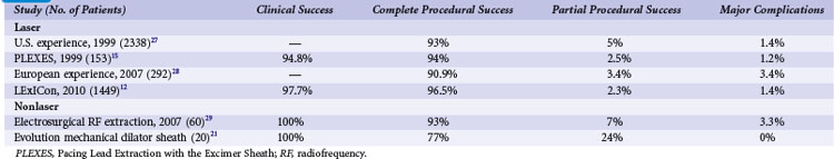
Lead extraction is indicated for patients with clinically significant thromboembolic events with evidence of clot on the lead, as well as in patients with superior vena cava (SVC) or subclavian vein stenosis or occlusion with symptoms. Lead extraction is also recommended in cases of bilateral occlusion for the creation of a conduit or for planned stent deployment (Fig. 26-6). When there is a contraindication to implantation on the contralateral side (e.g., arteriovenous fistula), extraction for creation of a conduit is recommended; otherwise, extraction is considered reasonable.

Creation of a Conduit

The rationale for creating a conduit is applicable to component failures. Consider a patient with a dual-chamber pacemaker and an atrial lead conductor coil fracture. This patient has a normal ventricular lead, an occlusion of the brachiocephalic vein, and the need to implant a new atrial lead. Although venoplasty through the total occlusion is possible, a more reasonable approach to maintaining access through the same vein entry site is to extract the failed atrial lead and to insert the new lead through the extraction conduit, or to remove both existing leads and start over with new leads. This same logic would apply to other situations, such as an upgrade requiring the addition of a new lead. For example, a patient with a dual-chamber ICD requires a cardiac vein implant for biventricular pacing. The alternatives are doing nothing, implanting the new lead through a contralateral vein or a transfemoral vein, or using a cardiac surgical approach. Venoplasty is also reasonable if the existing leads are all functional.

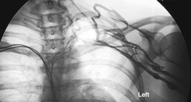
Sometimes, when there is severe stenosis with or without symptoms from the obstruction of flow, physicians have initiated balloon venoplasty and stenting without extraction of the leads. This is particularly difficult if either infection or reocclusion occurs, because extraction now becomes impossible without extensive open-heart surgery (Fig. 26-7). A more appropriate approach includes extraction, venoplasty, stenting, and reimplantation through the stent, as reported by Chan et al.7 in a subclavian occlusion that progressed to an SVC occlusion.

Implantation through a contralateral vein seems logical, especially if it is the implanter’s only skill-level option. The simplest alternative is to abandon the two original leads on the ipsilateral side and implant the new leads on the contralateral side. In the example of a conductor coil fracture, the patient has the risk of having two functioning and two nonactive leads in the heart and the risk of instrumentation of the superior veins on the opposite side. In the example of adding a left ventricular lead for biventricular stimulation, the patient has the risk of having three functioning leads and two nonactive leads in the heart and the risk of instrumentation of the superior veins on the opposite side. These risks are weighed against the risk of extracting the one atrial lead. The resultant risk of having a complication is the sum of the individual risk factors.

For these two examples, another approach would be to implant the new atrial lead or biventricular lead on the contralateral side and tunnel it across to the pulse generator and ventricular lead. In addition to the risk of instrumentation of the superior veins and tunneling, the patient with conductor coil fracture will have two functioning leads and one nonactive lead in the heart, and the patient with the biventricular lead will have three functioning leads in the heart.

Total bilateral occlusion of the superior veins further complicates the problem. The transfemoral approach is then the only approach available to the nonsurgeon implanter. The surgical implanter has the options of a transatrial or epicardial approach. The risks associated with these choices make the decision to extract the atrial lead easier. However, without extraction skills, the medical and surgical implanter may choose these alternatives.

The risk/benefit ratio is crucial to the rationale for extracting these leads. For example, the life-threatening risk associated with infection in effect forces an implanter to extract the lead and abandon the pocket. The risk of not creating a conduit to insert new leads is a potential risk for a future complication related to bilateral implants, nonactive leads, multiple implanted leads, and tunneling. This risk is obviously less than the life-threatening risk of infection. In the situation of lead failure, the alternatives presented provide an acceptable short-term solution and can be performed by implanters without lead extraction skills. Potential risks are not as compelling a reason for action as the immediate risks associated with lead extraction. However, physicians with experience in lead extraction may not be comfortable with abandoning two leads, creating two inactive leads, and leaving a total of four or five leads implanted. Tunneling of a lead from the opposite side, crossing over the sternum, is a potential source of infection and increased risk of lead fracture. Again, the risk of not extracting must be compared with the risk of extracting to help resolve these issues. To limit the risk of subclavian vein or SVC occlusion, it is considered reasonable to extract nonfunctional leads if a planned CIED implantation would require more than four leads on one side or more than five leads through the SVC.

image Risks and Outcomes

The risks associated with lead extraction are tamponade caused by intrapericardial vascular disruption of the SVC or heart; hemothorax from extrapericardial vascular tear outside the pericardial sac and into the thorax; arteriovenous fistula or dissecting hematoma (e.g., aortic arch); and failure to complete the lead extraction (Fig. 26-8). The latter is usually not considered a risk; however, a failed lead extraction may lead to additional procedures or may be a precursor for dangerous situations in the future.
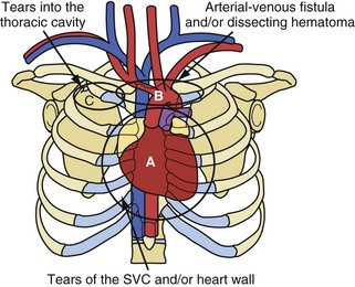
There are two situations in which the risk of tearing the SVC and heart is reduced but other risks could be increased. The first situation involves patients who have undergone an open-heart procedure; the pericardial space has been obliterated, and fibrous tissue reinforces the SVC and heart wall. In this situation, if an intrathoracic bleed occurs, quick surgical chest entry is difficult. The second situation involves an implant of short duration: less than 2 years for pacemaker leads and less than 1 year for ICD leads. The forces involved in freeing these leads usually are not sufficient to tear the SVC or heart.

Extraction centers from the continental United States and Hawaii voluntarily submitted data for a national registry between December 1988 and December 1999.911 The most recent published report, from 1996, included data from 226 centers, 2338 patients, and 3540 leads and demonstrated major complications in 1.4% of the cases (<1% for centers with >300 extraction procedures).10,11 The total U.S. data included 7823 extraction procedures and 12,833 leads (presented in 2000). Multivariate analysis of the data from 1994 through 1999 demonstrated four predictors of major complications (1.6%): (1) implant duration of oldest lead, (2) female gender, (3) ICD lead removal, and (4) use of laser extraction technique. Major complications were (1) death, 0.3%; (2) nonfatal hemopericardium or tamponade, 0.7%; (3) nonfatal hemothorax, 0.2%; (4) transfusion for bleeding/hypotension, 0.1%; (5) pneumothorax requiring a chest tube, 0.1%; and (6) other nonfatal events, 0.2% (including 4 arteriovenous fistulae, 2 pulmonary embolisms, 2 thoracotomies for defibrillator leads trapped in sheaths, 2 respiratory arrests, 2 strokes, 2 cases of renal failure, 1 anoxic encephalopathy, and 1 open-heart retrieval of a device fragment). In the more recent LExICon study of data from 13 centers in the United States and Canada, procedural success was 96.5%, with 97.7% clinical success.12 In LExICon, 1449 patients underwent laser-assisted lead extraction of 2405 leads. Failure to achieve clinical success was associated with body mass index (BMI) of 25 kg/m2 or less and low-extraction-volume centers. Procedural failure was higher in leads implanted for more than 10 years and when performed in low-volume centers. Major adverse events in 20 patients were directly related to the procedure (1.4%), including four deaths (0.28%). Major adverse effects were associated with patients with BMI less than 25 kg/m2. Overall, all-cause in-hospital mortality was 1.86%.

Risk of lead extraction depends on the extractor’s experience, duration of implant, and the patient’s age and condition. There is no ongoing national database or registry, and the risks depend on the individual, the assembled team, and the institution. The most reliable indicator of risk is the individual extractor’s personal statistics. It is important that each institution and individual keep track of complications and effectiveness.

Risks are caused by maturation of the encapsulating fibrous tissue, which is related to the duration of the implant and the patient’s age. With time, the tensile strength of the encapsulating fibrous tissue increases; it may calcify in 3 to 4 years in children and in 8 to 10 years in older adults. Sedentary patients increase their tensile strength more slowly, and the tissue takes longer to mineralize. In sedentary elderly patients, the tensile strength seems to decrease with time. The influences of duration and age are apparent in the extremes. Also, patients with calcium metabolism abnormalities can calcify at any age in a short duration. Although the properties of encapsulated fibrous tissue are known, it is difficult to apply general principles to a specific patient and assign a risk.

Potentially lethal complications requiring extensive surgical procedures include tear of the vein and heart wall causing tamponade, arterial tears causing arteriovenous fistula or dissecting hematoma; and tears into the thoracic cavity causing a hemothorax (see Fig. 26-8). The procedure-related complications are discussed in detail later. Time and surgeon experience are the two factors related to survival. Low blood pressure and poor tissue perfusion are time-dependent events. Being prepared for a cardiovascular emergency is the only way to meet time constraints. This includes having a cardiovascular surgeon immediately available, along with the proper instrumentation and experienced support personnel. A cardiovascular surgeon has the technical skill to manage these complications but may need direction from the extractor on the proper approach. Once a complication resulting in poor or no perfusion is recognized, the repair should begin immediately. The concern of needlessly subjecting the patient to extensive surgery and morbidity pales in comparison to that of applying the therapy late because of confusion or procrastination. Failure to recognize the complication in a timely fashion or the lack of access to qualified personnel may cause a lethal outcome.

image Clinical Considerations

Patient Information and Preparation

All patients presenting for an extraction procedure must have a standard history and physical examination with a detailed description and understanding of the indication for extraction. The initial indication for implantation should be known. Comorbidities that can affect outcome, such as kidney failure, should be noted. The procedure notes should be carefully reviewed for any problems observed during the implantation, such as difficult access. Also, if infection is present, detailed information on the time line, organism, susceptibilities, and antibiotic therapy is needed. Basic laboratory work is usually indicated and includes white blood cell count, hematocrit, hemoglobin, platelet count, blood urea nitrogen, creatinine, potassium, sodium, liver profile, and prothrombin time.

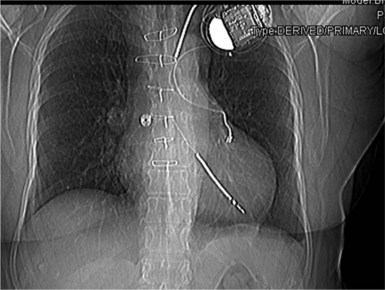
The patient’s blood should be typed and crossmatched for a possible blood transfusion. A current chest radiograph and electrocardiogram (ECG) are mandatory (Fig. 26-9). An echocardiogram is mandatory for two groups of patients before the procedure, even if transesophageal echocardiography (TEE) is routinely available in the procedure room: those with infection, to rule out vegetation in the right atrium; and those with heart failure, to define cardiac function.

A plan for antibiotics before and after the procedure should be formulated. Also, the need for temporary pacing while waiting for clearance to reimplant a new, permanent CIED should be established. The timing of reimplantation should also be determined before extraction. The operator must know all devices and leads present, including those abandoned. A preprocedural chest radiograph must be obtained and examined for any abandoned leads and unusual locations of lead placement. A lead thought to be in the left ventricle should be extracted in the operating room (OR) with surgical techniques. If there is any question as to the location of a lead, additional imaging using TEE or computed tomography (CT) may be required. It is imperative to know the initial implantation date because the age of the leads may dictate different preparations and approaches to extraction. The need for pacing during and after the procedure should be established. Dependent patients will need temporary pacing during the procedure using an electrophysiology catheter. After extraction is complete, a temporary active-fixation wire can be used until reimplantation of a CIED is possible.

The device to be extracted should be interrogated before the procedure and all parameters recorded, to serve as baseline characteristics of leads that will not be extracted. Therefore, these leads should be retested after extraction to ensure they have sustained no damage.

In noninfected patients, preprocedural antibiotics should be administered. Antibiotic therapy before the procedure can be given for infected patients when the infective organism and susceptibilities are established. The patient should be continuously monitored by ECG, arterial pressure line, oxygen saturation, and at times a Foley catheter. A reliable intravenous (IV) line should be placed. In addition, an arterial line should be used for patients needing continuous vasopressor support and for those sent to an intensive care unit. Patients are prepared from chin to midthigh for all transvenous and cardiac surgical approaches, including an emergency procedure, if needed. Transthoracic or TEE should be immediately available to assess for pericardial effusion or thrombus or vegetation dislocation. Because of the potential risks, extraction should be performed only in a facility with accredited cardiac surgery programs. A cardiothoracic surgeon must be physically present on site and be able to initiate emergency surgery. To facilitate the surgeon’s rescue efforts, the procedure room must be equipped with the necessary tools to perform cardiac surgery, such as thoracotomy or sternotomy instrument sets in addition to a sternal saw.

Anesthesia

The types of anesthesia available include local anesthesia, conscious sedation, managed anesthesia care (MAC), laryngeal mask anesthesia (LMA), and general endotracheal anesthesia. The rationale for using a specific form of anesthesia is based on type of procedure, physician comfort level with general anesthesia, perceived risk of a given type of anesthesia, and availability of general anesthesia.

General endotracheal anesthesia is the only type of anesthesia that is suitable for all procedures, and essential for some. General anesthesia consists of an “anesthesia package:” anesthesiologist, compliance with preoperative anesthesia protocols, anesthesia and monitoring machines, and general anesthetic agents and gases. It requires procedure room space, scheduling, and an anesthesia recovery room. In addition, the electrophysiologist and anesthesiologist must work as a team to manage the patient. The merits of placing a patient at any desired level of anesthesia and providing a satisfactory environment to perform any type of surgical procedure are universally accepted when presented in this abstract fashion. However, the practical demands of the anesthesia package and the fundamental questions relating to the safety of general anesthesia limit its use.

Many of the maneuvers performed during a lead extraction can reduce filling pressure. Traction on an atrial lead may block the SVC and reduce blood flow to the heart. Traction on a ventricular lead reduces the compliance of the chamber wall during diastole or, if strong enough, can pull the wall to the tricuspid valve, reducing blood flow. Immediate injections of a short-term α-adrenergic stimulant such as phenylephrine (Neo-Synephrine) or norepinephrine (Levophed) constrict the cardiovascular system, causing an increase in both filling pressure and systemic blood pressure. This may frequently be required throughout the case to compensate for these transient iatrogenic insults. Use of these agents does not reflect on the safety and efficacy of general anesthesia.

In conclusion, the risk of anesthesia is mostly related to the procedure and not the general anesthesia.

Procedure Room

Electrophysiology (EP) procedures are currently performed in general OR suites, device implantation procedure rooms, EP procedure rooms, catheterization laboratories, and fully equipped cardiovascular ORs. The room must be large enough to support the procedure. Small procedure rooms are sufficient for a device implantation but not large enough for a complicated lead extraction procedure. There should also be space to accommodate emergency procedures and/or a more extensive surgical EP procedure. Procedures should not be performed in smaller rooms without a contingency plan for an emergency.

The ideal procedure room should meet most of the requirements for an OR, especially those related to room cleaning, patient draping, gown and gloving, and instrument sterility. It should have the full “anesthesia package” including continuous monitoring of ECG, arterial pressures, and oxygen saturation. Essential specialty equipment includes high-quality fluoroscopy, echocardiography, pacemaker system analyzer (PSA), and other external EP devices to ensure pacing and defibrillation, as well as an excimer laser or dedicated electrosurgical unit for lead extraction. In addition, for minimally invasive cardiac procedures, lighted retractors, access to thoracoscopy equipment, and an emergency tray to open the chest should be available. Additional safety devices to help protect physicians and nurses include lead drapes for radiation protection, smoke evacuators, and chairs for sitting when appropriate during the procedure.

Pocket Management

Tissue debridement is the surgical removal of all inflammatory and damaged native tissues, leaving only normal native tissue behind. The need for tissue debridement in normal pockets seen on routine reimplantation is minimal. On opening an old pocket, the debridement goal is to remove any exuberant inflammatory tissue (fibrous tissue), leaving only thin, healthy fibrous tissue (biophysical interface) or normal native tissue behind. The fibrous tissue present is involved in the chronic remodeling inflammatory reaction; rarely is an acute inflammatory reaction present. This is important, because exuberant fibrous tissue masses, which are the result of an inflammatory reaction, can injure adjacent tissue, continuing the inflammatory reaction (recursive reaction). Also, if this tissue is contaminated by bacteria, it becomes a nidus for infection; bacteria adhere to the smooth surface of the exuberant encapsulating tissue, and the body’s immunodefense mechanisms cannot reach the bacteria. Leads entrapped in the encapsulating fibrous tissue may be under undue stress if the pulse generator is not placed back in the same position. Tissue debridement and freeing of the leads rectify this situation. Tissue debridement is best performed with an electrosurgical cautery tool.

Tissue debridement, even in infected cases, is rarely extensive and can be treated as described earlier. Occasionally, however, the pocket must be extensively debrided and then abandoned; this situation usually involves infection. If an acute inflammatory reaction is extensive, debridement is tedious because of the presence of acute and chronic inflammatory material and proximity of large blood vessels and important nerves. Knowledge of local anatomy is mandatory in this situation. In some cases, the inflammatory tissue can be more than 2.5 cm thick, extending above and below the pectoralis major muscle with fingers to the clavicle, and damaging a large amount of skin. In these cases, skin loss is significant, and reapproximation of skin edges is challenging. Once the debridement is complete, hemostasis is difficult to achieve, especially on the muscles. Wherever possible, muscle fascia should be reapproximated.

Tissue Closure

Healing by primary intention is the closing of an open wound by reapproximating tissue (muscle, subcutaneous tissue, and skin) using suture material. Suture holds the tissue in place until the tensile strength of the bonding fibrous tissue is sufficient to keep the tissue together. All initial and reimplanted pockets are closed in a conventional manner, reapproximating the tissue with suture material, and allowed to heal by primary intention. Chronic pockets are debrided of all exuberant inflammatory material before closure.

These pockets are debrided and closed using a closed-drainage system (Jackson-Pratt) placed in the debrided pocket, if necessary, applying suction to prevent the development of effusions or clot, and keeping normal tissue contiguous with normal tissue. A large defect resulting from loss of tissue during debridement may be challenging to close. In extreme cases, a major defect requires an experienced surgeon or a plastic surgeon, especially if a flap is needed. Alternatively, the pocket may be allowed to heal by secondary intention.

Another philosophy holds that pockets with debridement defects and infected pockets should be left open. Once the device (foreign body) is removed, the infection will heal, so allow it to heal by secondary intention. This rationale shortens the procedure and is an accepted method of healing. Infection is not an issue, and it avoids the need for acquiring surgical debridement and closure skills. Concerns such as morbidity, extensive healing time, long-term antibiotic therapy, constant professional care of the wound, and possible surgical intervention, including skin grafting, are considered the natural cost of healing the wound. Healing by primary intention, on the other hand, does not have these issues. Healing by secondary intention was previously used as the only way to heal an open wound. The healing stages were well documented: suppuration, granulation, closure of defect, and skin closure. This was the recommended method of treating contaminated wounds by surgeons until the 1980s. Since the 1970s, however, primary closure of debrided infected pockets has been successfully and almost exclusively used to manage device-related infections.

Training and Skills

Lead extraction is a fundamental skill that is required to manage device-related complications. Lead extraction, as with lead implantation, is a requisite skill with predictable and expected results. This was not always the case. From its inception in the early 1980s, the procedures evolved rapidly. The management of a device-related complication centered on the lead extraction procedure, overshadowing all other aspects of management. This was because unexpected tears in the SVC and heart wall sometimes occurred without warning, despite the rigid protocols followed. The technology and those rigid protocols have evolved into current procedures. The procedures are less stressful and have predictable results. Predictability allows an extractor to recognize an approach that has a potential for a poor result and change to an approach with a predictably good outcome. The 2009 HRS Expert Consensus document clearly indicates the minimum training needed for competency. Physicians in training should extract a minimum of 40 leads as the primary operator under supervision. To maintain skills a minimum of 20 leads annually should be performed. Supervising physicians should have extracted 75 leads with efficacy and safety consistent with accepted literature.1

Extraction techniques are much simpler now and are explained in detail. Unfortunately, some of the old, more complicated techniques are also needed to manage an occasional rare situation. Consequently, some of these techniques are also presented. Presentation of the extraction techniques and approaches from the medical and surgical electrophysiologist’s point of view has been challenging. The common ground is that most of the extractions involve transvenous leads. These are extracted using the techniques and approaches common to both groups. The alternative approaches are sometimes different for surgical and medical electrophysiologists. For example, in addition to the transvenous approaches, EP surgeons have the option of a transatrial or epicardial approach. Although not mainstream, these surgical approaches are essential to the management of certain complications. Although medical electrophysiologists cannot perform these procedures, they must be able to direct a non-EP surgeon enlisted to perform the procedure.

The new HRS Expert Consensus document was published after 1-year preparation.1 In the past, meaningful consensus was impossible because of the small number of practitioners and limited data. Now, however, the substantial growth and investment of the international HRS community in transvenous lead extraction permits significant consistency in tools, techniques, procedures, and expected benefits and risks.

image Lead Extraction Techniques

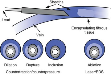
As discussed, segments of chronically implanted leads are encased in encapsulating fibrous tissue and bound to the vein and/or heart wall, bound to another lead, or both. Lead extraction is the removal of chronically implanted leads from these binding sites. Because the tensile strength of encapsulating fibrous tissue is greater than that of the surrounding tissue, leads cannot easily be removed without risking a tear or avulsion of the vein or heart wall. The word ablation best describes the removal, separation, and freeing of leads from encapsulating fibrous tissue. Ablation techniques include traction, countertraction, counterpressure, and tissue disruption, cutting locally with an instrument, laser, or electrosurgery unit. Lead extraction techniques are designed to free the lead from the encapsulating fibrous tissue (countertraction) or to free the encapsulating fibrous tissue (counterpressure) from the vein or heart wall.2 Telescoping sheaths are used remotely to apply countertraction and counterpressure at the selected binding sites. Lead extraction procedures are used to apply the sheaths and remove the lead in a safe and efficacious manner.

Traction, Countertraction, and Counterpressure

In the 1960s and early 1970s, transvenous leads were large, bipolar, and without fixation devices. These leads were usually isodiametric, implanted for 1 to 2 years, and removed by traction. With the introduction of tines and pulse generators lasting 4 to 6 years, leads were entrapped in encapsulating fibrous tissue with significant tensile strength. These leads could not be safely removed by traction alone.

Traction is the force exerted on the lead by pulling. Applying traction to the lead pulls directly on the binding site. Once the encapsulating tissue has a greater tensile strength than the venous or cardiac tissue, the tissue will tear or avulse. Disruption of the vein or heart wall can be lethal. Because the relative tensile strengths are not known, traction should be used with caution. However, traction is an acceptable extraction technique if applied judiciously. The force applied when pulling on a lead is related to lead size, lead tensile strength, how the lead is grasped, use of locking stylets, and most importantly the extracting physician’s catecholamine level. The catecholamine level is important in determining the actual traction force. When the extractor is relaxed and calm, the perceived force may be realistic; when upset, agitated, or mad, however, the force is much greater than perceived. Consequently, “giving it a little tug” is not wise.

Direct Traction

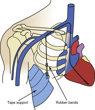
All current lead extraction procedures use some form of traction, or pulling force13 (Fig. 26-10). Pulling on leads was a successful method of extracting leads during the early years of pacing, when leads lacked efficient fixation devices and were implanted for short periods. Traction was applied manually for minutes or applied using various weights or elastic bands for days. Traction proved unsafe and had a high incidence of failure when applied to leads with efficient fixation devices and leads implanted for longer periods. The amount of traction required increases and becomes more dangerous as the duration of the implant and the tensile strength of the fibrous tissue increase. Leads with efficient passive-fixation devices may be difficult to remove 4 to 6 months after implantation. A failed previous attempt to extract a lead frequently damaged the lead, making future extraction attempts more difficult.
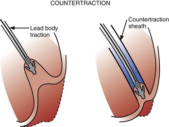
Again, traction must be applied judiciously to minimize the risk to the patient. The pulling force applied to the proximal portion of the lead is distributed to sites where fibrous tissue binds the lead or electrode and makes contact with the vein or heart wall. Multiple leads may be bound to the vein or heart wall and to each other. Because the pulling force is not focused, the distribution of force to the binding sites is unknown. The extractor may inadvertently tear a vein or the heart wall.

Traction, in some form, is integral to lead extraction. The physician must consciously limit the pulling force and apply a continuous, steady traction. Never jerk the lead, because impulse forces tear. Accidents are not predictable and frequently occur without warning, partly because it is impossible to judge accurately the level of force applied to the lead. To gauge the applied force, most direct-traction techniques apply sufficient force to feel the rhythmic tugging of the heart without producing arrhythmias, hypotension, or chest pain. These are crude and unreliable endpoints and do not reflect the tensile strength of the lead or the tissue. Breaking the lead, tearing a vein, or avulsing or tearing the heart wall are all potential complications. As stated, “just a little tug to see if the lead will come out” is not a consistently safe approach, and following the basic principles and guidelines acquired from practical experience minimizes the risk.

It is important to understand the difference between pulling from above and pulling from below. The mediastinal structures are not bound from below. If you pull upward on the heart, it will move, along with the lungs, diaphragm, and rest of the mediastinal contents, in that direction. If you pull downward, the superior veins and surrounding structures are bound to the musculoskeletal system and do not move downward. Assume that traction is applied from above to a lead implanted in the right ventricle. With continuous traction, the right ventricle starts to evaginate, decreasing the compliance of the wall and finally obstructing the flow of blood through the tricuspid valve. At the same time, the heart is pulled into the superior portion of the mediastinum. This maneuver is safe as long as the hemodynamic status is monitored and the process is reversible. Reversibility means that traction-induced deterioration in hemodynamic function is corrected on cessation of the traction, and the right ventricle, along with remainder of the mediastinal structures, returns to its normal position. Usually, the structures do return to normal. The danger is slippage of the lead body through a binding site. On release of the traction, the mediastinal forces pulling downward are insufficient to pull the lead back through the binding site. The forces required to elevate the mediastinum to this position are still applied to the heart. Any hemodynamic compromise persists, such as decreased blood flow through the right ventricle, creating a cardiovascular emergency. If the lead body cannot be released by manipulation, including use of a stiff stylet to help push it through the binding site, an emergency median sternotomy is required to restore manually the heart’s function.

Once a lead is freed from a distal binding site, the lead and associated fibrous material can then become wedged in a more proximal binding site. For example, a lead removed from the right ventricle can become wedged at a binding site in the atrium, SVC, or axillary-subclavian-brachiocephalic veins. With direct traction, the lead is freed from the binding sites, distal to proximal. The strongest binding site determines the outcome of an extraction attempt, regardless of its proximal or distal location.

Indirect Traction

Elevation of the mediastinum with traction is the main reason why traction from above is not as effective at pulling a lead through a binding site as is pulling from below. When traction is applied from below, the mediastinum is not pulled down (inferior) because the superior veins and surrounding tissues are bound to the musculoskeletal system. Traction forces applied to the binding sites are directed at freeing the lead, not at moving structures. The limit to the force applied to the axillary-subclavian-brachiocephalic veins is determined by the tensile strength of the lead. If the lead binding in these veins is extensive enough to require that type of force, the veins are probably occluded, atretic, or encapsulating sheaths. Disruption of these veins by tearing or avulsion is of no consequence. Binding sites in the SVC, however, must be treated in the same manner as in the heart. Disruption of the wall of the SVC has the same consequences as disruption of the heart wall.

The safety and efficacy (higher success rate) of applying indirect traction from below should now be apparent. Indirect traction is traction applied by an instrument, such as a snare passed into the heart, usually through a femoral vein. The lead is entrapped in the snare, and traction is applied by pulling or pushing. The difficulty is in grasping the lead in a way that allows sufficient traction to be applied. Only a few snares, such as the Dotter basket snare and Needle’s Eye-Snare (both Cook Vascular, Vandergrift, Pa.), or the Amplatz Gooseneck Snare (eV3/Vasocare, Seoul) have sufficient strength to support significant extraction forces. The lead must first be freed from the superior veins and then from the heart. The lead is pulled out of the superior veins and into the atrium or inferior vena cava (IVC). It can be regrasped, if necessary, and traction can be applied to the heart. The techniques for applying indirect traction are the same as for grasping and manipulating the leads in other approaches, such as applying countertraction sheaths, as described later. The risks eliminated by indirect traction are tearing of superior veins, wedging of the lead in the atrium or in a superior vein, and creating a low cardiac output caused by failure of the lead to return to its original position after traction. Indirect traction has the same potential for breaking the lead or tearing the heart wall as direct traction, if their tensile strength is exceeded.

Countertraction

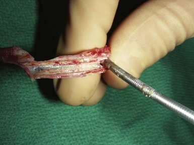
Countertraction is the technique used to free the lead from compliant, encapsulated fibrous tissue. Countertraction was first used to remove a lead from an implantation site in the right ventricle or atrium. Although the technique for extracting leads from the heart wall is discussed first, this is the last step in a normal lead extraction procedure, using any type of sheath. Extraction sheaths free leads from binding sites, proximal to distal. Once the sheath is passed over the lead and down to the implantation site, traction on the lead pulls the site to the sheath (Fig. 26-11). The traction force is countered by the circumference of the sheath. The countertraction sheath focuses the traction force at the tip of the sheath, limiting the excursion of the heart wall. This prevents compliance changes and blockage of the tricuspid valve, with possible perforation, tearing, and avulsion of the heart wall. The countertraction forces are limited by the tensile strength of the lead. At some point, the electrode is freed from the encapsulating fibrous tissue, allowing the heart wall to fall away and the electrode to be pulled out of the sheath.

The way countertraction actually frees the lead is postulated but not known. It is thought that the traction force wedges the lead against the countertraction sheath. The pulling force on the electrode tries to evaginate the encapsulating fibrous tissue. The electrode is then freed either by (1) a plastic deformation of the tissue that allows it to slide out of the encapsulating tissue as the countertraction sheath peels the tissue off the electrode or (2) an actual disruption or bursting of the encapsulating tissue that frees the electrode, or both. For a passive-fixation electrode, the tines are removed intact with the electrode; for an active-fixation electrode, the fixation mechanism is ideally retracted or unscrewed before countertraction is applied. In some cases, continued “unscrewing” of an active-fixation lead results in complete lead removal without the need for countertraction, because of the absence of significant binding at other sites along the lead.14 If the helix will not retract, the electrode and fixation mechanism are removed together. The same scenario likely applies to removal of electrodes from the atrial wall.

Countertraction is also used to free the lead from the encapsulating fibrous tissue at binding sites along the vein and heart wall (see Fig. 26-11). This is possible only if the encapsulating fibrous tissue still has plastic qualities (compliant). The tissue at the binding site is pulled against or into the sheath and is removed by evagination, peeling, or tissue rupture. Countertraction can be performed with either the inner or the outer sheath.

Counterpressure

Counterpressure was the term given to the removal technique used for noncompliant, encapsulating fibrous tissue (mineralized tissue). A sheath larger than the solid encapsulating tissue is used, and the tissue is pulled into the sheath. The encapsulating fibrous tissue is usually attached to the vein, tricuspid valve, or heart wall. The sheath counters the traction force applied to the tissue mass by converting this force into a pressure concentrated locally between the edge of the sheath and the vein wall (counterpressure sheath). This local action peels the calcified mass off the vein or heart wall. The encapsulating tissue is included with the lead inside the sheath (inclusion). The force applied is limited by the tensile strength of the lead and the wall. Because the magnitude of the counterpressure force actually focused on the wall is unknown, application of force is subjective. Counterpressure is potentially dangerous and should be approached with caution. Some believe that mineralization of tissue may lead to a higher risk associated with lead extraction.

The inability to pass a binding site safely using counterpressure is the primary reason for abandoning this approach and changing to a transfemoral or transatrial approach. These approaches allow the lead to be pulled out of the superior veins from below, through the binding site.

It is unknown whether the lead is being removed by countertraction or counterpressure (Fig. 26-12). In the past, counterpressure was used to describe the removal of tissue from all sites other than the electrode implantation site. In most cases, removal is still credited to counterpressure; compliant tissue is removed primarily by countertraction, and noncompliant tissue by counterpressure. Not discussed are leads bound to one another by the calcified encapsulating tissue. Separation of the two leads is safe, and the traction force is limited only by the tensile strength of the lead.

Extraction Instruments

Lead extraction instruments are separated into mechanical sheaths, powered mechanical sheaths, and snares.

Mechanical Sheaths

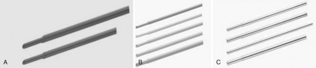
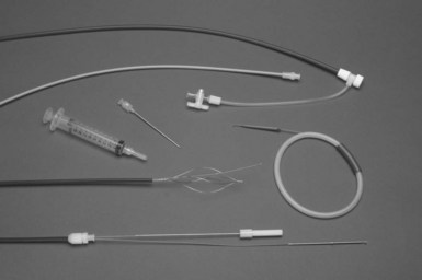
Mechanical sheaths are telescoping sheaths made of Teflon, polypropylene, or stainless steel (Fig. 26-13). These telescoping sheaths are designed to pass over the lead, which acts as a rail guiding the sheaths through the veins and down to the heart wall. Countertraction and counterpressure are applied as the sheaths move down the lead from one binding site to another. The outer sheath also acts as a workstation, facilitating the free movement of the inner sheath and lead by eliminating binding, and it protects the surrounding vascular structures. The leading edges of the sheaths are beveled. The rotation of the beveled tips facilitates maneuvering past obstructions and through the narrow channels along the tortuous paths surrounding the lead body. This is especially true in the superior veins. For the sheaths to pass down the lead in a true course, the lead must be stiff enough to act as a guide rail. The lead is stiffened by pulling it taut. The lead must be stiff enough to resist bending or kinking as the sheaths are passed over it (lead stiffness > sheath stiffness). The telescoping action of the sheaths allows the suppler inner sheath to track over the lead. The larger outer sheath is then advanced using the combination of taut lead and inner sheath as the guide rail. The lead is made taut by traction (pulling on it). A locking stylet is inserted, and a suture is usually tied to the lead, acting as both an extender and a traction handle.

As described earlier, experience and judgment are required to avoid tearing and avulsing vein and heart wall tissues. Also, if the traction force exceeds the lead’s tensile strength, it can cause lead disruption and breakage. Insertion of a locking stylet adds some stiffness to the lead and focuses the traction force to a locking site near the electrode. Except for simple cases, in which leads have been implanted for a short duration, a locking stylet should be inserted. Despite these provisions, when there is poor tensile strength of the lead, it is more difficult and more time-consuming to free the lead from its binding site. The forces involved frequently exceed the tensile strength of the lead, unless there is a good locking stylet and suture, resulting in lead disruption and breakage.

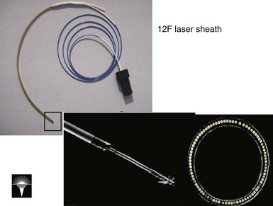
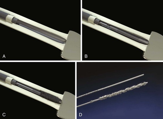
Excimer Laser Sheath

The development of the excimer laser was a milestone for lead extraction (Fig. 26-14). The excimer laser generated a high-energy 308-nanometer laser beam known to disrupt tissue (both cells and hydrated proteins) by an explosive vaporization of intracellular water. The rapid vaporization helped to cool the site. These were appealing properties for lead extraction. Unfortunately, the laser does not ablate heavily mineralized tissue. If the laser could not be maneuvered through this tissue in a grinding manner, counterpressure techniques were required.

The development of the excimer laser sheath was a technical achievement. It required expertise in polymer chemistry and optic fibers to develop a small-diameter, flexible sheath capable of withstanding the excessive forces applied during a lead extraction. At the time, optic fibers were bundled circumferentially inside a cylindrical metal housing, which protected the optic fibers from the applied forces generated while maneuvering the tip through the encapsulating tissue. Initially, the only sheath meeting all the clinical requirements was a 12-French (12F) sheath that was interchangeable with the 12F/16F mechanical Teflon sheaths. In time, larger 14F and 16F sheaths were perfected. These sizes were sufficient to manage all sizes of pacing leads up to the largest ICD leads. The last iteration of the laser sheath was to place a 15-degree bevel at the tip. It is the largest angle permitted by the circular configuration of the optic fibers at the electrode.

The laser is controlled by a foot switch. By design, the laser is on for 10 seconds and off for 5 seconds. The sound caused by the rapid pulsing of the laser furnishes a unique sound indicating the laser is on. The laser beam is a light cone that ablates tissue up to a distance of 1 mm. The water vapor generates bubbles that are clearly visible on echocardiography. Although the bubbles and other particulate debris are filtered out in the lungs, there are no apparent clinical sequelae. The cutting action of the laser can disrupt the SVC or the atrial wall if the lead is embedded in the wall. Because there is no way to know when a lead is embedded in the wall, the same emergency precautions apply to the laser as to the mechanical sheaths.

The laser sheath technique was evaluated prospectively in two clinical trials. The Pacing Lead Extraction with the Excimer Sheath (PLEXES) trial included only the initial version of the 12F sheath.15 Although there have been substantial improvements in the 12F sheath, including an outer sheath, better mechanical properties to prevent crushing of the optical fibers, lubrication, a flexible distal and a more still proximal segment, and certainly better understanding of how to use the tool, the PLEXES trial was a dramatic success. This randomized clinical trial compared mechanical extraction tools with laser-assisted lead extraction and was used to support the clinical release of this technology. The complete lead removal rate was 94% in the laser group and 64% in the nonlaser group (P = .001). Failed nonlaser extraction was completed with the laser tools 88% of the time. The mean time to achieve a successful lead extraction was significantly reduced for patients randomized to the laser tools: 10.1 ± 11.5 minutes versus 12.9 ± 19.2 minutes for the nonlaser techniques (P < .04). There was only one death, but it was in the laser group; and there were two other potentially life-threatening bleeding episodes in the laser group.

After the trial with the 12F sheaths, a second, nonrandomized cohort trial was done with 14F and 16F sheaths.16 This was particularly important, because implantable defibrillator leads required the 16F sheaths, and many of the bipolar leads (almost all) were better approached with the 14F sheath. In contrast to other, nonlaser sheaths, upsizing of the laser sheath to pass over (include) the fibrosis or calcification is frequently an effective maneuver. In this study, 863 patients underwent extraction of 1285 leads. Expanding the number of research sites from fewer than 10 to 52 gave a broader view of this tool in general practice. The patients treated with the 14F device tended to have older leads than patients in the 12F population; the 16F population, composed mostly of defibrillator patients, was younger, had more recent leads, and was more often male than the 12F population. Clinical success (extraction of entire lead or of lead body minus distal electrode) was observed in 91% to 92% of cases for all device sizes. The overall complication rate was 3.6%, with 0.8% perioperative mortality. The incidence of complications was independent of laser sheath size.

Ultimately, a cohort comparison trial of defibrillator and pacemaker leads extracted with laser assistance was done at the Cleveland Clinic.17 ICD extraction results were compared with the results for a matched cohort of patients undergoing extraction of ventricular pacemaker leads from a national registry and with the experience with pacemaker lead extraction at the Cleveland Clinic. Successful complete extraction of ventricular nonthoracotomy implantable defibrillator leads, in the absence of major complications, was achieved in 96.9% of attempts to extract leads from 161 patients. Clinical success was achieved in 98.1% of patients. There were three major complications, including one death. ICD lead extraction was done at an experienced center with equal risk and no significant difference in procedure or fluoroscopy time.

The total investigational experience with laser sheaths was also reported (October 1995–December 1999), including 2561 pacing and defibrillator leads in 1684 patients at 89 U.S. sites. Of these leads, 90% were completely removed, 3% partially removed, and 7% failures. Major perioperative complications (tamponade, hemothorax, pulmonary embolism, lead migration, death) were observed in 1.9% of patients, with in-hospital deaths in 13 (0.8%). Minor complications were seen in an additional 1.4% of patients. Multivariate analysis showed that implant duration was the only preoperative independent predictor of failure, and female gender was the only multivariate predictor of complications. Success and complications were not dependent on laser sheath size. At follow-up, various extraction-related complications were observed in 2% of patients. The learning curve showed a trend toward fewer complications with experience.18 A similar experience was observed in Europe.19

The LExICon study was the most recent study evaluating the most recent iteration of the laser-assisted lead extraction system.12 In this study, 2405 leads were extracted in 1449 patients from 13 centers. The procedural success was 96.5% with 97.7% clinical success. Major adverse events in 20 patients were directly related to the procedure (1.4%) including four deaths (0.28%). Major adverse effects were associated with patients with BMI less than 25 kg/m2. Overall, all-cause in-hospital mortality was 1.86%.

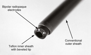
Electrosurgical Dissection Sheaths

The EDS has two bipolar electrodes positioned at the tip of the bevel (Fig. 26-15). The sheath is connected to an interface plate inserted on a conventional electrosurgery unit (Valley Lab Force V; PEMED, Denver), placed in a bipolar cutting configuration, and activated with a foot switch. The interface plate is attached to the front panel of the electrosurgical unit to ensure that the EDS is connected in a bipolar configuration. The interface also has an attachment to pulse the electrosurgical unit 80 times per minute. A plasma arc is generated between the electrodes. The plasma arc extends out from the electrodes and vaporizes the tissue to a depth of about 1 mm. On continuous discharge, desiccated tissue debris shunts the arc between the electrodes, preventing it from cutting. Also, on continuous discharge, if one of the electrodes touches a conductor coil, a parallel, alternate current (AC) circuit is created consisting of the EDS electrode in contact with the conductor coil, the conductor coil down to an electrode in the heart, and back to the other EDS electrode. An AC current applied to the heart in a unipolar configuration can fibrillate the heart. To ensure cutting and avoid fibrillating the heart, the EDS is operated in a pulsed mode at 80 pulses/min. In the pulsed mode, if a conductor coil is touched, it paces the heart.

The EDS is a conventional Teflon mechanical sheath with two tungsten electrodes embedded in the polymer. Placement of the EDS electrodes in a bipolar configuration at the tip of the inner telescoping sheath endows the EDS sheath with properties of mechanical sheaths; it can maneuver through tortuous veins and can apply countertraction and counterpressure. These sheaths are currently available in sizes 7F, 9F, 11F, and 13F (circumference of inner sheath).

The electrodes and sheath are radiopaque, allowing visualization during fluoroscopy. The electrodes’ position is continuously adjusted by rotation of the sheath. For example, around a curve, the electrodes are placed on the inner curvature passing down the lead. Also, the electrodes are rotated away from skeletal muscle and nerves to avoid stimulation. Stimulation of skeletal muscle or the phrenic nerve does not harm the patient and is not an issue with general anesthesia. However, skeletal muscle and diaphragmatic contractions can be discomforting and even frightening if the patient is awake. At present, this is the only clinical issue associated with the EDS. The same emergency precautions applicable to mechanical and laser sheaths also apply to the EDS.

Clinical evaluation of the EDS has been formally published only in an observational study from five centers, involving 265 patients with extraction of 459 leads.20 During the investigation, only the 9F and 11F sheaths were used, excluding almost all ICD leads from consideration for extraction. As in all extraction series, some of the leads came out easily and others were more difficult to remove, and the techniques consisted more of an approach than of universal use of one tool to remove all leads. In this case, 542 leads were potentially presented for extraction, but about 15% were removed with direct traction, yielding 459 for which the EDS was employed. The laser tool was used in fewer than 3% of the leads. The average implant duration of the patient’s oldest lead was 8.4 ± 5.0 years; 31% of patients had leads implanted for longer than 10 years. Major complications occurred in 2.6% of patients, including cardiac tamponade in 4 patients (1 surgical repair, 1 after switching to a femoral approach), 1 hemothorax, 1 arteriovenous fistula (surgical repair), and 1 death that was associated with the mechanical removal of an oversized SVC lead for which the EDS was not used. For the 459 leads with attempted removal by the EDS, 99.4% were removed (95.9% completely, 3.5% subtotally with ≤4 cm of lead remaining), and only 0.6% were not removed.
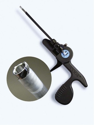
Overall, the experience with the EDS has been good. The application of 7F and 13F sheaths with intermittent pulsing of the electrosurgical energy has been useful in removing smaller and larger leads and in making the sheath more powerful in cutting through the fibrosis.

Evolution Sheath

A newer mechanically powered lead extraction sheath set is the Evolution mechanical dilation sheath (Cook Vascular). A rotating inner sheath with a threaded-barrel metal tip is designed to function as a dilating drill (Fig. 26-16). This bores through the encapsulating fibrous tissue as it advances down the lead through the binding sites. The outer sheath is a conventional, beveled plastic sheath. The rotation of the inner sheath is powered by a pistol-grip handle squeezed by the operator (mechanical power). Multiple inner diameter (ID) sizes (7, 9, 11, and 13 French) are available. The initial experience with the Evolution had mixed results.21 There was a high procedural and clinical success rate. The Evolution is particularly useful as a rescue when heavy calcifications are encountered while using the laser sheath. There was a tendency toward wrapping of adjacent leads and also collateral damage to the leads not intended for extraction. The system seems to work best in single-lead systems when collateral damage or wrapping is not an issue. No complications resulted from use of the Evolution.

Locking Stylets

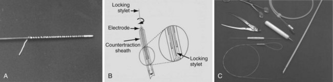
Locking stylets were developed after the mechanical sheaths. From the beginning, the major pacing companies (Medtronic, Cordis, Pacesetter [St. Jude Medical], and Boston Scientific [Guidant, CPI]) all attempted to make a universal locking stylet (one size stylet to fit all leads). These initial attempts were unsuccessful because of breakage of the locking mechanism. The tensile strength was inadequate to withstand the traction forces. Cook Pacemaker (now Cook Vascular) took another approach, abandoning the concept of a universal stylet. Their first-generation locking stylet came in various sizes to fit a variety of conductor coil diameters (Fig. 26-17). The conductor coil had to be measured before selecting the locking stylet. This locking mechanism was a small wire welded to the tip of the stylet and wrapped around the lead. Once the lead was passed down the conductor coil to the electrode, it was rotated counterclockwise, bundling the free wire and causing it to bind against the conductor coil. The greater the traction, the greater was the binding force. The locking stylet bound to the inner conductor coil, ideally at the distal electrode, functioned as a lead extender for applying traction and focused the traction force at the binding site. Focusing the traction force helped maintain the integrity of the lead but did not prevent lead disruption if excessive force was applied or if the lead had poor tensile strength. Also, the locking stylet was conductive, and the heart could be paced during parts of the lead extraction procedure if needed. This was the first effective locking stylet.

Cook’s second-generation locking stylet (Wilkoff Locking Stylet) had a different locking mechanism (Fig. 26-18). These stylets had a small flange at the tip designed to stay flat against the stylet until the preloaded thin cylinder was advanced; the cylinder deflected the flange to lock into the conductor coil. This was an efficient locking stylet that was easy to implant and that could be removed by rotating the stylet, breaking the flange. However, it could not be used if the conductor coil diameter was 0.016 inch or less, and it could not be used with extendable/retractable screw-in leads. Spectranetics made the first near-universal locking stylet (Lead Locking Devices 1, 2, 3 and e) to fit all leads. It used a long wire mesh that bundled and bound the stylet to the conductor coil. This type of locking stylet was efficient and functioned well. Ultimately, the Spectranetics LLD-EZ was developed that works for all but a few leads with extremely small IDs to the inner coil.

Cook’s most recent, third-generation locking stylet (Liberator Locking Stylet) is a true universal locking stylet (one size fits all). It uses a wire coil that is compressed by a reloaded cylinder, expanding the wire coil and binding it against the conductor coil (Fig. 26-19). Both the Liberator and the LLD-EZ are extremely versatile and provide an advantage over the alternative for either advancement down the conductor coil in locking.
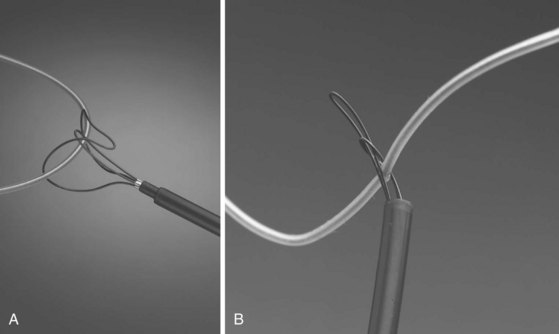
As mentioned earlier, the goals for inserting the locking stylet were to provide a lead extender and to focus the traction force at the tip to help maintain lead integrity. Although these goals were met, the insulation still has to be secured with a suture. Without traction to the insulation, it can “snow plow” and tear more easily, making passage of the extraction sheaths difficult, and in some cases preventing their passage. Therefore, traction should be applied to both the suture (insulation) and the locking stylet (conductor coil) to be most effective.

A new tool, the Bulldog lead extender, is designed for leads that have no conductor coil, or when the conductor coil is severely damaged. It mechanically locks onto the conductors to the shocking and pacing electrodes and produces an excellent mechanism for improving the tensile properties of the lead. The Bulldog can also be used to lock onto the cables to the shocking coils in leads with a cylindrical conductor coil, and a locking stylet is still used in the cylindrical coil.

Snares

Snares are used to grasp leads and to remove tissue in the bloodstream from the vein or transatrial entry site. The vein entry sites typically used are the subclavian, internal jugular, and femoral veins. Only a few snares are safe to maneuver in the cardiovascular system or have the tensile strength to support the forces involved in a lead extraction procedure. The Dotter snare, Needle’s Eye Snare (Cook), and Amplatz Goose Neck Snare are discussed as examples of the types of snares available. The Dotter snare, together with a deflection catheter, is prepackaged in the femoral workstation. Before the availability of powered sheaths, lead breakage and the need to use a femoral approach were common. The only substitute for a snare is a cardiac surgical procedure. Consequently, facility with snares still is a requisite skill. Also, there are still extractors who do not use powered sheaths, relying only on the mechanical sheath extraction, and snares are integral to these procedures.

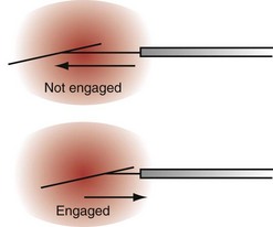
With the Dotter, needle-eye, and gooseneck snares, a reversible loop is created around the lead body to pull the proximal end of the lead out of the superior veins into the IVC, without placing traction on the electrode-myocardial interface (Fig. 26-20). A loop must be created and bound to the lead body. The binding of the loop must be reversible. Irreversibly binding the lead, or inability to remove the loop from around the lead, may result in dangerous traction maneuvers being performed in desperation while trying to extract the lead and snare. Failure to extract the lead subjects the patient to more invasive procedures to remove both lead and snare.

Creating a reversible loop using two snares is a complicated maneuver requiring practice to perfect. One technique is to use a Cook deflecting wire guide and a Dotter basket snare. The Cook deflecting wire guide is wrapped more than 360 degrees around the lead body. Next, the tip of the deflection catheter is passed into the Dotter basket snare. When the basket snare is pulled into the workstation, the basket closes, grasping the deflection catheter and completing the loop. The loop is pulled into the workstation, tightly binding the lead body to the workstation, and traction is applied. If needed, the loop is relaxed and repositioned on the lead body. This sequence is repeated until the lead is pulled out of the superior veins, through the right atrium, and into the IVC. The reversible loop is then released, and the deflection catheter is removed. This same process can be done with a gooseneck snare and tip deflecting guidewire or a deflectable EP catheter and gooseneck snare.

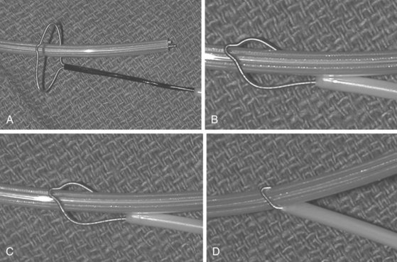
The Cook Needle’s Eye Snare is a more efficient method of grasping the lead in a reversible manner (Fig. 26-21). This snare has a wire loop that is passed over the lead body. A small, wire-loop tongue is then passed over the opposite side of the lead and into the larger wire-loop tongue. Pulling this apparatus into the workstation binds the lead reversibly for safe, indirect traction. Also, the binding forces are more diffuse, resulting in less lead breakage.

The Amplatz Goose Neck Snare is a radiopaque noose that is slipped over the free proximal end of a lead (Fig. 26-22). In situations where the proximal end is floating in the SVC, heart, or a pulmonary vein, the free end can be lassoed and extracted. The gooseneck snare can also be used to grasp the end of a tip-deflecting guidewire or deflectable EP catheter draped over the midportion of the lead. This is an excellent technique to reposition a lead so that the end can be grasped after being pulled down from the subclavian/jugular system, or to grasp the lead reversibly for extraction.

image Lead Extraction Approaches

Extraction procedure approaches involve transvenous and cardiac surgical extraction techniques. Transvenous approaches are usually performed through the lead implant site, but any other vein suitable for the extraction instruments may be used. Suitable veins include the axillary-subclavian-brachiocephalic, external jugular, internal jugular, and femoral-iliac. Cardiac surgical approaches to intravascular leads are transatrial, right ventriculotomy, and open-heart surgery (OHS). All epicardial lead extractions are cardiac surgical procedures.

In the past, procedure approaches were separated into superior (SVC) and inferior (IVC) vena cava approaches. An SVC approach is defined as the passage of lead extraction instruments through the vein entry site and down the SVC to the heart. The IVC approach was defined as a transfemoral approach using remote extraction instruments inserted in a femoral vein and passed through the IVC into the heart. This classification was based on the two types of extraction tools available at that time. Currently, a more descriptive classification is to separate the approaches into transvenous and cardiac surgical procedures. Transvenous procedures are further divided into procedures through the vein entry site and those through a remote vein, usually the femoral or internal jugular veins. Cardiac surgical procedures are separated into the individual cardiac surgical approaches: transatrial, ventriculotomy, and OHS.

The approach selected depends on the experience, extraction skills, and extraction instruments available to the physician; the reason for the lead extraction; and situations that arise during lead extraction. For example, a transvenous lead implanted in the right ventricle with a conductor coil fracture is ideally extracted from the vein entry site, using a powered sheath. A change in approach must be considered if this same lead is found to be attached to the lateral wall in the distal half of the SVC by a calcified mass of encapsulating tissue. A determination to continue the approach or a decision to choose a new one depends on experience and extraction skills. Two safe approaches are through the femoral vein using snares and a transatrial cardiac surgical approach. If this same lead is found to have large, solid thrombi measuring 4 × 8 cm, it is best extracted through a transatrial approach or OHS.

Extracting leads is dangerous if it is not properly performed. Procedures with these inherent dangers can be made safe only by knowledge of the pathology, pathophysiology, extraction skills, and available approaches. Using a nonmedical comparison, flying an airplane has inherent, potentially life-threatening risks, but with proper training, experience, and “flying by the numbers,” it is safe. A lead extraction performed in an organized, step-by-step manner, following known principles and proven guidelines, is similar. The antithesis is “flying by the seat of your pants” or extracting a lead in a reckless, cavalier fashion, hoping for the best.

While reading this section, remember that life-threatening complications are rare. During the evolution of lead extraction, the EP community and surgeons helped define the procedures, techniques, and approaches to minimize and ultimately eliminate complications of all types. The material presented represents more than 25 years of experiences that form the basis for current lead extraction procedures.

Transvenous Approaches

Lead Vein Entry Site

A transvenous approach through the lead vein entry site is a natural approach. After the pocket debridement procedure, which includes freeing the leads to near the vein entry site, it is natural to continue and remove the lead from this site. In some situations, the lead is broken or cut and retracted into the axillary-subclavian-brachiocephalic veins. If this lead can be grasped by an instrument passed into the vein and exteriorized, it is then considered to be a lead passing through the vein entry site, for purposes of this discussion. The lead can be extracted by direct traction, if applicable, or by a mechanical or powered telescopic sheath. Most extractors currently use this natural approach. Working through the vein entry site is efficient and subjects the patient and physician to the least amount of radiation, with the fewest maneuvers and least time required. Radiation exposure is related to the amount of fluoroscopy time. Also, once the lead is removed, a new lead may be readily inserted through the conduit created during the extraction.

As previously discussed, direct traction is applied by pulling directly on the lead manually, with a suture or locking stylet or both. Indirect traction refers to grasping the lead with a snare and applying traction by the snare. Some leads are easily removed by direct traction.

The vein entry site is not always easily accessed. In some situations, the lead is entrapped in calcified encapsulating tissue and cannot be entered using the nonmetal extraction sheaths, with or without power. The cause of the calcified encapsulating tissue at the vein site is usually related to introducer technique. If the introducer needle scores the clavicle or first rib, elevating the periosteum, the periosteum will re-form about the lead, entrapping it in a bone sheath. If the introducer needle passes through the costoclavicular ligament, this damaged tissue will mineralize and entrap the lead. Natural maturation of a thrombus into fibrous tissue, which mineralizes with time, also may create this problem. Usually, however, the problem is associated with the periosteum and costoclavicular ligament, and metal sheaths are used.

Telescoping stainless steel sheaths look dangerous but are safe and effective if properly used (see Fig. 26-13, C). The principle is simple: keep the sheaths “tracking true” over the lead, and apply the force needed to destroy this tissue. A combination of pushing and rotation of the beveled tip is most effective. Once the metal sheaths break into the vein, they are removed and replaced by plastic extraction sheaths. “Tracking true” is a principle used for all sheath maneuvers. All sheath maneuvering must be performed under fluoroscopy, to ensure that the sheaths are tracking over the lead. Any kinking or other deviation of the vein course is dangerous. It creates a false passage that may be extravascular and may damage nearby structures.

Complications

Telescoping sheaths use a combination of countertraction, counterpressure, and tissue ablation (powered sheaths) to maneuver past the intravenous and intracardiac binding sites to the heart wall. These techniques, used with various types of sheaths, have already been described in detail. The maneuvering of these sheaths over the lead and down to the heart is separated into the three anatomic regions: axillary-subclavian-brachiocephalic veins, SVC, and intracardiac. This is the most dangerous portion of the procedure, and a detailed understanding of the issues unique to these three regions is essential for a safe and successful lead extraction.

Complications associated with lead extraction involving loss of vein or heart wall integrity are tearing (including bone spicules), avulsion, rupturing, penetration, embedding of lead into the wall, and exclusion of the lead from the vascular space. The sequelae of these events are related to the anatomy of the region. The presence of fibrous tissue must be included when considering the anatomy of the region. Inflammatory reaction and the resultant fibrous tissue formation increase the tensile strength of the tissue. For example, a dissecting arterial hematoma or pericardial effusion is rarely seen in patients with previous OHS, because scar tissue has the positive effect of reinforcing the tensile strength of the surrounding tissue. The forces required to tear aorta, subclavian, or innominate arteries or heart wall reinforced with scar tissue exceed those normally applied during lead extraction.

The axillary veins are surrounded by soft tissue on both sides, and the loss of vein wall integrity causes a low-pressure extravasation of blood, which is usually of no consequence. The subclavian-brachiocephalic veins are surrounded by structures that, if damaged, could lead to life-threatening consequences. On the left side, the vein is contiguous with the subclavian artery and aorta. During an inflammatory reaction, if the lead becomes embedded in the vein wall and involves the wall of the subclavian artery, innominate artery, or aorta, passage of extraction sheaths over the lead could tear these contiguous vessels, causing an arteriovenous fistula or a dissecting hematoma or both. The arteriovenous fistula could cause high-output heart failure, and the dissecting hematoma could cause major blood loss. If the hematoma ruptures into the left thoracic cavity, the resultant hemorrhage could be lethal. Immediate surgical intervention is required for a dissecting hematoma. An emergency median sternotomy provides satisfactory surgical exposure. If time permits, a minimally invasive approach may be preferable, especially for correction of a small arteriovenous fistula.

Also on the left side, it is common to maneuver in and out of the subclavian-brachiocephalic veins, causing low-pressure extravasation. After vein occlusion and organization, frequently all that is left is an atretic, encapsulating fibrous tissue sheath (sometimes mineralized). For a single lead, the telescoping sheaths may be larger than the circumference of this fibrous tissue sheath, and the entire capsule may be included, along with the lead in the telescoping sheaths. If two or more leads are present, the circumference of the telescoping sheaths will remove at least one wall of the encapsulating sheath entirely or in segments. The resulting extravasation is not clinically significant with reasonable venous pressures. In exception cases in which the venous pressure reaches a systemic level of 70 to 90 mm Hg, a dissecting hematoma would ensue, causing the same clinical scenario as described earlier.

The right-sided vein is contiguous with the right pleura. If the encapsulating fibrous tissue and embedded lead constitute the inferior vein wall, a tear in the pleura will result in hemorrhage into the right thoracic cavity. This can be lethal without immediate surgical intervention. The area is difficult to reach through a median sternotomy or an anterior thoracotomy. Because of the time constraints, a median sternotomy with elevation of the clavicle and right anterior chest wall is radical but provides adequate exposure. In less urgent situations, the patient may be repositioned for a less extensive approach. Venous bleeding into the right chest, if not massive, can be insidious, and may not be recognized until the onset of cardiovascular collapse. It is not painful, and the signs and symptoms associated with blood volume depletion can be masked by anesthesia or by increased catecholamines compensating for the low filling pressures. With sufficient blood loss, compensation is no longer possible, blood pressure falls, metabolic acidosis develops, and cardiovascular collapse ensues.

The SVC passes from the brachiocephalic veins to the right atrium. Along the upper half of the SVC, the lateral surface is contiguous with the right pleura, and the lower half is within the pericardium. Loss of integrity of the upper half results in blood loss into the right thoracic cavity; in the lower half, it causes cardiac tamponade. The sequelae of blood loss into the right chest are identical to those described for the right subclavian-brachiocephalic veins. In an emergency, a median sternotomy provides adequate exposure to either the upper or the lower half of the SVC.

Cardiac Tamponade

Bleeding into the pericardial space causes a cardiac tamponade (pathologic cardiac compression). Small amounts of blood (~200 mL), accumulating rapidly in the pericardium, will cause symptoms. Rapid accumulation increases ventricular wall compliance, decreasing filling of the ventricles and the resultant stroke volume. Rapidly accumulating blood in the pericardial space clots, whereas a slow accumulation does not. Clot formation can localize the compression, and some parts of the heart are more sensitive to compression than others. A 1-cm tear causes immediate tamponade with instantaneous decrease in systolic pressure. A precipitous drop of systolic blood pressure with failure to recover within 2 to 3 minutes is an emergency requiring immediate surgical intervention. The patient and operating field are then prepared for a median sternotomy. The TEE is usually definite, but if it is not, and no other cause is apparent, a tamponade must be considered the cause. The time constraints force a decision to be made within 2 to 3 minutes to prevent irreversible brain damage. A median sternotomy is used to decompress the pericardium, manually remove the clot, and surgically repair the tear. Needle aspiration and tube drainage are ineffectual for removing clot.

A tear in the lower half of the SVC measuring 2 mm is an example of a slow-onset pericardial effusion. A slow onset of tamponade, while maintaining a blood pressure, allows more time to confirm the tamponade and to make the decision of whether to use a less invasive pericardial drainage procedure. Pericardial drainage tubes inserted into a pericardial effusion are therapeutic in relieving the compression, with immediate restoration of blood pressure. It provides a drainage system for monitoring bleeding and in some cases for blood replacement, cell saver, or direct reinfusion, and it buys time to set up for a corrective surgical procedure. A rushed insertion of a percutaneous pericardial tube without an effusion being present can be a disaster. This is especially true in an enlarged heart if the diaphragm is penetrated and torn during insertion of the tube, creating the need for surgical correction. The safest approach is through a small, subxyphoid incision that opens the pericardium under direct vision. Although the incision is slightly larger than for a percutaneous approach, it is safe.

Other conditions mimicking a tamponade are mechanical occlusion of the SVC, lead traction applied to the RV wall decreasing compliance and filling, tachyarrhythmia, and metabolic acidosis from poor perfusion. Reflex therapeutic actions for a drop in blood pressure during lead removal include pausing lead extraction maneuvers (including lead traction), immediate IV administration of a vasopressor (phenylephrine or norepinephrine), cardioversion of arrhythmias, and administration of sodium bicarbonate in patients with low cardiac output. These reflex actions are used throughout any procedure, at any time, to compensate for transient decreases in filling pressure from the causes mentioned. They should be considered routine maintenance and not emergency treatment.

Loss of Wall Integrity

The right atrium and right ventricle are also contained within the pericardium, and loss of wall integrity from a penetration, tear, or tissue avulsion can cause cardiac tamponade as described earlier. The treatment is the same. There are four mechanisms for tearing the wall not previously discussed: bone spicule laceration of the right atrium, chronic penetration of the right ventricle, fibrous tissue between the electrode and the heart wall, and size disparity between the electrode and the sheath.

The first mechanism involves a variation of the encapsulating tissue seen only in some extreme cases of mineralization. An example is a large silicone lead (>10F) that was in place for more than 20 years and making contact with the inferolateral atrial wall. The encapsulating tissue was completely mineralized, with “bonelike spicules” embedded within the wall. With any traction on the encapsulating sheath, the spicules act as a knife blade, causing a surgical incision in the wall. This scenario is rare. It has not been seen with any of the silicone leads manufactured since the late 1970s. To be safe, a lead bound to the lateral wall of the atrium should be removed with caution.

A second mechanism involves the application of countertraction in removing a lead from the ventricular wall. For example, an ICD lead implanted for 12 years is attached to the thin anterior wall of the right ventricle. The tissue at the electrode removed during the extraction contains epicardial fat, indicating a defect in the ventricular wall and suggesting that the electrode had penetrated the wall, most likely during implantation. Unless the lead has perforated and is positioned within the pericardial space, it is undetectable. Fortunately, this is a rare occurrence.

A third mechanism that could tear the heart wall involves a band of tissue attached to the extracted electrode that is still connected to the heart wall. Continued traction on the freed lead could avulse the tissue or tear the heart wall. When this is seen, the sheath (preferably powered) is maneuvered until the band is cut. With mechanical sheaths, care must be taken with the force used to break the band. This mechanism is readily seen on fluoroscopy, and once recognized, care is taken.

A fourth mechanism in loss of wall integrity is caused by a large size disparity between the extraction sheath and the lead. Although this mechanism has been reported, it is rare. If an attempt is made to apply countertraction with a sheath larger than the lead, the electrode and the ventricular wall both will be pulled into the sheath, potentially tearing the wall. Once the ventricular wall tissue is pulled into the sheath, it becomes a form of direct traction and not countertraction. For this to occur, the size difference between the lead and the sheath must be sufficient to accommodate the electrode-tissue mass. The cardiac tissue being pulled into the sheath cannot be seen. The only protection is avoiding large size differences between the lead and the sheath. Unfortunately, this is not always possible. For example, the smallest laser sheath is 12F with a 16F outer sheath, so extracting a 4F lead involves a sizable mismatch, and care must be taken. Also, a 9F sheath may need to be changed to an 11F sheath to include calcified encapsulating tissue, resulting in a size mismatch during countertraction.

Remote Vein Sites

A remote vein site is a vein other than the lead vein entry site. A remote vein site requires a remote instrument, such as some type of snare, to manipulate the lead. The femoral vein was the first remote vein site used. During the early evolution of lead extraction, removing leads with mechanical sheaths from the superior veins through the vein entry site was frequently a failure and potentially dangerous. A more difficult approach through a femoral or other remote vein site was more effective and safer. The problems were related to maneuvering the long, telescoping mechanical sheaths from the femoral vein to the heart wall and the complexities associated with grasping the lead body. With the advent of powered sheaths, the safety and efficacy of removing leads from the vein entry site improved dramatically.

Although most of the leads can be removed from the vein entry site, there are still dangerous situations and lead breakages. In these cases, a remote vein or a transatrial surgical procedure are the only options. In many cases, the femoral vein approach is still the best remote vein approach. However, other remote veins (contralateral external jugular, internal jugular, or axillary-subclavian-brachiocephalic) may be more suitable and easier to use. Some physicians have developed combination approaches, applying mechanical sheaths from both the vein entry site and a remote vein site. This combination constitutes a safe and efficacious approach, as developed by Bongiorni et al.,22 and has become increasingly popular, particularly when the need for reimplantation through the original venous access is no longer required. For cases such as lead breakage, use of a snare to grasp the lead and the subsequent removal of the lead using direct traction or a powered sheath is advantageous. Powered sheaths are not long enough in moderately tall patients for a femoral approach. Consequently, these approaches are confined to the superior veins, via both remote and vein entry sites.
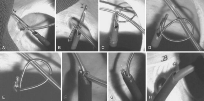
The techniques used involve manipulation of snares to grasp the lead body. Although these techniques are simple in principle, they can be difficult to achieve in a timely fashion. Initially, there were no snares designed for lead removal, so a suitable off-the-shelf snare designed for other purposes (“off-label”) had to be found. The criteria for such a snare included the tensile strength to withstand the forces applied during lead extraction, safety of the surrounding tissue when maneuvering the snare, and reversibility of the grasping mechanism. The Dotter snare was the first snare to meet the tensile strength requirements. When the Dotter snare was combined with the Cook deflection snare, the safety and reversibility requirements were met. Later, two snares were manufactured meeting these requirements: Cook Needle’s Eye Snare and Amplatz Goose Neck Snare, which are still in use (see earlier discussion). Angled, pigtailed angiographic catheters, endsnares, bioptomes, and every conceivable catheter has been used to good effect for some patients. Thus the principles of use are more important rather than the precise tool.

A classification of remote vein approaches deviates from the historic use of the IVC (femoral vein) approach. A natural classification is to employ the removed vein (e.g., femoral approach, internal jugular approach). Remote vein site use is discussed here based on this natural classification. The goal is the same as for the vein entry discussion: to define the approach, procedure techniques, and expected results in relation to the pathology and pathophysiology associated with the implanted lead.

Femoral Approach

As discussed earlier, before the advent of powered sheaths, the femoral approach was used extensively. The indications for its use were failure to extract leads from the superior veins by any technique, lead breakage, and avoidance of the application of excessive force with the mechanical sheaths. The techniques used have not evolved significantly over the past 15 years. The transvenous approach through a femoral vein requires a special sheath set (e.g., Byrd WorkStation) that functions as an introducer, as a workstation for manipulation of snares, and as countertraction sheaths (Fig. 26-23). The set consists of an introducer needle, a guidewire, a 16F workstation, an 11F tapered dilator, an 11F telescoping sheath, a Cook deflection snare, and a Dotter basket snare. The workstation serves many functions. Initially, it acts as a protective sheath. The outer sheath prevents the insertion, withdrawal, and manipulation of the inner sheath and snares from damaging the veins or heart. To prevent clot formation, the workstation has a valve (Check-Flo) to continuously irrigate the sheath. The workstation and snares form a reversible loop to pull the proximal portion of the lead out of the superior veins; the workstation also acts as the outer telescoping countertraction sheath.

The safe insertion and removal of the workstation is a prerequisite for an extraction procedure through a femoral vein. The workstation is 16F and must be inserted with care. Fluoroscopic monitoring is mandatory. Once the guidewire is passed into the heart, the workstation with its tapered dilator must be maneuvered through the iliac vein and IVC and into the right atrium. The route can be circuitous, especially from the left side. In rare cases, the curvature may be too sharp for the stiff dilator to follow the guidewire. Forcing the dilator in this situation is unsafe, and the approach should be abandoned. A torn retroperitoneal iliac vein or IVC is a serious complication. Once the workstation is inserted, irrigation fluids are run continuously through the Check-Flo valve to prevent clotting.

Pulling leads down and out of the superior veins has been remarkably successful. Many leads freed from the heart cannot be pulled up through these same veins. The lead binding forces caused by the circumferential bands of fibrous tissue are the same in both directions. The hypothesis for this difference is the free upward mobility of the mediastinal structures and the inability to pull the superior veins downward. The mediastinum is easily pulled upward, compressing the veins. This compression of the fibrous tissue bands around the lead as they bunch together is postulated to increase the binding strength. The superior veins cannot be pulled downward, and irreversible lead slippage through the binding sites is not an issue. Cardiac function is not influenced by pulling leads downward out of the superior veins. Failure to extract the proximal lead from the superior veins using this technique is rare and usually caused by other complicating factors, such as thrombosis of the superior veins and excessive fibrosis around the lead caused by a previous extraction attempt that left the conductor coil exposed or pulled the lead taut against the heart and vein wall. Such leads are removed using approaches (e.g., transatrial) reserved for more complicated extractions.

To apply countertraction through the workstation, the proximal end of the lead must be entangled in the basket snare. This is accomplished by placing the basket snare close to the lead and rotating it slowly. The lead will flip into the basket. The basket closes when the 11F sheath is advanced over the snare. For most leads, the snare and lead are pulled into the 11F sheath. The workstation and 11F sheaths are then worked in a telescoping fashion to a point near the electrode. At this point, countertraction is applied to extract the electrode, as previously described. Removal of the workstation must be carefully performed. Once the lead is extracted, clot and debris may be attached to the end of the tubing. If this material dislodges in the femoral vein entry site, it can act as a nidus, forming a thrombus or initiating thrombophlebitis and its sequelae. To prevent this complication, blood is aspirated during the withdrawal of the workstation. If the entry site does not bleed freely after withdrawal of the workstation, a surgical exploration of the vein is recommended. Bleeding is controlled by applying pressure over the vein entry site after withdrawal of the sheath and during Valsalva maneuvers induced by the anesthesiologist. A suture or staple is required to close the skin. A potential complication is thrombophlebitis and pulmonary embolus. Concern about this complication was the incentive for the workstation. Postoperatively, anti-embolic stockings (pneumatic, if possible) and subcutaneous heparin (5000 units twice daily) are the only precautions taken.

The technique for grasping the lead in a reversible loop using a Cook deflection catheter and a Dotter snare or with other tools is more complicated. Once mastered, it is an effective method of performing precision extraction. For example, a patient may have six leads in the heart. Two new leads are connected to a pacemaker and are to be saved. Four leads are abandoned and are to be extracted. The abandoned atrial and ventricular leads can be extracted, leaving the newly implanted leads intact. The IVC approach allows this level of precision.

Combined Approaches

To some degree, most remote approaches are a combination of a lead vein entry site and a femoral vein approach. An extensive effort may have been used to free the lead from the superior veins; this was always in preparation for the intended use of the remote approach. From its inception, some physicians championed the use of a combined approach. Initially, the vein entry approach and femoral vein approach were combined, sometimes with two teams working, one for each approach. The goal was to free the lead by applying the extraction techniques from both approaches and removing the lead in an opportunistic fashion (the resultant easiest approach). The electrophysiologist might work from the vein entry site and a radiologist from the femoral vein site. Although successful, this approach was overkill for most lead extractions, especially once powered sheaths became available.

A combination of the vein entry site approach and a right internal jugular approach is used by some physicians as their primary approach to lead extraction. They use snares and/or a grasping instrument passed through the internal jugular vein into the SVC to pull the lead out of the axillary-subclavian-brachiocephalic veins and, if necessary, to pull the lead into the atrium. Mechanical sheaths are then applied to complete the extraction (to date, powered sheaths are not used).23

The techniques used with remote approaches can be applied through the vein entry site. A lead may have broken or been cut, retracting into the axillary-subclavian-brachiocephalic veins, floating in the SVC or heart, or migrating into the pulmonary veins. If the lead can be reached and grasped inside the axillary-subclavian-brachiocephalic veins by a surgical instrument, it can be pulled out of the vein entry site and secured with a suture or a locking stylet, or both. The lead can then be extracted using the vein entry approach. If the lead cannot be grasped by an instrument but a snare can be passed into the SVC from the vein entry site or a contralateral vein, the lead is grasped and removed using mechanical and/or powered sheaths. Before powered sheaths became available, the femoral approach was common, but now it is rarely used. Surgical electrophysiologists have the transatrial approach as a viable alternative. Regardless of the frequency of use today, the transfemoral approach is a requisite procedure for managing device-related complications.

Surgical Approaches

The surgical approach to lead extraction is a cardiac surgical procedure. The three procedures used for transvenous endocardial implants are the transatrial approach, right ventriculotomy, and an open-heart procedure using cardiopulmonary bypass (CPB). These procedures should be performed only by experienced cardiac surgeons. The technical and patient management skills of an experienced cardiac surgeon negate the normal risk associated with the complexity of the surgical procedure. The transatrial procedure is a general procedure that can be used for both extraction and implantation of leads. It can be used instead of the transvenous remote approach for lead extraction. It has the added advantage of being an implantation site that bypasses the SVC and IVC. The right ventriculotomy is a technique for removing leads from the right ventricle in special situations. Currently, an open-heart procedure is reserved for removing thrombotic material (usually infected) from the right atrium and ventricle and for removing leads implanted in the left ventricle.

Transatrial Approach

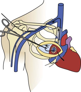
The transatrial approach, first described by Byrd in 1985, is a surgical EP procedure suitable for intracardiac implantation, explantation, and ablation procedures.25 The only disadvantages are the morbidity associated with surgical thoracic pain and the need for a medical electrophysiologist to work with a cardiac surgeon. It is a primary approach for noninfected patients who are candidates for a transatrial lead implantation. Younger patients with occlusion of one brachiocephalic vein or with SVC syndrome have the old leads extracted through a transatrial approach, followed by implantation of new leads. The advantage of the transatrial approach is the ability to remove leads that are not accessible or removable by the SVC or IVC approach. Most of the transatrial extractions are failures of the IVC approach. Rarely, failure of an SVC approach will lead directly to a transatrial approach (e.g., when the workstation cannot be passed through the femoral veins into the heart). Infected patients who are candidates for transatrial lead implants will have the leads extracted by an SVC or IVC approach and the transatrial implantation done after the infection is properly treated.

The transatrial approach is performed as originally described, through a limited surgical incision on the right anterior chest wall (Fig. 26-24). The right atrium is exposed by removal of the third or fourth right costal cartilage (determined by fluoroscopy). The pericardium is opened and suspended, and a pledgeted purse-string suture is placed in the right atrium. If the pericardium has been obliterated from a previous procedure or disease process, a small region of the lateral wall of the right atrium is dissected free. Using fluoroscopy, a pituitary biopsy instrument is inserted through the purse-string. The lead body is grasped in the atrium and pulled out. The lead is then cut, extracting the proximal and distal segments separately. The proximal portion of the lead can usually be pulled out by direct traction. The only limitation to the force employed is the tensile strength of the lead. In occasional cases where the tensile strength is insufficient, telescoping powered sheaths may be required. The distal segment is extracted by inserting a locking stylet, advancing telescoping powered sheaths to the wall, and removing the electrode from the wall using countertraction. This procedure is repeated for each lead to be extracted. On completion of the lead extraction, the atriotomy site is used to insert new leads, or to perform another EP procedure, or the purse-string suture is tightened, tied, and abandoned.

After transatrial extraction, patient management is more involved than for the transvenous extraction techniques. The pericardium must be drained, in most cases by a closed-drainage system such as a chest tube, if the pleural space is free; by a mediastinal tube, if the pleural space is obliterated; or by a Jackson-Pratt closed-drainage system, if both the pleural and pericardial spaces are obliterated. The thoracic cavity is occasionally entered and a chest tube inserted to drain both the pericardium and the pleura. These drainage tubes are removed in 2 to 3 days. The procedure-related morbidity increases the hospital stay by 1 or 2 days, compared with a transvenous procedure. Patients must be managed in an intensive care setting until the thoracic pain sequelae are controlled.

Right Ventriculotomy

A right ventriculotomy is a cardiovascular surgical procedure. This approach is rare. Because this procedure is virtually unknown, a cursory discussion of indications for use is in order. Initially, it was reserved for infected broken leads retained in the right ventricle that were not reachable by the other approaches. These are fragile leads that break near the ventricular wall or within a fibrous tissue tunnel along the ventricular wall and are impossible to grasp using transvenous or transatrial techniques. It is also used with lead penetration requiring lead removal and a repair of the heart wall, and possibly for an emergency tear in the ventricular wall caused by an attempt to remove a lead. In the latter, the defect is turned into a ventriculotomy site for lead extraction.

The first ventriculotomy procedures were performed by Byrd in the late 1980s. The heart is exposed through a median sternotomy incision. The heart is then elevated on a pad, exposing the right ventricle. The tip of the electrode is localized by fluoroscopy and by using needles for triangulation of the electrode. A purse-string suture is placed around the electrode, and a ventriculotomy incision is made to the electrode. The electrode is grasped with a clamp and pulled out of the heart. Because the lead segment is being pulled in the direction of the tines, the tines slip out of the embedding scar without resistance. At present, the procedure is performed through a minimally invasive incision on the anterior surface of the left chest through the fifth intercostal space. The purse-string and extraction techniques are the same. This is a stressful procedure for the surgeon, and number of procedures performed to acquire some comfort level is unknown. As long as a need exists for right ventriculotomy, attempts will continue to perfect it.

Open-Heart Procedure

The concept of using CPB to perform an open-heart procedure is intuitively obvious. It is a standard cardiac surgical procedure involving a median sternotomy incision. Right-sided leads were initially removed by direct traction and under direct vision during OHS. Transvenous and transatrial lead extraction techniques evolved to eliminate the need for an OHS procedure.

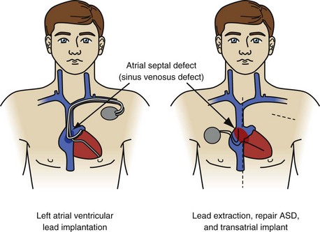
Implantation of leads in the left atrium and ventricle is a notable exception (Fig. 26-25). Leads are implanted into the left ventricle in two ways: through a congenital atrial or ventricular septal defect or retrograde through the aortic valve. All physicians consider the presence of left-sided leads and embolic symptoms an urgent indication for lead removal. Most consider their presence, with the potential for a complication, to be an urgent indication for lead extraction. A few physicians believe that, in the absence of complications (cerebrovascular accident, coronary artery occlusion, infarction of another organ, or sequelae of peripheral embolus), leaving the leads intact is an acceptable option. The rationale is that the extraction procedure is more dangerous than the presence of left-sided leads. The views on lead removal are just as varied. Some believe the leads should be removed; the chambers debrided of vegetation; and congenital defects, if present, repaired with CPB. Others think it is safe to extract the leads using the established right-sided techniques and protecting the brain from emboli by compressing the carotid arteries when necessary. If the second approach has any merit, it would be in removing newly implanted leads. If the newly implanted leads are proved to be free of vegetation and the procedure is monitored using TEE, it is probably safe to extract using direct traction. Specific data are not available on the numbers of physicians using these opinions.

Byrd’s experience is based on the management of 10 consecutive patients, most of whom had chronic leads and were symptomatic. Symptoms included transient ischemic attack and/or cerebrovascular accident, and vegetation was apparent in symptomatic patients. All but one patient had leads passing from right to left through an atrial septal defect. One patient had a lead that was implanted through the subclavian artery and passed retrograde through the aortic valve into the left ventricle. The procedure included establishing CPB, using cardioplegia to stop ventricular contraction, cross-clamping the aorta as needed, extracting the leads using conventional tools, debriding the chambers, repairing the septal defects, continuously observing the left atrium and ventricle by TEE, and carefully restarting the heart with protection from inadvertent ejection of embolic material. A new transatrial right-sided implantation was performed off bypass with the use of fluoroscopy and the conventional approach. With this technique, all leads were removed, the congenital defects repaired, and the new devices implanted without sequelae. Developing a safe technique that does not use an OHS procedure would be difficult.

image Special Situations

The most challenging development in lead extraction is related to the placement of leads into the coronary sinus (CS) and cardiac veins. Cardiac resynchronization therapy (CRT) has made this a common and evolving issue. The short-term outcomes thus far when extracting these leads have not been noteworthy, partly because of the short implantation durations and the simple, extraction-friendly design of the leads. Smooth, thin, single diameter, and unipolar with good tensile properties are all characteristics that enhance the safe and quick removal of the leads. However, because of concerns with their stability in the cardiac veins, these leads have been developed into more complicated shapes, with multipolar design and fixation mechanisms, and are unlikely to be easily removed.

To evaluate extraction tools and a novel lead design technique for reducing the barriers to extraction of complicated leads, Wilkoff et al.26 used a sheep model with atrial defibrillator leads placed in the CS to the great cardiac vein. The leads, originally designed for atrial defibrillation in the Metrix atrial defibrillator (InControl, Redmond, Wash.), were modified but kept their pigtail configuration for lead stability. Three configurations—one without modification of the defibrillation coil, one with medical adhesive backfill under the defibrillation coil, and one covered with an expanded polytetrafluoroethylene polymer (ePTFE)—were implanted in sheep and were subjected to extraction at either 6 or 14 months. The model proved to be excellent for developing profound fibrosis, and the unmodified leads were almost impossible to remove without hemopericardium. The medical adhesive backfill was much better, and there was almost no trouble removing the ePTFE-covered leads. The study also demonstrated that laser sheaths were dangerous in the CS, because the sheath approximated the size of the vein; a special 7F electrosurgical extraction sheath was relatively much safer to use. During the procedures, the electrosurgical sheath was rotated away from the pericardial and toward the myocardial surface.

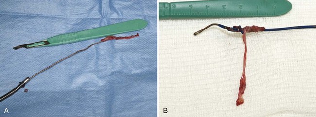
Although CS lead extraction has not yet become a clinical issue, implanters should make sound decisions now that promote the extraction of these leads. From the sheep experience, implanters should avoid construction that allows for tissue ingrowth. In some cases, significant ingrowth around CS leads clearly makes extraction more difficult. This is particularly true in patients with the new Starfix (Medtronic) leads, which are designed to increase fibrosis to affix the lead to the intended vein position. The ingrowth of the fibrotic tissue into any lead, but especially in this lead, will make the long-term extraction experience much more challenging and potentially life threatening (Fig. 26-26).

References

1 Wilkoff BL, Love CJ, Byrd CL, et al. Transvenous lead extraction. Heart Rhythm Society Expert Consensus on Facilities, Training, Indications, and Patient Management (endorsed by American Heart Association). Heart Rhythm. 2009;6:1085-1104.

2 Byrd CL, Schwartz SJ, Hedin N. Intravascular techniques for extraction of permanent pacemaker leads. J Thorac Cardiovasc Surg. 1991;101:989-997.

3 Byrd CL. Advances in device lead extraction. Curr Cardiol Rep. 2001;3:324.

4 Byrd CL. Extraction of transvenous pacing leads. Am Heart J. 1992;124:1667-1668.

5 Chamis AL, Peterson GE, Cabell CH, et al. Staphylococcus aureus bacteremia in patients with permanent pacemakers or implantable cardioverter-defibrillators. Circulation. 2001;104:1029-1033.

6 Chua JD, Wilkoff BL, Lee I, et al. Diagnosis and management of infections involving implantable electrophysiologic cardiac devices. Ann Intern Med. 2000;133:604-608.

7 Chan AW, Bhatt DL, Wilkoff BL, et al. Percutaneous treatment for pacemaker-associated superior vena cava syndrome. Pacing Clin Electrophysiol. 2002;25:1628-1633.

8 Suga C, Hayes DL, Hyberger LK, et al. Is there an adverse outcome from abandoned pacing leads? J Interv Card Electrophysiol. 2000;4:493-499.

9 Smith HJ, Fearnot NE, Byrd CL, et al. Five-years experience with intravascular lead extraction. U.S. Lead Extraction Database. Pacing Clin Electrophysiol. 1994;17:2016-2020.

10 Fearnot NE, Smith HJ, Goode LB, et al. Intravascular lead extraction using locking stylets, sheaths, and other techniques. Pacing Clin Electrophysiol. 1990;13:1864-1870.

11 Byrd CL, Schwartz SJ, Hedin NB, et al. Intravascular lead extraction using locking stylets and sheaths. Pacing Clin Electrophysiol. 1990;13:1871-1875.

12 Wazni O, Epstein LM, Carrillo RG, et al. Lead extraction in the contemporary setting: the LExICon study: an observational retrospective study of consecutive laser lead extractions. J Am Coll Cardiol. 2010;55:579-586.

13 Bilgutay AM, Jensen MK, Schmidt WR, et al. Incarceration of transvenous pacemaker electrode: removal by traction. Am Heart J. 1969;77:377-379.

14 Karagoz T, Celiker A, Hallioglu O, Ozme S. Unusual extraction of an active fixation ventricular pacing lead with outer coil fracture in a child. Europace. 2003;5:185-187.

15 Wilkoff BL, Byrd CL, Love CJ, et al. Pacemaker lead extraction with the laser sheath: results of the Pacing Lead Extraction with the Excimer Sheath (PLEXES) trial. J Am Coll Cardiol. 1999;33:1671-1676.

16 Epstein LM, Byrd CL, Wilkoff BL, et al. Initial experience with larger laser sheaths for the removal of transvenous pacemaker and implantable defibrillator leads. Circulation. 1999;100:516-525.

17 Saad EB, Saliba WI, Schweikert RA, et al. Nonthoracotomy implantable defibrillator lead extraction: results and comparison with extraction of pacemaker leads. Pacing Clin Electrophysiol. 2003;26:1944-1950.

18 Byrd CL, Wilkoff BL, Love CJ, et al. Clinical study of the laser sheath for lead extraction: the total experience in the United States. Pacing Clin Electrophysiol. 2002;25:804-808.

19 Kennergren C. Excimer laser assisted extraction of permanent pacemaker and ICD leads: present experiences of a European multi-centre study. Eur J Cardiothorac Surg. 1999;15:856-860.

20 Love C, Byrd C, Wilkoff BL, et al. Lead extraction using a bipolar electrosurgical dissection sheath: an interim report. Europace. 2001;3:223-228.

21 Hussein AA, Wilkoff BL, Martin DO, et al. Initial experience with the Evolution mechanical dilator sheath for lead extraction: safety and efficacy. Heart Rhythm. 2010;7:870-873.

22 Bongiorni MG, Di Cori A, Zucchelli G, et al. A modified transvenous single mechanical dilatation technique to remove a chronically implanted active-fixation coronary sinus pacing lead. Pacing Clin Electrophysiol. 2011;34:e66-e69.

23 Bongiorni MG, Giannola G, Arena G, et al. Pacing and implantable cardioverter-defibrillator transvenous lead extraction. Ital Heart J. 2005;6:261-266.

24 Fischer A, Love B, Hansalia R, et al. Transfemoral snaring and stabilization of pacemaker and defibrillator leads to maintain vascular access during lead extraction. Pacing Clin Electrophysiol. 2009;32:336-339.

25 Byrd CL, Schwartz SJ. Transatrial implantation of transvenous pacing leads as an alternative to implantation of epicardial leads. Pacing Clin Electrophysiol. 1990;13:1856-1859.

26 Wilkoff BL, Belott PH, Love CJ, et al. Improved extraction of ePTFE and medical adhesive modified defibrillation leads from the coronary sinus and great cardiac vein. Pacing Clin Electrophysiol. 2005;28:205-211.

27 Byrd CL, Wilkoff BL, Love CJ, et al. Intravascular extraction of problematic or infected permanent pacemaker leads: 1994-1996. U.S. Extraction Database, MED Institute. Pacing Clin Electrophysiol. 1999;22:1348-1357.

28 Kennergren C, Bucknall CA, Butter C, et al. Laser-assisted lead extraction: the European experience. Europace. 2007;9:651-656.

29 Neuzil P, Taborsky M, Rezek Z, et al. Pacemaker and ICD lead extraction with electrosurgical dissection sheaths and standard transvenous extraction systems: results of a randomized trial. Europace. 2007;9:98-104.