162 Systemic lupus erythematosus
Salient features
History
• Fatiguability and tiredness: suggests anaemia
• Joint symptoms: particularly small joints (90% of patients)
• Gangrene of the digits: vasculitis
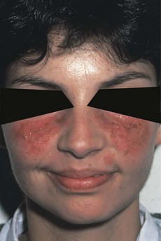
• Skin rash (butterfly rash on face; Fig. 162.1) in sun-exposed areas, livedo reticularis, alopecia, Raynaud’s phenomenon
• Hypertension, oedema (suggesting renal involvement)
• Fever, enlargement of lymph nodes
• Bleeding from gums excessive menstrual bleeding, purpura (caused by thrombocytopenia)
• Neuropsychiatric symptoms, seizures
Examination
• Butterfly rash: follicular plugging, scales, telangiectasia and scarring affecting the bridge of the nose and cheeks (the patient may be cushingoid because of steroids)
• Tell the examiner that you would like to examine the urine for proteinuria.
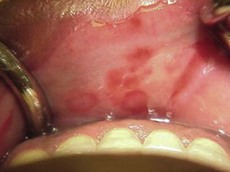
Fig. 162.2 Oral mucosal ulcerations: generally painless and can present on other cutaneous tissues.
(With permission from Powers 2008.)
Note: SLE principally affects skin, joints, kidney and serosal membranes.
Advanced-level questions
What is the histology of the skin rash in systemic lupus erythematosus?
• Liquefactive degeneration of the basal layer of the epidermis together with oedema at the dermoepidermal junction. Immunofluorescence microscopy shows deposition of immunoglobulin and complement along the dermoepidermal junction.
• Oedema of the dermis accompanied by infiltrates of perivascular mononuclear cells.
What are the criteria for diagnosis of systemic lupus erythematosus?
Any four of the following 11 criteria are required to make a diagnosis:
• Arthritis: non-erosive arthritis
• Serositis: pleuritis, pericarditis
• Renal involvement: proteinuria, cellular casts (nephritis is more common in patients with anti-native DNA)
• Neurological involvement: seizures, psychosis
• Haematological involvement: haemolytic anaemias, leukopenia, thrombocytopenia
• Anti-nuclear antibody (ANA): seen in over 95% of patients
• Immunological disorder: positive for LE cell (a specific type of cell found in SLE), anti-DNA antibody, false-positive syphilitic serology.
What do you know about drug-induced lupus erythematosus?
• Procainamide is responsible for the majority of the cases. Other causes include hydralazine, quinidine, isoniazid.
• Drugs causing lupus-like syndrome do not seem to aggravate primary SLE.
• Patients with drug-induced lupus usually present with skin and joint manifestations.
• Although multiple organs are affected, nephritis and CNS features are not ordinarily present.
• Anti-histone antibodies are characteristic of drug-induced lupus but are not specific for this syndrome; anti-native DNA is almost never detected.
• Clinical manifestations and many laboratory features return to normal after the offending drug is withdrawn.
How useful is the detection of anti-nuclear antibodies in the diagnosis of systemic lupus erythematosus?
What are the patterns of lupus nephritis?
The WHO’s morphological classification of lupus nephritis describes five patterns:
How would you manage a patient with systemic lupus erythematosus?
• Avoidance of sunlight provocation. In Britain, patients should avoid being outside from 11 am to 3 pm from March to September and should wear protective clothing, including hats; patients should holiday in temperate latitudes or during the winter. In addition, sun block creams such as titanium dioxide (rather than barrier cream) are essential
• Avoidance of drug provocation: penicillin, sulphonamides
• Encourage the patient to join the Lupus Society
• Educate the patient that no therapy is curative and that medical treatment is largely empirical, selected on the basis of specific manifestations:
| Disease manifestation | Treatment |
|---|---|
| Serological abnormalities and minor cytopenia unaccompanied by symptoms | No treatment |
| Rash and mild systemic symptoms | Antimalarial drugs |
| Mild or moderate arthritis, fever, pleuropericarditis | NSAIDs or low-dose prednisolone |
| Malaise, weight loss and lymphadenopathy | Low-dose steroids |
| High fever, active inflammatory glomerulonephritis, severe thrombocytopenia, severe haemolytic anaemia and most neurological disturbances | High-dose prednisolone (>60 mg per day for 4–6 weeks) |
| Rapidly progressive renal disease | Intravenous administration of bolus doses of 1000 mg methylprednisolone |
| Lupus nephritis with high histological activity score | Oral or intravenous cyclophosphamide or mycophenolate mofetil with high-dose steroids (N Engl J Med 2000;343:1156–62) |
| Thromboembolism with antiphospholipid antibody | Long-term anticoagulation |
| Severe disease | Experimental treatments |
What do you know about the pathogenesis of systemic lupus erythematosus?
• Pathogenic autoantibodies are the primary cause of tissue damage in patients with lupus including:
• T lymphocyte help is critical in the pathogenesis of lupus. The effect on the T cell depends on the interaction between molecules on the surface of the cell with the antigen presented on the surface of the antigen-presenting cell.
• B cells acting as an antigen-presenting cells also interact with T cells, costimulation requiring interaction between CD40 and the CD40 ligand. The interaction stimulates the T cell to produce cytokines, some of which act on the B cell to promote antibody formation, to stimulate cell division, to switch antibody production from IgM to IgG, and to promote changes in the secreted antibody so that it binds more strongly to the driving antigen. (Note: CD20 and CD22 are present on B cells and interleukin-10 is produced by B cells).
• Plasmacytoid dendritic cells (producing interferon) have been found in the inflamed skin of lupus. They can internalize immune complexes in SLE, which, in turn, triggers the cells to secrete interferon-α through activation of toll-like receptor (TLR).
Promising therapies base on these pathlogical changes include:
• rituximab: antibody against CD20 on the surface of all mature B cells; non-specific
• abetimus sodium: depletes only B lymphocytes that produce anti-double-stranded DNA antibodies and forms complexes with anti-double-stranded DNA antibodies, which can then be cleared from the circulation; more specific.
Cervaera R, the European Working Party on SLE. Systemic lupus erythematosus: clinical and immunologic patterns of disease expression in a cohort of 1000 patients. Medicine. 1993;72:113.
Hay EM, Snaith ML. Systemic lupus erythematosus and lupus-like syndromes. BMJ. 1995;310:1257-1261.
Mills JA. Systemic lupus erythematosus. N Engl J Med. 1994;330:187.
Tan EM, Cohen AS, Fries JF, et al. The 1982 criteria for classification of SLE. Arthritis Rheum. 1982;25:1271.