Popliteal Swellings
Popliteal swellings are not uncommon. The majority are easily diagnosed on clinical examination alone.
Causes
History
Superficial
Sebaceous cyst
A sebaceous cyst will present as a firm swelling in the skin. It may be tender if it becomes inflamed and there may be a discharge from it.
Lipoma
This presents as a soft lobulated swelling in the subcutaneous tissue.
Varix of the short saphenous vein
This presents as a soft swelling behind the knee, usually associated with varicose veins of the short saphenous system.
Deep
Lipoma
Deeper lipomas are difficult to feel because of the tough overlying fascia. They do not always feel soft and lobulated, as superficial ones do.
Lymphadenopathy
There may be several palpable lumps within the popliteal fossa. The patient will usually draw attention to a lesion distally, either on the leg or on the foot, usually on the lateral margin of the foot or the back of the leg. Other groups of nodes may be enlarged.
Semi-membranosus bursa
The patient complains of a swelling behind the knee that interferes with knee movement, particularly flexion.
Baker’s cyst
A Baker’s cyst is a pulsion diverticulum of the knee joint caused by chronic disease within the joint. The patient will usually give a history of a painful knee joint and may have osteoarthritis or rheumatoid arthritis. Occasionally the cyst ruptures and the patient complains of severe pain in the calf, which has to be carefully distinguished from venous thrombosis.
Popliteal artery aneurysm
These often get quite large before they are noticed. The patient may be aware of a pulsatile swelling behind the knee but often notices it when crossing the legs, the upper of the crossed limbs moving with each pulse.
Bony
Exostoses
Exostoses may grow in the region of the epiphyseal cartilage of the femur. There is a well-defined bony swelling in the popliteal fossa.
Osteogenic sarcoma
This may affect either the lower end of the femur or the upper end of the tibia. There is a diffuse swelling around the knee joint. Pain may be a presenting symptom. There may also be general malaise and weight loss. Occasionally pulmonary metastases have occurred at presentation, and there may be cough and haemoptysis.
Examination
Superficial
Sebaceous cyst
There will be a small, well-defined swelling in the skin with a punctum. If it is infected, the surrounding skin will be red and there may be discharge.
Lipoma
There will be a soft, lobulated swelling in the popliteal fossa.
Varix of the short saphenous vein
This is not uncommon. There will be a soft, compressible dilatation at the termination of the short saphenous vein. There may be an expansile cough impulse. A fluid thrill will be palpable when the short saphenous vein lower down the leg is tapped.
Deep
Lipoma
A deep lipoma in the fat of the popliteal fossa may be difficult to define accurately. It does not always have the soft, lobulated appearance of a more superficial lipoma.
Lymphadenopathy
There may be a number of discrete glands palpable or they may be firm and matted together. Check for a lesion distally on the leg or foot, e.g. malignant melanoma. Check for lymphadenopathy elsewhere.
Semi-membranosus bursa
The swelling lies above the level of the knee joint line, slightly to the medial side of the popliteal fossa. It tends to be firm in consistency and transilluminates.
Baker’s cyst
Baker’s cysts occur more often in elderly patients with longstanding arthritis or in younger patients with rheumatoid arthritis. The lump is below the level of the knee joint and deep to gastrocnemius. It may transilluminate. Pressure over the lump may reduce it into the knee joint. Examination of the knee joint reveals changes consistent with arthritis, i.e. limited movements, crepitus and occasionally an effusion.
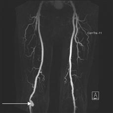
Popliteal artery aneurysm
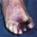
An expansile, pulsatile mass will be palpable in the popliteal fossa. They are often bilateral. Check the distal circulation. Popliteal aneurysms may thrombose with distal ischaemia, or may throw off small emboli causing ischaemic toes or ischaemic ulcers on the tips of the toes. Palpate the abdomen: there is an association between popliteal aneurysms and abdominal aortic aneurysms.
Bony
Exostoses
A bony mass may be felt in the popliteal fossa.
Osteogenic sarcoma
The overlying skin may be reddened, with dilated subcutaneous veins. There may be tenderness. The swelling is usually smooth until it spreads into the surrounding tissues, when it becomes irregular. The swelling feels firm but is not usually bony hard.
General Investigations
■ FBC, ESR
Hb ↓ lymphadenopathy, e.g. part of generalised lymphadenopathy associated with reticulosis. WCC ↑ reticulosis. ESR ↑ reticulosis, osteogenic sarcoma with secondaries.
■ US
Lipoma. Semi-membranosus bursa. Baker’s cyst. Popliteal artery aneurysm.
■ Knee X-ray
Exostoses. Osteoarthritis (Baker’s cyst). Rheumatoid arthritis (Baker’s cyst). Osteogenic sarcoma – bone destruction, grows out of cortex, elevating periosteum with reposition of subperiosteal bone (Codman’s triangle), radiating spicules of bone (‘sunray’ spicules).
■ CXR
Secondary deposits from osteogenic sarcoma. Hilar lymph nodes with generalised lymphadenopathy.