Leg Swellings
Swelling of the lower limb may be unilateral or bilateral. Bilateral swellings are usually due to medical conditions such as cardiac, renal or hepatic failure. Unilateral swellings are commonly due to trauma, venous disease or lymphatic disease.
Causes
Local
Acute Swelling
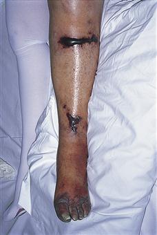
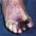
Figure 42 Phlegmasia cerulea dolens.
The painful, swollen, blue leg results from a massive ilio-femoral venous thrombosis. It may lead to venous gangrene unless the resulting high tissue pressure is relieved by thrombectomy. In this patient, blood-filled blisters and gangrene of the toes have already developed. This is a relatively rare presentation of deep vein thrombosis.
History
Bilateral leg swelling is usually due to systemic conditions, e.g. cardiac failure, while unilateral leg swelling is more often due to local causes, e.g. DVT, cellulitis. Pain will be associated with trauma, DVT, infection or complications of varicose veins. Other causes of swelling of the lower limb may be painless, although the limb may be uncomfortable if the oedema becomes tense. Evidence of the following should be sought in the past medical history: trauma to the limb, recent pregnancy (DVT), abdominal or pelvic malignancy, previous surgery or radiotherapy to lymph nodes, thyroid disease, congestive cardiac failure, renal failure, liver failure, malnutrition, poliomyelitis in childhood, nerve lesions. With primary lymphoedema, the leg may have been swollen since birth or oedema may have developed at puberty (lymphoedema praecox) or in the third decade (lymphoedema tarda).
Examination
The presence of fracture, contusion or haematoma should be obvious. With cellulitis, the limb will be red, swollen, hot and tender. A puncture wound may be visible. DVT may be suspected if the limb is tender and swollen, particularly the calf, with a positive Homans’ sign, i.e. pain in the calf on dorsiflexion of the foot. Tenderness may be present over the femoral vein with femoral vein thrombosis. The limb may be pale and swollen to the groin (phlegmasia alba dolens) or tense and purplish and extremely painful (phlegmasia cerulea dolens) with iliofemoral thrombosis. Bilateral swelling with dilated collateral veins on the abdominal wall suggests IVC thrombosis. Swelling and tenderness over the joints may suggest rheumatoid arthritis. Lymphoedema pits in the early stages but in the later stage, it becomes non-pitting, when the skin and subcutaneous tissue become thick and eventually hyperkeratotic and warty. Previous scars in the groin, a mass of malignant nodes or signs of previous radiotherapy will suggest lymphoedema as a possible cause.
In the presence of an arteriovenous fistula, there will be dilated veins which do not collapse on elevation of the limb. The limb will be warmer than the opposite limb; palpation will reveal a thrill and auscultation will reveal a continuous machinery bruit.
Localised swelling of the limb may be due to a tumour. There may also be associated distal swelling due to venous or lymphatic obstruction.
Swelling due to neurological damage, e.g. nerve lesions or poliomyelitis, will result in wasting, which should be apparent. Examination of the abdomen may reveal hepatomegaly or an abdominal mass, which could obstruct the venous outflow. It is appropriate to carry out a rectal examination for a pelvic tumour, which may be causing back pressure on the venous and lymphatic system or may have resulted in a ‘frozen’ pelvis.
General Investigations
■ FBC, ESR
A large haematoma associated with trauma or fracture may result in a low Hb. WCC ↑ will suggest infection. Haematoma may be associated with a reduced platelet count.
■ Urinalysis
Proteinuria may suggest a renal cause.
■ U&Es and creatinine
Raised urea and creatinine will be associated with renal failure.
■ LFTs
May indicate impaired liver function with associated hypoalbuminaemia.
■ Blood glucose
Cellulitis or other infections of the leg may be associated with diabetes.
■ CXR
Findings suggestive of cardiac failure are cardiomegaly, pulmonary oedema and pleural effusions. Pulmonary oedema may be associated with fluid overload associated with renal failure. Secondary deposits may be seen in association with sarcoma of the limb.
■ Limb X-ray
May show fracture, tumour or gas in the tissues associated with gas gangrene.
Specific Investigations
■ Clotting screen
There may be abnormal clotting associated with coagulopathy, resulting in spontaneous haematoma.
■ US
Haematoma or soft-tissue sarcoma.
■ US or CT pelvis
May show an abdominal or pelvic mass causing extrinsic pressure on the veins.
■ Duplex Doppler
Will show DVT or arteriovenous fistula.
■ Arteriography
Will confirm the presence of an arteriovenous fistula.
■ Lymphangiography
May demonstrate the cause of lymphoedema, e.g. hypoplasia or obstruction.