Groin Swellings
These are common clinical problems, especially hernias. They are therefore common in clinical examinations.
Causes
Above The Inguinal Ligament
Below The Inguinal Ligament
History
Sebaceous cyst
The patient will complain of a mobile lump on the skin. It may be red and inflamed and discharging.
Lipoma
The patient will present with a soft, painless swelling.
Hernias
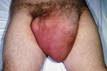
A patient with a groin hernia will present with a lump that disappears on recumbency or may be pushed back (reducible). The patient may present with a tense, tender lump that will not reduce and is accompanied by signs and symptoms of intestinal obstruction. Femoral hernia is more common in females. With hernias, there is occasionally a history of sudden straining or trauma, following which a lump may become manifest.
Imperfectly descended testis
An imperfectly descended testis may present as a groin swelling. The patient, or, if in a young child, the mother, will have noticed absence of a testis from the scrotum. Enlargement and pain may indicate malignant change, which is more common in an imperfectly descended testis.
Lipoma of the cord
The patient will have noticed a soft swelling in the groin. This is often mistaken for a hernia.
Hydrocele of the cord
This may present as a lump in the inguinal region which does not reduce.
Hydrocele of the canal of Nuck
This is similar to a hydrocele of the spermatic cord but presents in the female. It represents a cyst forming in the processus vaginalis.
Lymph nodes
Lymph nodes may present as swellings below the inguinal ligament. They may be discrete and firm, tender and red or matted to form a mass. The patient may have noticed a lesion on the leg. Care must be taken to elicit a full history with inguinal lymphadenopathy, as the nodes drain not only the tissues of the leg but also the penis, the scrotal skin, the lower half of the anal canal, the skin of the buttock and the skin of the lower abdominal wall, up to and including the umbilicus. In the female, they drain the labia, the lower third of the vagina and the fundus of the uterus, via lymphatics accompanying the round ligament down the inguinal canal. A careful history should therefore be taken, of any anorectal disease, e.g. bleeding PR, or gynaecological disease, e.g. bleeding PV to suggest a carcinoma of the uterus.
Saphena varix
A saphena varix is normally associated with varicose veins lower down the leg. The patient will present having noticed a small, soft, bluish mass in the lower part of the groin.
Femoral artery aneurysm
A pulsatile expansile mass suggests a femoral aneurysm. Check for a history of arterial surgery at the groin or arteriography via the femoral artery, which may suggest the presence of a false aneurysm.
Imperfectly descended testis
An imperfectly descended testis may descend into the upper thigh but its descent is arrested by the attachment of Scarpa’s fascia to the deep fascia of the thigh.
Neuroma of the femoral nerve
This is rare and may be associated with anaesthesia or paraesthesia on the anterior aspect of the thigh and inability to extend the knee.
Synovioma of the hip joint
This is rare. The patient complains of a lump deep in the groin, which may interfere with hip movement.
Obturator hernia
This is rare. The sac passes through the obturator canal and may present in the groin deep to pectineus. A lump is rarely palpable due to the overlying pectineus. The diagnosis is not usually made until obstruction or strangulation occurs.
Examination
Sebaceous cyst
This is attached to the skin and a punctum is usually seen at the point of attachment. Sebaceous cysts are firm, spherical and mobile on the underlying tissue. They may be hot, red and tender when inflamed.
Lipoma
The lump will be soft, lobulated, mobile, being fixed neither to the skin nor to the underlying tissue. They may be distinguished from hernias, in that they are not reducible and do not have a cough impulse.
Hernias
A hernia may be reduced and has an expansile cough impulse. They may be irreducible. Irreducible hernias may be: (1) incarcerated – imprisoned in the sac because of adhesions between contents and the wall of the sac; (2) obstructed – small bowel is caught in the sac and intestinal contents cannot pass on; (3) strangulated – the arterial blood supply is cut-off and gangrene of the contents ensues. In the last case, the lump would be tender, the overlying skin may be red and the patient will be pyrexial with a tachycardia. There will be signs of intestinal obstruction.
It is important to distinguish between an inguinal and a femoral hernia. An inguinal hernia lies above and medial to the pubic tubercle, a femoral hernia below and lateral. An inguinal hernia may be either direct or indirect. An indirect hernia comes down the inguinal canal from the deep inguinal ring. A direct hernia comes through the posterior wall of the canal, through Hesselbach’s triangle (base – inguinal ligament; lateral border – inferior epigastric artery; medial border – the lateral border of rectus abdominis muscle). Distinction between direct and indirect hernia is made by reducing the hernia and exerting pressure over the deep inguinal ring, asking the patient to cough. If the hernia sac appears medial to the fingers, it is direct. If the hernia appears only after removing the pressure over the deep inguinal ring, then it is indirect.
Imperfectly descended testis
An imperfectly descended testis may be in an ectopic position (root of the penis, upper thigh or perineum) or along the normal line of descent. An imperfectly descended testis cannot be felt in the inguinal canal – it is usually too flabby and atrophic and cannot be palpated through the tough overlying external oblique aponeurosis. However, should it become malignant, then the hard and irregular testis can be palpated in the inguinal canal. An imperfectly descended testis may also be palpable in the upper thigh below the inguinal ligament. The scrotum will be empty on that side. The testis cannot descend more than a few centimetres into the upper thigh, as its descent is prevented by the attachment of Scarpa’s fascia to the deep fascia of the thigh below the inguinal ligament. An imperfectly descended testis may also be palpable at the root of the penis or in the perineum.
Lipoma of the cord
This can be confidently diagnosed only at surgery, although there will not be an expansile cough impulse as with a hernia.
Hydrocele of the cord
This is rare. There will be a smooth, palpable swelling along the line of the cord. It does not have a palpable expansile cough impulse. If gentle traction is exerted on the testis, a hydrocele of the cord will be felt to move down the canal. It may transilluminate.
Hydrocele of the canal of Nuck
A swelling, similar to a hydrocele of the cord in the male, occurring in the female is called a hydrocele of the canal of Nuck. Findings will be similar to those of a hydrocele of the cord in the male, except there is nothing to exert traction on!
Femoral hernia
This is more common in females. It lies below and lateral to the pubic tubercle. There will be an expansile cough impulse. It may be reducible. Strangulation of a femoral hernia is not uncommon, particularly one of the Richter’s type. A tense, tender, irreducible swelling will be found below and lateral to the pubic tubercle.
Lymph nodes
Lymph nodes are palpable below the inguinal ligament. Classically, they are arranged into groups: (1) superficial, with subdivision into horizontal and vertical groups; and (2) deep. In practice, it is difficult to distinguish between the groups. Lymph nodes in the groin may be palpable as discrete nodules or they may be hard, irregular and matted together. This type is usually associated with malignant disease. Tender, fluctuant lymph nodes with erythema of the overlying skin are usually associated with lymphadenopathy due to an infective condition. It is important to examine all sites that are drained by these nodes, namely: (1) the skin of the leg, including examination under the toe nails; (2) the skin of the buttock; (3) the skin of the lower abdominal wall up to and including the umbilicus; (4) the skin of the scrotum, penis and glans penis; (5) the labia and lower third of the vagina; (6) the lower half of the anal canal; (7) the fundus of the uterus. It is therefore necessary not only to examine superficial structures but also to carry out a digital rectal examination and a bimanual vaginal examination.
Saphena varix
This is a soft, compressible dilatation at the termination of the saphenous vein. It has a cough impulse and disappears on recumbency. A fluid thrill is felt if the veins lower down the leg are percussed.
Femoral artery aneurysm
A femoral artery aneurysm presents as an expansile pulsatile mass in the line of the femoral artery. Look for a scar in relationship to it, which may suggest a false aneurysm.
Imperfectly descended testis
An imperfectly descended testis may be palpable in the upper thigh below the inguinal ligament. The scrotum will be empty on that side. The testis cannot descend more than a few centimetres into the upper thigh, as its descent is prevented by the attachment of Scarpa’s fascia to the deep fascia of the thigh below the inguinal ligament.
Neuroma of the femoral nerve
This is rare. It will be palpable along the course of the nerve (lateral to the femoral artery). Test the integrity of the femoral nerve (sensation on the anterior aspect of the thigh; extension of the knee joint).
Synovioma of the hip joint
This is rare. There may be a palpable thickening deep in the groin in relation to the hip joint. Hip joint movements may be limited.
Obturator hernia
This is rare. In a very thin patient, a lump may be felt deep in the medial aspect of the groin. More commonly, obturator hernias present with intestinal obstruction and the diagnosis is made at laparotomy.
Psoas abscess
This is rare. It used to be associated with spinal TB, a cold abscess of a vertebral body discharging down the psoas sheath and presenting as a soft fluctuant swelling below the inguinal ligament. Most psoas abscesses nowadays are related to perforation of a hollow viscus retroperitoneally, e.g. the right colon into the psoas sheath.
General Investigations
The diagnosis of most groin swellings is made on history and physical examination.
■ FBC, ESR
Hb ↓ lymph node tumours. WCC ↑ infection in the lymph nodes, strangulated hernia. ESR ↑ lymph node tumours, infection, e.g. TB spine.
■ US
Lipoma, imperfectly descended testis, femoral artery aneurysm, psoas abscess.
■ Hip X-ray
Osteoarthritis with synovioma.
■ AXR
Intestinal obstruction associated with obstructed/strangulated hernia.