Swelling in the neck in a 58-year-old man

Published on 10/04/2015 by admin

Filed under Surgery

Last modified 22/04/2025

Print this page

rate 1 star rate 2 star rate 3 star rate 4 star rate 5 star
Your rating: none, Average: 0 (0 votes)

This article have been viewed 2080 times

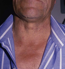
Problem 5 Swelling in the neck in a 58-year-old man

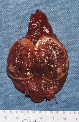
The patient undergoes a right thyroid lobectomy and makes an uneventful recovery.

Answers

A.1 There is a smooth surfaced swelling at the base of the neck on the right side. You notice that the swelling elevates on swallowing. The swelling is in the anterior triangle of the neck and about 4 cm in maximal diameter. The trachea appears to be displaced to the left. The overlying skin is normal. This may well be a swelling arising in the right lobe of the thyroid gland, as it elevates on swallowing. Thyroid swellings elevate on swallowing because they are enveloped by the pretracheal fascia which attaches the thyroid to the laryngopharynx.

A.2 You will want to know about the following:

A.3

A.4 The differential diagnosis of this thyroid mass – in order of most to least common – is:

Even when it is thought that the nodule is a true solitary nodule, further investigation will show that 50% of cases are in fact dominant nodules within a multinodular goitre. Around 8% of the population will have a palpable goitre, and of those subjected to ultrasound of the thyroid, by the age of 50, 50% of the population will have demonstrable ultrasound identified thyroid nodules. Of all these thyroid nodules, only 5% will be malignant. Overall, thyroid cancer is rare, representing only 1% of all malignancies.

A.5

A.6 The cytological aspirate shows sheets of follicular cells with minimal colloid. There are many clusters of bland epithelial cells which form well-defined circular follicular structures in the centre and to the right of the picture. This is a typical microfollicular pattern. There is, however, no clear evidence of malignancy. When follicular structures are identified on a fine needle aspirate of a thyroid nodule, the differential diagnosis includes a hyperplastic (adenomatous or colloid nodule) follicular adenoma, or follicular carcinoma. A reliable distinction between these is not possible on cytological appearances from an FNA because the distinction between follicular adenoma and follicular carcinoma can only be made histologically by demonstrating invasion of the capsule of the tumour or by vascular invasion.

A.7 The ultrasound scan shows a well-circumscribed, solitary, solid lump in the lower pole of the right lobe of the thyroid gland. The remainder of the thyroid gland appears to be normal. This solid mass lesion, in the context of an otherwise normal thyroid gland, is therefore likely to be a tumour, but still most likely benign.

A.8 You should tell the patient that he has a growth in the thyroid gland. The gland appears to be functioning normally but the lump is solid and solitary and might be a cancer, although you favour a benign or innocent growth (80–90%). The only way you will be able to tell exactly what the lump is will be to perform an operation and remove the lump. This will involve removal of the right half of his thyroid gland. The pathologist will then assess the excised lump and provide a definitive diagnosis. This process will take at least 48 hours following surgery. Once a definitive diagnosis is reached, you will be able to discuss further treatment with the patient should it be required. At this stage it is probably unnecessary to have a detailed discussion on thyroid malignancy, since the lump is most likely benign.

A.9 A detailed process of consent is essential. The operation and the anaesthetic, the likely inpatient stay and the postoperative recovery phase should all be discussed in detail.

A.10 The right lobe of thyroid has been opened longitudinally, there is an encapsulated tumour 4 cm in diameter which occupies most of the lobe. The tumour is pinkish red and vascular with areas of haemorrhage. Gross examination does not show any evidence of capsular invasion. Histologically the lesion is shown to be composed of follicular cells and there is invasion through the capsule of the nodule. There is no evidence of invasion in the blood vessels. It was concluded the lesion was an invasive follicular carcinoma.

A.11 You will need to explain to the patient that an invasive follicular carcinoma has been found in the excised half of the thyroid. As a consequence further surgery, treatment and long-term follow-up are required. It is important to emphasize at the start that these tumours have an excellent prognosis (85% 10 year survival).

You therefore recommend to the patient the following measures:

Further Information

, www.british-thyroid-association.org. The website of the British Thyroid Association, with a number of useful links

, www.endocrinesurgeons.org.au. The website of Australian Endocrine Surgeons

, www.aace.com. The website of the American Association of Clinical Endocrinologists, with clinical guidelines for the management of thyroid carcinoma

, www.thyroidmanager.org. A site covering all aspects of thyroid disease

Yeung M.J., Serpell J.W. Management of the solitary thyroid nodule. The Oncologist. 2008;13:105-122.