Chapter 14 Supraventricular Arrhythmias, Part I Premature Beats and Paroxysmal Supraventricular Tachycardias
Please go to expertconsult.com for supplemental chapter material.
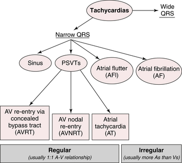
General Principles: Triggers and Mechanisms of Tachyarrhythmias
This chapter and the next focus on disturbances of cardiac rhythm with a rapid rate, namely: supraventricular and ventricular tachyarrhythmias (Fig. 14-1).
Key Pathophysiologic Concept
For any rapid, abnormal heart rhythm to occur, two major factors have to be present:


Tachyarrhythmias, both supraventricular and ventricular, in turn, usually start with premature beats that initiate arrhythmias by either focal or reentrant mechanisms (Fig. 14-2).

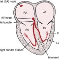
The sinus or sinoatrial (SA) node (see Chapter 13) is the physiologic (natural or intrinsic) pacemaker of the heart. The SA node normally initiates each heartbeat, producing (normal) sinus rhythm. However, pacemaker stimuli can arise from other parts of the heart, including the atrial muscle or pulmonary vein area, the atrioventricular (AV) junction, or the ventricles.
Ectopic beats are often premature; that is, they come before the next sinus beat is due. Examples include atrial premature beats (APBs), AV junctional premature beats (JPBs), and ventricular premature beats (VPBs). Ectopic beats can also come after a pause (delay) in the normal rhythm, as in the case of AV junctional or ventricular escape beats (see Chapter 13). Ectopic beats originating in the AV junction (node) or atria are referred to as supraventricular (i.e., literally coming from above the ventricles).
This chapter and Chapter 15 describe the major supraventricular arrhythmias, and Chapter 16 deals with ventricular tachycardias.
Atrial and Other Supraventricular Premature Beats
APBs∗ result from ectopic stimuli and are beats arising from loci in either the left or right atrium, or interatrial septum, but not the SA node itself. The atria, therefore, are depolarized from an ectopic site. After an atrial or junctional depolarization, the stimulus may spread normally through the His-Purkinje system into the ventricles. For this reason, ventricular depolarization (QRS complex) is generally not affected by APBs or JPBs. The major features of APBs are listed in Box 14-1 and are depicted in Figures 14-3 to 14-6.
BOX 14-1 Major Features of Atrial Premature Beats
• The atrial depolarization (P′ wave) is premature, occurring before the next sinus P wave is due.
• The QRS complex of the atrial premature beat (APB) is usually preceded by a visible P wave that usually has a slightly different shape or different PR interval from the P wave seen with sinus beats. The PR interval of the APB may be either longer or shorter than the PR interval of the normal beats. In some cases the P wave may be subtly hidden in the T wave of the preceding beat.
• After the APB a slight pause generally occurs before the normal sinus beat resumes. This delay is due to “resetting” of the sinoatrial (SA) node pacemaker by the premature atrial stimulus. This slight delay contrasts with the longer, “fully compensatory” pause often (but not always) seen after ventricular premature beats (VPBs) (see Fig. 16-9).
• The QRS complex of the APB is usually identical or very similar to the QRS complex of the preceding beats. Remember that with APBs the atrial pacemaker is in an ectopic location but the ventricles are usually depolarized in a normal way. This sequence contrasts with the generation of VPBs, in which the QRS complex is abnormally wide because of abnormal depolarization originating in the ventricles (see Chapter 16).
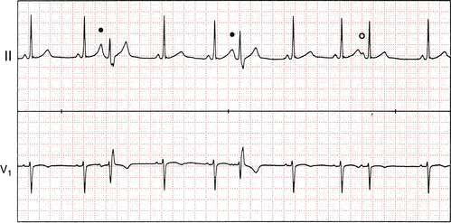
• Occasionally, APBs result in aberrant ventricular conduction, so that the QRS complex is wider than normal. Figures 14-5 and 14-6 show examples of such APBs causing delayed (aberrant) depolarization of the right and left ventricles, respectively.
• Sometimes when an APB is very premature, the stimulus reaches the atrioventricular (AV) junction just after it has been stimulated by the preceding beat. Because the AV junction, like other cardiac tissues, requires time to recover its capacity to conduct impulses, this premature atrial stimulus may reach the junction when it is still refractory. In this situation the APB may not be conducted to the ventricles and no QRS complex appears. The result is a blocked APB. The ECG shows a premature P wave not followed by a QRS complex (see Fig. 14-3B). After the blocked P wave, a brief pause occurs before the next normal beat resumes. The blocked APB, therefore, produces a slight irregularity of the heartbeat. If you do not search carefully for these blocked APBs, you may overlook them.
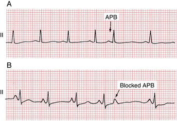
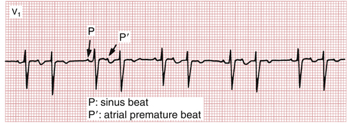
Figure 14-4 Sinus rhythm with atrial bigeminy. Each sinus beat is coupled to an atrial premature (early) beat followed by a slight postectopic pause. This sequence is one of the causes of group beating pattern and must be distinguished from second-degree atrioventricular (AV) heart block in which the sinus P waves come “on time” and one is not conducted (see Chapter 17).
 ) is conducted with normal ventricular activation. Notice how the first two premature P waves come so early in the cardiac cycle that they fall on the T waves of the preceding sinus beats, making these T waves slightly taller.
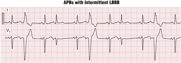
APBs may occur frequently or sporadically. Two APBs occurring consecutively are referred to as an atrial couplet. Sometimes, as shown in Figure 14-4, each sinus beat is followed by an APB. This pattern is referred to as atrial bigeminy.
Clinical Significance
APBs, conducted and blocked, are very common. They may occur in people with normal hearts or with virtually any type of organic heart disease. Thus, the presence of APBs does not imply that an individual has cardiac disease. In normal people these premature beats may be seen with emotional stress, excessive intake of caffeinated drinks, or the administration of sympathomimetic agents (epinephrine, isoproterenol). APBs may also occur with hyperthyroidism. APBs may produce palpitations; in this situation, patients may complain of feeling a “skipped beat” or an irregular pulse. APBs may also be seen with various types of structural heart disease. Frequent APBs are sometimes the forerunner of atrial fibrillation or flutter (see Chapter 15) or other supraventricular tachyarrhythmias discussed later.
Paroxysmal Supraventricular Tachycardias
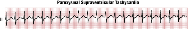
Premature supraventricular beats (Box 14-2) may occur singly or repetitively. A sudden run of three or more such consecutive nonsinus beats constitutes an episode of paroxysmal supraventricular tachycardia (PSVT). Episodes of PSVT may be brief and nonsustained (i.e., lasting from a few beats up to 30 sec). Sustained episodes (greater than 30 sec) may last minutes, hours, or longer.
The major types of PSVT are shown in Figure 14-7.
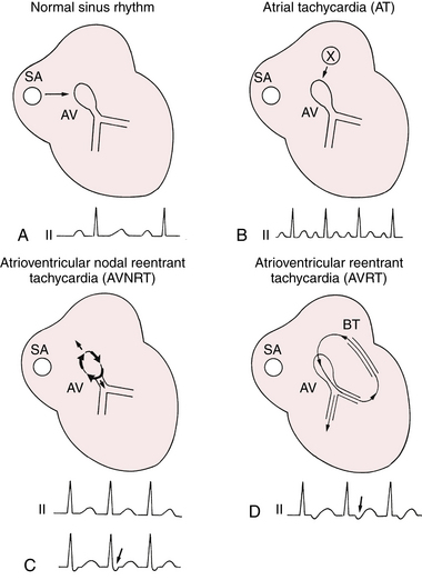
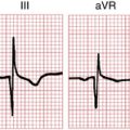
Figure 14-7 The three major types of paroxysmal supraventricular tachycardias (PSVTs). A, The reference is normal sinus rhythm. B, With (unifocal) atrial tachycardia (AT), a focus (X) outside the sinoatrial (SA) node fires off automatically at a rapid rate. C, With atrioventricular (AV) nodal reentrant tachycardia (AVNRT) the cardiac stimulus originates as a wave of excitation that spins around the AV nodal (junctional) area. As a result, retrograde P waves may be buried in the QRS or appear just after the QRS complex (arrow) because of nearly simultaneous activation of the atria and ventricles. D, A similar type of reentrant (circus-movement) mechanism may occur with a manifest or concealed bypass tract (BT) of the type found in Wolff-Parkinson-White syndrome (see Chapter 12 and text). This mechanism is referred to as atrioventricular reentrant tachycardia (AVRT). Note the negative P wave (arrow) in lead II, somewhat after the QRS complex.
Atrial Tachycardia
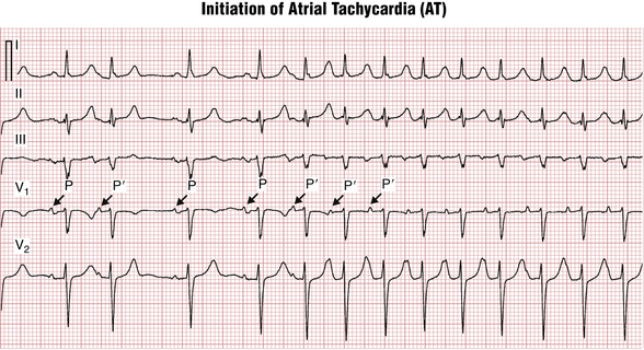
Classical atrial tachycardia (AT) is defined as three or more consecutive APBs coming from a single atrial focus and having identical nonsinus P wave morphology (Fig. 14-8). The arrhythmia focus can be located in either the left or right atrium, or proximal pulmonary veins, and fires off “automatically” in a rapid way. A variant, discussed later, is multifocal AT, in which the P waves vary because they come from different “firing” sites.
Conduction
Conduction occurs over the AV node and His-Purkinje system and usually produces a narrow QRS tachycardia. However, if the atrial rate is high, or the AV node is not normal, different degrees of delay and block can occur in any part of the conduction system (similar to that with isolated APBs), including PR prolongation, QRS aberration, and dropped (nonconducted) P waves that are not followed by QRS complexes (i.e., they produce functional second-degree AV block) with a regular or irregular heart rate. Also, if there is a preexisting bundle branch block or rate-related interventricular conduction delay (IVCD), the QRS during AT will remain wide and can be confused with ventricular tachycardia.
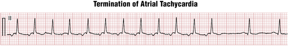
Termination
Termination occurs when the ectopic atrial focus stops firing (either spontaneously or after administration of an antiarrhythmic drug). The last P wave of the tachycardia usually conducts to the ventricles producing QRS complex at the end of the run. Therefore, AT almost always terminates with a QRS complex (Figs. 14-9 and 14-10). This is an important feature for differential diagnosis of other types of PSVTs (see later discussion).

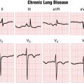
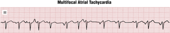
Multifocal Atrial Tachycardia
As noted, a special variant is related to multiple sites of atrial stimulation and is called multifocal atrial tachycardia (MAT) (Fig 14-11). This tachyarrhythmia is characterized by multiple ectopic foci stimulating the atria. The diagnosis of MAT requires the presence of three or more consecutive (nonsinus) P waves with different shapes at a rate of 100 or more per minute. MAT contrasts with classic (unifocal) AT, which involves only a single atrial focus and produces one repetitive, nonsinus P wave. The PR intervals of P waves during MAT also vary. MAT is usually seen in patients with chronic lung disease. Because the ventricular rate is irregular and rapid, this arrhythmia is most likely to be mistaken for AF.
AV Nodal Reentrant (Reentry) Tachycardia
AV nodal reentrant (reentry) tachycardia (AVNRT) is a supraventricular arrhythmia, usually paroxysmal, resulting from the reentry in the AV node area. Normally, the AV node behaves as a single conductor connecting the atria and His-Purkinje-ventricular electrical network. However, in some people it can behave as two functional conduction channels with different electrical properties (so-called dual pathways). One AV nodal pathway has fast and the other has slow conduction speeds.
The mechanism of AVNRT initiation is presented in Figure 14-12. During sinus rhythm the atrial signal engages both “fast” and “slow” pathways. It reaches the His bundle through the fast pathway first and from there conducts to the ventricles. At the same time it turns around and goes “up” the slow pathway, colliding with the more slowly conducting down-going signal. The surface ECG registers sinus rhythm with a normal PR interval; there is no evidence of the “slow” pathway existence (beats 1 and 2 in Fig. 14-12).
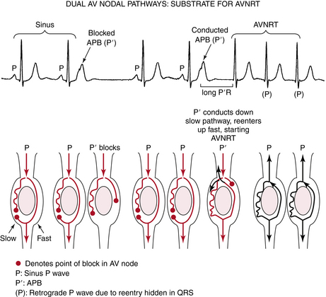
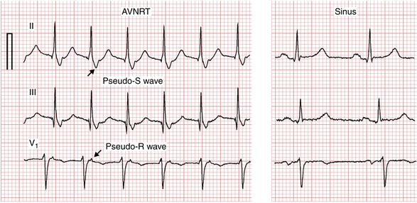
Because the arrhythmia circuit operates within the AV node (between the atria and ventricles), the activation spreads nearly simultaneously up the atria and down the ventricles with every reentrant rotation of the signal. As a result, the P waves can be completely hidden in the QRS complex (Fig. 14-13) or appear just after it. Because of retrograde (bottom-to-top) activation of the atria, the retrograde P waves are negative in leads II, III, and aVF, sometimes producing subtle but distinctive “pseudo-S” waves and positive in leads V1 and aVR where they are referred to as “pseudo-R′” waves that are absent during sinus rhythm (Fig. 14-14).
Termination
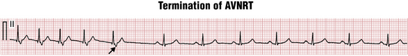
Unlike focal AT, which terminates when the ectopic focus stops firing, AVNRT is a self-perpetuating rhythm that will continue indefinitely unless a block develops in some part of the circuit. The block can occur in either fast (up-going) or slow (down-going) AV nodal pathway. The slow AV nodal pathway is more susceptible to vagal influences (see Fig. 14-15) or AV nodal blocking drugs (e.g., adenosine,∗ beta blockers, calcium channel blockers, or digoxin). The block in the slow pathway usually occurs just before the signal activates the ventricles from the bottom of the circuit; therefore, the last arrhythmia deflection on the electrocardiogram is a retrograde P wave (Fig. 14-15).
Clinical Considerations
One of the most commonly reported symptoms at the onset of AVNRT is the sensation of a “flip-flop” in the chest (resulting from the initiating APB), followed by rapid, regular palpitations. The arrhythmia may start with the person suddenly changing position or may be associated with psychological or physiologic stress, pregnancy, or sympathomimetic drugs. Patients may complain of pulsations in the neck due to simultaneous atrial and ventricular contraction, producing “cannon A waves” on inspection of the jugular venous waves during the neck examination. AVNRT can also produce dizziness, lightheadedness, and rarely, syncope.
Atrioventricular Reentrant (Bypass Tract–Mediated) Tachycardia
Atrioventricular reentrant (bypass tract–mediated) tachycardia (AVRT), the third most common cause of PSVT, involves an accessory atrioventricular bypass tract (see Chapter 12), which provides the substrate for reentry. Clinicians refer to two types of bypass tracts: manifest and concealed. Manifest bypass tracts can conduct the electrical signal in both directions: from the atria to the ventricles and in reverse. During sinus rhythm, this produces the classic triad (“signature”) of WPW: delta wave, short PR interval, and wide QRS (see Chapter 12).
Initiation and Conduction
During sinus rhythm, retrograde conduction through the bypass tract usually does not occur because the signal gets to the ventricular end of the bypass tract through the normal conduction system while the atrium around it is still refractory from the preceding sinus beat (Fig. 14-16A).
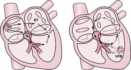
Figure 14-16 Atrioventricular reentrant tachycardia (AVRT) initiation by a ventricular premature beat (VPB). A, During sinus (SA) rhythm there is no retrograde conduction across the bypass tract (BT) or atrioventricular (AV) node because of refractoriness from the previous SA beat. B, A VPB (star) firing close to the bypass tract conducts to the atrium via the bypass tract, while simultaneously blocking in the His-Purkinje system. This sequence initiates a narrow complex tachycardia in which the impulse travels down (antegrade conduction) the AV node–His–bundle branch system and reenters the atrium by going up (retrograde conduction) the bypass tract. AVRT may also be initiated by atrial premature beats (APBs). See also Figure 14-17.
An early VPB that occurs close to the ventricular entrance of the concealed bypass tract can block the His-Purkinje system while conducting back to the atria through the bypass tract (Fig. 14-16B). In fact, a narrow complex tachycardia initiated by a VPB is highly suggestive of AVRT (see Figs. 14-16 and 14-17). Since the atria and ventricles activate one after another “in sequence” with AVRT as opposed to “in-parallel” as during AVNRT, the interval between the QRS and the P waves is longer in the former and P waves are often visible superimposed on the middle of the T wave or in the ST segment.
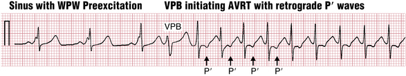
Figure 14-17 Atrioventricular reentrant tachycardia (AVRT) initiated by a ventricular premature beat (VPB). During sinus rhythm (first three beats) a short PR interval, wide QRS complex, and a delta wave are present, consistent with the classic Wolff-Parkinson-White (WPW) preexcitation pattern. A VPB (beat 4) initiates a narrow complex tachycardia. Retrograde P waves are visible as negative deflections in the middle of the T wave (black arrows). This finding corresponds to the mechanism depicted in Figure 12-5.
AVRT (as well as other PSVTs) also can produce QRS alternans—a periodic change in the QRS shape, occurring with every other beat (Fig. 14-18). This interesting pattern may be due to subtle conduction variations that occur at rapid rates.
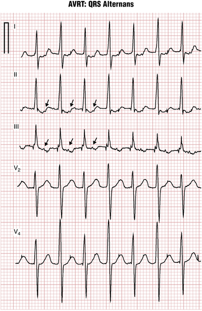
Figure 14-18 QRS alternans during atrioventricular reentrant tachycardia (AVRT). Note the periodically alternating (an “ABAB” pattern) QRS amplitudes, which are best seen in leads V2 and V4. Retrograde P waves are seen in the middle of T wave in leads I, II, and III (arrows). Alternans during paroxysmal supraventricular tachycardia (PSVT) should not be confused with alternans during sinus tachycardia (see Chapter 11), where it is usually an indicator of pericardial effusion and often cardiac tamponade (compare with Fig. 11-3). Also, alternans may occur with other types of PSVT (and, sometimes, ventricular tachycardia), so it is not specific for AVRT.
Clinical Considerations
The first episode of AVRT usually presents in childhood or young adulthood in contrast to AVNRT, which is predominantly seen in young to middle-aged female subjects. AVRT occurs more frequently in men. The accessory bypass tracts can be located on the left or right side of the heart (see Chapter 12). The symptoms, including palpitations and lightheadedness, as well as shortness of breath, are similar to AVNRT, discussed earlier.
Differential Diagnosis and Treatment of PSVT
The differential diagnosis of PSVT can be difficult, even for seasoned cardiologists. P waves may not be clearly visible even if present because they are hidden in T waves or ST segments, especially in a single monitor lead. Sometimes it is impossible to tell the exact mechanism of the arrhythmia (especially when initiation and termination of it are not recorded) unless an invasive electrophysiologic study is performed. A summary of major diagnostic findings is presented in Table 14-1. More detailed discussion on the differential diagnosis of PSVT is available in selected bibliographic references.
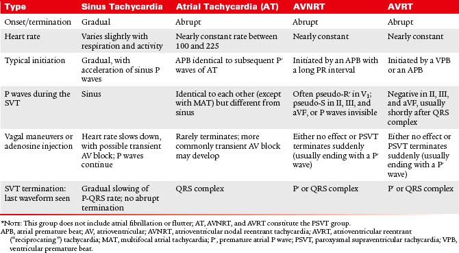
The response of PSVT to CSM (or other vagal maneuvers) or adenosine injection is summarized in Table 14-1.
Management of an Acute PSVT Episode
The management of the first acute PSVT episode should start with vagal maneuvers (e.g., the Valsalva maneuver, CSM—see Fig. 14-15) followed by adenosine injection. Many patients find ways to terminate the arrhythmias on their own (e.g., by coughing, deep breathing, using the Valsalva maneuver, squatting, facial immersion in cold water, or with CSM).
If adenosine is not available or is contraindicated, intravenous beta blockers or calcium channel blockers can be used, but may produce hypotension. These longer acting drugs can be beneficial in patients with extremely high sympathetic tone when the arrhythmia terminates with adenosine but immediately restarts. Digoxin can also be used. Rarely, synchronized external cardioversion is required to terminate PSVT.
∗ The terms atrial premature contractions, atrial premature beats, atrial premature depolarizations, and atrial extrasystoles are used synonymously. Most cardiologists prefer the designations premature beat, depolarization, and complex because not every premature stimulus is associated with an actual mechanical contraction of the atria or ventricles.
∗ Adenosine depresses the electrical activity in both the SA and AV nodes. Its very rapid action appears to be mediated by increasing an outward potassium current, hyperpolarizing the cells of both nodes. As a result, SA node automaticity is decreased, as are automaticity and conduction in the AV node area. Adenosine also is a vasodilator (accounting for its transient blood pressure lowering and facial flushing effects).