CHAPTER 9 Subscapularis
After the deltopectoral surgical approach is completed, the next step in performing unconstrained shoulder arthroplasty is management of the subscapularis musculotendinous unit. Various techniques for obtaining anterior access to the glenohumeral joint during shoulder arthroplasty have been described, including complete and partial subscapularis tenotomy, lesser tuberosity osteotomy, and access to the joint solely through the rotator interval. In many patients in whom shoulder arthroplasty is performed the subscapularis lacks normal excursion, thereby creating loss of external rotation. In such cases, surgical techniques must address this soft tissue contracture to allow sufficient postoperative external rotation and minimize the incidence of postoperative dehiscence of the subscapularis. This chapter details our preferred technique for handling the subscapularis, including accessing the glenohumeral joint and addressing loss of external rotation caused by subscapularis contracture.
TECHNIQUE FOR HANDLING THE SUBSCAPULARIS
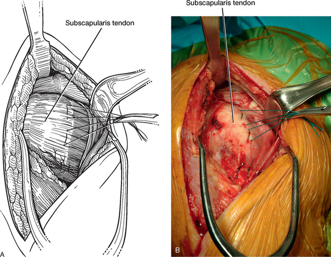
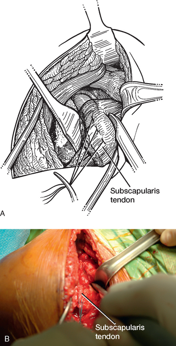
After the conjoined tendon is retracted medially with a narrow Richardson retractor, the subscapularis is readily visualized. In some cases a hypertrophic subscapularis bursa may be present, and a bursectomy is necessary to allow adequate visualization of the subscapularis tendon. To minimize hemorrhage, we perform bursectomy with the needle tip electrocautery. Once the subscapularis tendon is adequately visualized, two stay sutures of no. 1 nonabsorbable braided suture are double-passed through the tendon approximately 15 mm lateral to the musculotendinous junction, one in the superior half and one in the inferior half of the tendon (Fig. 9-1).
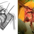
The glenohumeral joint is opened initially through the rotator interval. The tips of large curved Mayo scissors are passed tangential and just superior to the subscapularis tendon and used to puncture the rotator interval tissue for access to the glenohumeral joint. Once the glenohumeral joint is entered, the scissors are spread to enlarge the arthrotomy (Fig. 9-2). Usually after the Mayo scissors are spread, synovial fluid egresses from the joint.
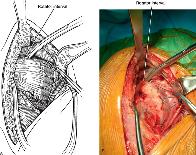
Figure 9-2 A and B, Arthrotomy of the glenohumeral joint through the rotator interval with large Mayo scissors.
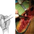
The next step involves elevation of the subscapularis from its humeral insertion to allow further access to the glenohumeral joint. We prefer performing a complete subscapularis tenotomy because it allows unhindered access to the glenohumeral joint and sufficient release of subscapularis contracture. Additionally, tenotomy does not risk disruption of the existing proximal humeral osseous anatomy, as may occur with lesser tuberosity osteotomy. The location of the tenotomy is critical to allow adequate repair after insertion of the prosthesis is completed. We perform the tenotomy along the anatomic neck of the humerus while leaving a small amount of tendon on the lesser tuberosity to use in later subscapularis closure. The shoulder is placed in neutral to slight external rotation during this portion of the procedure. To identify the anatomic neck of the humerus, a no. 10 scalpel blade on a long handle is used to further open the rotator interval from the initial arthrotomy site extending laterally along the superior border of the subscapularis tendon (Fig. 9-3). Once the anatomic neck of the humerus is identified by observing the lateral extent of the articular surface, the scalpel blade is directed inferiorly to transect the superior two thirds of the subscapularis tendon along the anatomic neck of the humerus (Fig. 9-4A). At the inferior third of the subscapularis, we switch to the needle tip electrocautery and complete the tenotomy. The needle tip passes between the previously placed anterior humeral circumflex ligation sutures and cauterizes these vessels (Fig. 9-4B). As the subscapularis tenotomy is completed, the shoulder is progressively externally rotated to allow visualization of the inferior humeral capsular attachment (Fig. 9-4C and D). This capsule is released from the humerus with the needle tip electrocautery while keeping the electrocautery in contact with the humerus (Fig. 9-5).
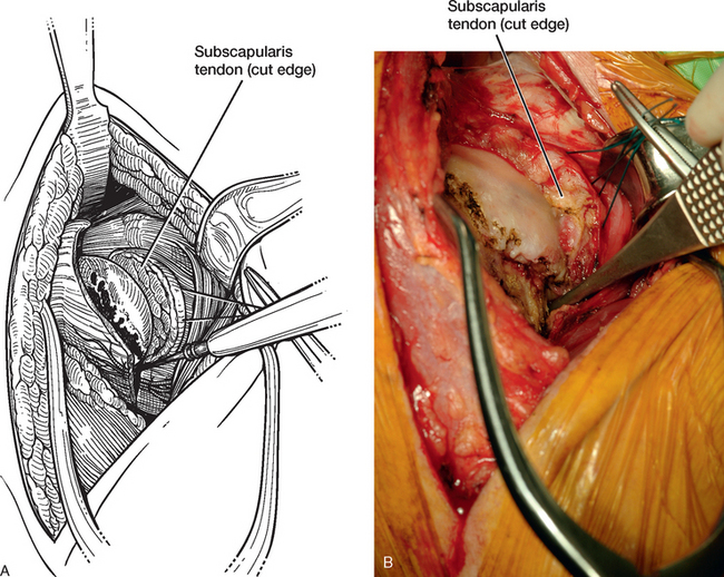
Figure 9-5 A and B, Release of the medial joint capsule from the humerus with the needle tip electrocautery.
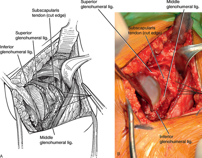
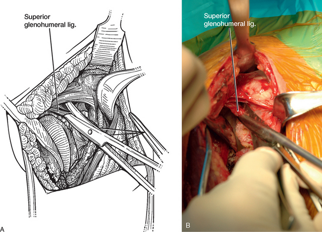
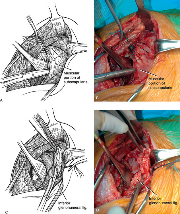
A humeral head retractor (we prefer the Trillat type of retractor) is placed in the glenohumeral joint and the humeral head is retracted posteriorly. To facilitate insertion of this retractor, the shoulder is initially externally rotated and moved into internal rotation as the retractor is pushed posteriorly between the humeral head and the glenoid. With the humeral head retracted posteriorly and traction applied to stay sutures in the subscapularis tendon, the glenohumeral ligaments are readily visualized (Fig. 9-6). To increase subscapularis excursion, circumferential release of the subscapularis tendon is performed. The superior glenohumeral ligament is first released with large curved Mayo scissors along the superior aspect of the subscapularis tendon (Fig. 9-7). The middle glenohumeral ligament, which may be absent in up to a third of patients, is released parallel to the anterior glenoid rim with large curved Mayo scissors (Fig. 9-8). Anteriorly and inferiorly the tissue plane between the capsuloligamentous structures and the primarily muscular inferior portion of the subscapularis is identified and established with large curved Mayo scissors. Once this plane is established, the inferior glenohumeral ligament is divided with the scissors. Risk of injury to the axillary nerve is avoided because the muscular portion of the subscapularis isolates it from the path of the scissors (Fig. 9-9). After release of the glenohumeral ligaments, the intra-articularly located subscapularis recess is readily accessible. This recess should be routinely inspected for loose bodies. We remove all loose bodies (Fig. 9-10). In most cases these loose bodies are osseous and may be anticipated by identification on preoperative imaging studies. Finally, blunt dissection with a Cobb elevator is used to release any adhesions that remain anterior to the subscapularis (Fig. 9-11). After release of the subscapularis, which effectively lengthens the contracted tendon, increased excursion should be possible. A sponge is used to tuck the subscapularis into the subscapularis fossa, and it is held with a small glenoid rim retractor (Fig. 9-12).
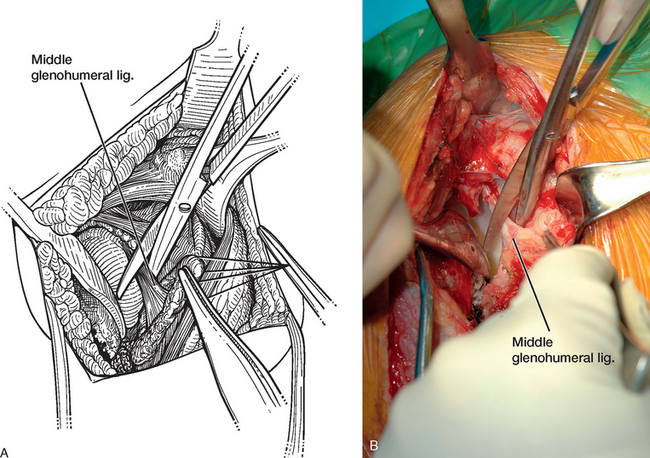
Figure 9-8 A and B, Transection of the middle glenohumeral ligament during release of the subscapularis tendon.

Figure 9-10 A and B, Loose body identified in the subscapularis recess after release of the glenohumeral ligaments.

