206 Subhyaloid haemorrhage
Salient features
Examination
• A large, solitary subhyaloid haemorrhage (there may be no fluid level if the patient is lying flat) (Fig. 206.1)
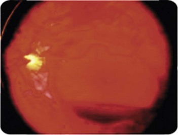
Fig. 206.1 Funduscopic photograph of a subhyaloid haemorrhage.
(With permission from Field, Heran 2010.)
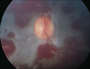
Note: When the subhyaloid (preretinal) haemorrhage extends into the vitreous humour it is called Terson syndrome (Fig. 206.2).
Advanced-level questions
What is the cause of subarachnoid haemorrhage?
• Aneurysms are the cause of subarachnoid haemorrhage in 85% of cases (Lancet 2007; 369:306–18). Intracranial aneurysms are not congenital, as was once believed, but develop in the course of life.
• The best estimate of the frequency of aneurysms for an average adult without specific risk factors is 2.3% (95% confidence interval, 1.7–3.1); this proportion increases with age.
• Saccular aneurysms arise at sites of arterial branching, usually at the base of the brain, either on the circle of Willis itself or at a nearby branching point.
• Most intracranial aneurysms will never rupture. The rupture risk increases with the size of aneurysm, but paradoxically, most ruptured aneurysms are small (<1 cm); the explanation is that 90% of all aneurysms are small and even though only a small fraction of these rupture, this total outnumbers the greater fraction of the large aneurysms that rupture. The case fatality after aneurysmal haemorrhage is 50%; one in eight patients with subarachnoid haemorrhage dies outside hospital.
What is the management of subarachnoid haemorrhage?
• Rebleeding: occlusion of the aneurysm. Endovascular obliteration by means of platinum spirals (coiling) is the preferred mode of treatment, but some patients require a direct neurosurgical approach (clipping).
• Cerebral ischaemia: the risk is reduced with oral nimodipine and probably by maintaining circulatory volume.
• Hydrocephalus might cause gradual obtundation in the first few hours or days; it can be treated by lumbar puncture or ventricular drainage, dependent on the site of obstruction.


