Chapter 287 Strongyloidiasis (Strongyloides stercoralis)
Diagnosis
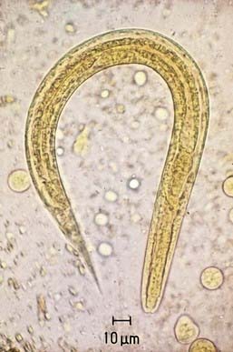
Intestinal strongyloidiasis is diagnosed by examining feces or duodenal fluid for the characteristic larvae (Fig. 287-1). Several stool samples should be examined either by direct smear, Koga agar plate method, or the Baermann test. Alternatively, duodenal fluid can be sampled by the enteric string test (Entero-Test) or aspiration via endoscopy. In children with the hyperinfection syndrome, larvae may be found in sputum, gastric aspirates, and rarely in small intestinal biopsy specimens. An enzyme-linked immunosorbent assay for IgG antibody to Strongyloides may be more sensitive than parasitologic methods for diagnosing intestinal infection in the immunocompetent host. The utility of the assay in diagnosing infection in immunocompromised subjects with the hyperinfection syndrome has not been determined. Eosinophilia is common.
Biggs B, Caruana S, Mihrshahi S. Management of chronic strongyloidiasis in immigrants and refugees: is serologic testing useful? Am J Trop Med Hyg. 2009;80:788-791.
Keiser PB, Nutman TB. Strongyloides stercoralis in the immunocompromised population. Clin Microbiol Rev. 2004;17:208-217.
Knopp S, Mgeni AF, Khamis IS, et al. Diagnosis of soil-transmitted helminths in the era of preventive chemotherapy: effect of multiple stool sampling and use of different diagnostic techniques. PLoS Negl Trop Dis. 2008;2:e331.
Marcos LA, Terashima A, DuPont HL. Strongyloides hyperinfection syndrome: an emerging global infectious disease. Trans R Soc Trop Med Hyg. 2008;102:314-318.
Segarra-Newnham M. Manifestations, diagnosis, and treatment of Strongyloides stercoralis infection. Ann Pharmacother. 2007;41:1992-2001.


