Stroke

Published on 06/06/2015 by admin

Filed under Pediatrics

Last modified 22/04/2025

Print this page

rate 1 star rate 2 star rate 3 star rate 4 star rate 5 star
Your rating: none, Average: 0 (0 votes)

This article have been viewed 3078 times

79 Stroke

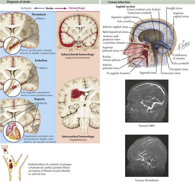
Stroke refers to acute vascular events involving the brain or brainstem. Childhood stroke occurs at a rate approximating that of childhood brain tumors. It is among the top 10 causes of death in children and is a significant cause of morbidity among survivors. Pediatric stroke can be subdivided into perinatal stroke, occurring from 28 weeks of gestation to 1 month of age, and childhood stroke, occurring from 1 month to 18 years. Important subtypes of stroke include arterial ischemic stroke (AIS), watershed infarction, intracerebral hemorrhage (ICH), and cerebral sinus venous thrombosis (CSVT). AIS is usually defined as an acute neurologic deficit of any duration consistent with focal brain ischemia conforming to an arterial distribution. Transient ischemic attacks (TIAs) are defined as focal deficits in a vascular territory lasting less than 24 hours, with some authors including the caveat that there must be no magnetic resonance imaging (MRI) evidence of infarction (Figure 79-1).

Etiology and Pathogenesis

Estimates of the incidence of stroke in children range from two to 13 per 100,000 children per year. As opposed to adults in which ICH accounts for about 15% of stroke, ICH accounts for about half of pediatric stroke. The incidence of perinatal stroke is approximately one per 4000 live births, with 80% of these secondary to AIS. One-quarter of all strokes in children take place during the perinatal period. The incidence of cerebral venous sinus thrombosis is approximately 0.3 to 0.7 per 100,000 per year, with almost half of these occurring in the perinatal period. Strokes are more common in boys than in girls, with 55% to 60% of all childhood strokes occurring in boys. In the United States, strokes are more common among African American children, even when controlling for the presence of sickle cell disease (SCD).

Unlike adults, in whom hypertension, diabetes, and atherosclerosis predominate as risk factors for ischemic stroke, children presenting with AIS have much more varied etiologies. Approximately 50% of children presenting with stroke have an obvious underlying cause at the time of presentation, with arteriopathy, congenital heart disease, and sickle cell anemia representing some of the most common causes. In an additional 20% to 40%, an underlying cause can be found with further investigation. Somewhere between 10% and 30% are cryptogenic. Factors associated with pediatric AIS include inherited or acquired prothrombotic states, cardiac disease, arteriopathies or vasculopathies, trauma, and infections (Box 79-1). In older teenagers, traditional risk factors for adults, such as hypertension, diabetes, high cholesterol, and smoking, may also play a role. Cocaine or sympathomimetic medications are additional risk factors. Watershed strokes can be seen after cardiac arrest or other causes of shock, near drowning, and cardiac surgery.

Box 79-1 Etiologies of Pediatric Arterial Ischemic Stroke

Arteriopathy

Hematologic

Cardiac

Infectious

Rheumatologic

Metabolic

Other

CADASIL, cerebral autosomal dominant arteriopathy with subcortical infarcts and leukoencephalopathy; CNS, central nervous system; MELAS, mitochondrial encephalopathy with lactic acidosis and strokelike episodes.

ICH in children is also etiologically distinct from ICH in adults. Unlike adults, in whom hypertension and amyloid angiopathy are the most common causes, childhood ICH is most commonly caused by ruptured vascular malformations (e.g., arteriovenous malformations, cavernomas, and aneurysms), hematologic abnormalities, and brain tumors (Box 79-2 and Figure 79-2).

The major risk factors for CSVT include dehydration, head and neck infections, hypercoagulable states, malignancies, congenital heart disease, inflammatory bowel disease, oral contraceptives, and the use of certain chemotherapeutics.

AIS occurs when occlusion of an artery with a thrombus or embolus prevents blood flow to the region supplied by that artery. In watershed infarctions, global or near-global hypoperfusion preferentially damages areas on the border of two different vascular territories. The end result is a complex chain of events in cerebral neurons and support cells in which lack of oxygen and glucose leads to mitochondrial dysfunction, ion pump failure, calcium influx, and glutamate-induced neurotoxicity, eventually producing cell necrosis or apoptosis. The extent of ischemic damage to the region is determined by oxygen demand, collateral blood flow, and time to reperfusion. After an acute ischemic insult, there is a core area of infarction in which cells have sustained irreversible injury. This core is surrounded by an area with reversible ischemia known as the ischemic penumbra. If no further improvement of blood flow takes place or if additional insults occur, the tissue within the penumbra will eventually infarct over a period of hours to days, potentially causing additional neurologic impairment. In TIAs perfusion is restored before infarction can take place, and neuronal dysfunction induced by ischemia is reversed.

In ICH, blood extravasates into the brain parenchyma (intraparenchymal hemorrhage) into the ventricles (intraventricular hemorrhage), or into the subarachnoid space (subarachnoid hemorrhage). Intraparenchymal hemorrhage causes neuronal damage through a combination of mechanical damage and chemical irritation. Secondary ischemic stroke can occur as well because of mechanical deformation of cerebral vasculature by mass effect, vasospasm, and altered pressure dynamics leading to decreased cerebral perfusion pressure. Intraventricular hemorrhage can lead to hydrocephalus.

In CSVT, obstruction of venous blood by clot formation alters blood flow dynamics and overall venous drainage is decreased. Increased cerebral venous pressure causes resistance to cerebral perfusion and can cause venous infarction or hemorrhage. All contribute to increasing intracranial pressure (ICP).

Clinical Presentation

The clinical presentation of a child with stroke depends on the location, type, and size of the stroke and the age and developmental stage of the child. The diagnosis is often missed or delayed in children because of a low index of suspicion on the part of care providers. In addition, children are much more likely than adults to present with headaches, seizures, or altered mental status as the first sign of stroke. Any child with acute onset of a focal neurologic deficit should be investigated for possible stroke, but other situations that should be investigated for possible stroke include seizures in the newborn, seizures in a child of any age occurring after cardiac surgery, and changes in mental status associated with headache.

Approximately 75% percent of AIS in children occurs in the anterior circulation coming from the internal carotid arteries. Older children with ischemic strokes most commonly present with acute hemiparesis, hemisensory loss, or aphasia but may also present with isolated seizures or hemichorea. Children with posterior circulation strokes affecting the vertebrobasilar distributions may present with ataxia, vertigo, visual field cut, or brainstem signs. Children with watershed strokes can present with proximal greater than distal weakness.

Intraparenchymal hemorrhage classically presents with focal neurologic signs or seizures accompanied by severe headache, emesis, and loss of consciousness but may be impossible to distinguish from AIS based on clinical features alone because presentations vary (see Figure 79-2). Children with CSVT may present acutely with headaches, emesis, seizures, altered mental status, or focal neurologic signs but can also present more insidiously with symptoms of increased ICP such as chronic headache, blurry vision, or diplopia secondary to sixth cranial nerve palsies.

Evaluation

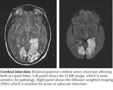
The initial evaluation of a child with suspected stroke should focus on confirming the diagnosis and ruling out common stroke mimics. The initial history and physical examination should assess the child’s ability to maintain a natural airway and adequate perfusion. Initial testing should include a complete blood count, electrolytes, blood glucose, prothrombin time, international normalized ratio, partial thromboplastin time, toxicology screen, and electrocardiography (ECG). Brain imaging should be obtained as soon as is possible. In most cases, unless MRI can be performed immediately, head computed tomography (HCT) should be performed first. HCT is fast and widely available and allows for the quick assessment of hemorrhage or mass lesions. In AIS, HCT results may be normal early in symptom evolution, but subtle findings of AIS, such as hyperdense middle cerebral artery (MCA) sign or blurring of the gray-white matter border can sometimes be seen. However, MRI is usually required to confirm the diagnosis of AIS. The most sensitive MRI sequence for detecting acute ischemic stroke is diffusion-weighted imaging with apparent diffusion coefficient; abnormalities on these sequences usually last 7 to 10 days from the acute stroke (Figure 79-3).

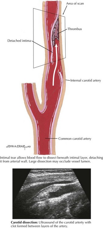
Additional testing should attempt to identify the etiology of the stroke (Box 79-3). Imaging of the vessels of the head and neck with MR angiography (MRA) or computed tomography angiography (CTA) should be performed to look for arterial dissection (Figure 79-4) or other arteriopathy. In certain cases, conventional catheter angiography may be necessary.
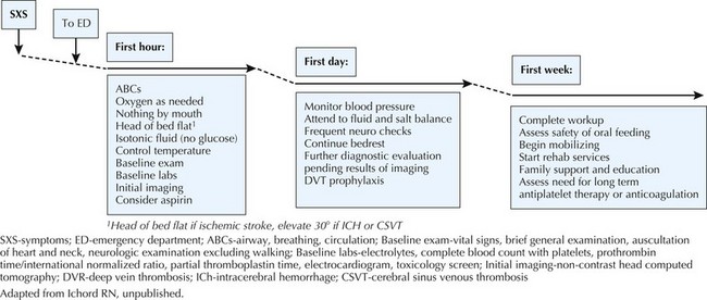
In children with ICH, MRA, CTA, or conventional angiography should be performed to evaluate for vascular malformations. If such a lesion is suspected but is not found in the acute setting, arterial imaging may need to be repeated at intervals because overlying hemorrhage may obscure the detection of vascular malformations. In children with suspected or confirmed CSVT, MR venography is the modality of choice to evaluate the dural sinuses for clot and residual blood flow.

In children with either AIS or CSVT, an evaluation for prothrombotic states should be performed. Hematology should be consulted regarding the most up-to-date recommendations, but currently testing includes protein C and S activity, factor V Leiden mutation, antithrombin III level, antiphospholipid antibodies (anticardiolipin antibodies, β2 glycoprotein, diluted Russell’s viper venom time), prothrombin gene mutation, lipoprotein (a), and homocysteine. In the appropriate clinical setting, testing for infectious causes of vasculopathy, including varicella, HIV, and syphilis, is appropriate. Evaluation of the heart for cardiac sources of thrombi should be performed in all children with AIS. Transthoracic echocardiography is usually the initial cardiac imaging modality, but if there is a strong suspicion for a cardiac source, transesophageal echocardiography should be considered, even when the transthoracic test results are normal. A “bubble study” (agitated saline contrast) during the ECG is used to detect any abnormal arterial–venous connections. More extensive testing may be appropriate if the initial evaluation does not demonstrate a clear cause or if an underlying metabolic, infectious, or inflammatory disease is suspected.

Management

The phrase “time is brain” has been used in adult stroke to emphasize that a stroke is a “brain attack” that should be recognized and treated emergently. Unfortunately, treatment of childhood stroke is often delayed by more than 24 hours because parents and physicians often do not recognize childhood stroke immediately. The goal of therapy in acute ischemic stroke is to save as much of the penumbra as possible by restoring perfusion, minimizing demand, and avoiding additional insults such as hyperglycemia. Thrombolytics have not been studied in children younger than 18 years of age, so therapy is primarily supportive.

An overview of initial therapy is summarized in Figure 79-5. In the acute setting, the child should lie supine with the head of the bed flat and isotonic intravenous fluids given to maximize perfusion. Hypertension is an adaptive response to ischemia, and thus outside of extreme hypertension, blood pressure should not be aggressively reduced. In most cases, aspirin should be given after hemorrhage has been excluded with neuroimaging; however, in many centers, anticoagulation with a heparinoid is started after hemorrhage is excluded. In certain cases of AIS such as arterial dissection, cardioembolism, or a known prothrombotic disorder, anticoagulation should strongly be considered (see Figure 79-5). Fever and seizures should be treated to minimize additional metabolic demands, and normoglycemia should be maintained. After the neonatal period, dextrose-free fluids are preferred to minimize the risk of hyperglycemia.

After the child has been stabilized, attention should be directed at identifying the cause of the stroke and minimizing risk factors for recurrence. Speech, physical, and occupational therapy and other rehabilitation services should be involved early in the child’s hospitalization course so a recovery plan can be initiated in a timely manner.

In the acute management of patients with ICH and CSVT, the head of the bed should be elevated to at least 30 degrees to promote venous drainage and thereby lower ICP. The management of ICH is sometimes surgical, and prompt consultation with a neurosurgeon is advised. The need for acute lowering of ICP should be continuously reassessed. In CSVT, anticoagulation should be strongly considered, even in the presence of associated hemorrhage. In neonatal CSVT, the data for anticoagulation is less clear, and the approach should be highly individualized.

Special Considerations

Perinatal Arterial Ischemic Stroke

The perinatal period is the most common period in childhood for ischemic strokes, and the stroke rate in neonates approaches that in the elderly. The most common presentation of perinatal stroke is seizure during the first few days of life, but perinatal stroke may present only with lethargy or poor feeding or may be completely asymptomatic. Many infants who have perinatal stroke are only diagnosed later in childhood upon evaluation of early hand preference, developmental delay, or emerging hemiparesis.

The pathophysiology of perinatal ischemic stroke is incompletely understood but is thought to be secondary to intrinsic hypercoagulability, dehydration, and shifts from the fetal to the mature circulation that lead to clot formation. The majority of strokes are in the anterior circulation, and there is a predilection for the MCA territory. There is a moderate preponderance of left-sided stroke in the perinatal period, presumably secondary to altered flow dynamics from the ductus arteriosus. Watershed infarcts are common, both in the classic distribution between the MCA and anterior cerebral artery territories and in a watershed area unique to the perinatal period between the territory of the long circumferential and paramedian penetrating branches of the basilar artery. This leads to a linear-shaped infarction affecting structures in the brainstem governing feeding and respiratory drive and thus can present solely with unexplained apnea or poor feeding.

Risk factors for perinatal stroke include complications during pregnancy or delivery, congenital heart disease, and thrombophilias in both the infant and the mother. However, in about 50% of cases of perinatal stroke, no specific cause can be identified. Evaluation of the neonate with suspected stroke should include the same elements as that of the older child, although infectious causes and maternal prothrombotic states should also be investigated. The placenta should be evaluated for pathology when possible. Management of a neonate with suspected stroke is different from that of an older child in that hypotonic and dextrose-containing fluids are acceptable. In addition, aspirin is rarely used. The prognosis depends on the extent and location of the injury, with complete MCA territory strokes and bilateral basal ganglia infarction portending a poor prognosis in almost all cases. Cerebral palsy is a common sequela, occurring in 30% to 50% of survivors of perinatal stroke. Because most perinatal stroke is secondary to factors unique to the perinatal period, recurrence is rare.

Metabolic Stroke

Unlike AIS, which is caused by hypoperfusion, metabolic strokes are usually caused by inherited defects in metabolic pathways that lead to an inability to compensate for metabolic stresses. In the setting of metabolic stress, neuronal energy production is unable to keep up with metabolic demand, leading to a final common pathway of cell death. Metabolic strokes or strokelike episodes are most commonly associated with mitochondrial disease such as MELAS (mitochondrial encephalopathy with lactic acidosis and strokelike episodes). However, they can be seen in a variety of metabolic diseases in which energy production is impaired, including organic and amino acidurias, disorders of fatty acid oxidation, and carnitine transport disorders. Clues to the diagnosis of metabolic stroke include a history of developmental regression, decompensation in the setting of acute illness, recurrent episodes in the absence of any vascular or hematologic pathology, unexplained acidosis, and nonvascular distribution of infarct. MRI findings suggestive of metabolic stroke include bilateral basal ganglia or bilateral occipital lobe infarcts. Further evaluation of a child with suspected metabolic stroke should include urine organic acids, serum amino acids, an acylcarnitine profile, lactate, pyruvate, and ammonia. MR spectroscopy (MRS) can also be very helpful, with lactate peaks over the basal ganglia suggestive of metabolic stroke. However, an MRS can be delayed because lactate might be elevated in the setting of any acute infarction. Management of a child with metabolic stroke depends on the underlying disease, but the emphasis should be on minimizing metabolic stress. Conventional measures to decrease thrombus formation or increase cerebral perfusion are unlikely to be effective because the origin of the “stroke” is not thrombus but is instead neuronal energy failure.

Suggested Readings

Adams RJ, McKie VC, Hsu L, et al. Prevention of a first stroke by transfusions in children with sickle cell anemia and abnormal results on transcranial Doppler ultrasonography. N Engl J Med. 1998;339:5-11.

Amlie-Lefond C, Bernard TJ, Sebire G, et al. Predictors of cerebral arteriopathy in children with arterial ischemic stroke: results of the international pediatric stroke study. Circulation. 2009;119:1417-1423.

deVeber G, Andrew M, Adams C, et al. Cerebral sinovenous thrombosis in children. N Engl J Med. 2001;345:417-423.

Elbers J, Benseler SM. Central nervous system vasculitis in children. Curr Opin Rheumatol. 2008;20:47-54.

Fullerton HJ, Johnston SC, Smith WS. Arterial dissection and stroke in children. Neurology. 2001;57:1155-1160.

Fullerton HJ, Wu YW, Zhao S, et al. Risk of stroke in children: ethnic and gender disparities. Neurology. 2003;61:189-194.

Hutchison JS, Ichord R, Guerguerian AM, et al. Cerebrovascular disorders. Semin Pediatr Neurol. 2004;11:139-146.

Jordan LC. Stroke in childhood. Neurologist. 2006;12:94-102.

Jordan LC, Hillis AE. Hemorrhagic stroke in children. Pediatr Neurol. 2007;36:73-80.

Lynch JK. Cerebrovascular disorders in children. Curr Neurol Neurosci Rep. 2004;4:129-138.

Lynch JK, Han CJ. Pediatric stroke: what do we know and what do we need to know? Semin Neurol. 2005;25:410-423.

Kirton A, deVeber G. Therapeutic approaches and advances in pediatric stroke. NeuroRx. 2006;3:133-142.

Nelson KB, Lynch JK. Stroke in newborn infants. Lancet Neurol. 2004;3:150-158.

Roach ES, Golomb MR, Adams R, et al. Management of stroke in infants and children: a scientific statement from a Special Writing Group of the American Heart Association Stroke Council and the Council on Cardiovascular Disease in the Young. Stroke. 2008;39:2644-2691.

Shellhaas RA, Smith SE, O’Toole E, et al. Mimics of childhood stroke: characteristics of a prospective cohort. Pediatrics. 2006;118:704-709.

Scott RM, Smith ER. Moyamoya disease and moyamoya syndrome. N Engl J Med. 2009;360:1226-1237.

Zimmer JA, Garg BP, Williams LS, et al. Age-related variation in presenting signs of childhood arterial ischemic stroke. Pediatr Neurol. 2007;37:171-175.