Streptococcal Infections

Published on 09/02/2015 by admin

Filed under Allergy and Immunology

Last modified 22/04/2025

Print this page

rate 1 star rate 2 star rate 3 star rate 4 star rate 5 star
Your rating: none, Average: 0 (0 votes)

This article have been viewed 3957 times

Streptococcal Infections

Etiology

Most streptococci that contain cell wall antigens of the Lancefield group A (Table 17-1) are known as Streptococcus pyogenes. Members of this species are almost always beta-hemolytic streptococci. S. pyogenes is the most common causative agent of pharyngitis and its resultant disorder, scarlet fever, and the skin infection, impetigo. The most common type of bacteria causing necrotizing fasciitis is S. pyogenes.

Table 17-1

Lancefield Streptococcus Classifications

Lancefield Group Examples of Bacterial Species in the Group Comments
A Streptococcus pyogenes Strains most pathogenic for human beings can cause strep throat, rheumatic fever, scarlet fever, acute glomerulonephritis, and necrotizing fasciitis.
B Streptococcus mastitis
Streptococcus agalactiae
Strains from mastitis in cows and from normal milk, including strains from the human throat and vagina
Can cause pneumonia and meningitis in neonates and older adults, with occasional systemic bacteremia
C Streptococcus equii
Streptococcus dysgalactiae
Strains from various lower animals, including cattle, and from the human throat
Can cause pharyngitis and other pyogenic infections similar to group A streptococci
D Streptococcus faecalis (now Enterococcus faecalis)
Other nonenterococcal group D strains include Streptococcus bovis and Streptococcus equinus.
Strains from cheese and humans
Many former group D streptococci have been reclassified and placed in the genus Enterococcus.
E   Strains from certified milk
F Streptococcus anginosus (Lancefield classification) or Streptococcus milleri group (European system) Strains mainly from the human throat, associated with tonsillitis; minute hemolytic
G Streptococcus canis is an example of a GBS Group B streptococcus (GBS) which is typically found in animals, but does not cause infection except in newborns at birth Strains can cause infection in human beings (a few strains from monkeys and dogs).
Note: This is not exclusively beta-hemolytic.
H, K, O   Nonpathogenic strains occasionally from normal human respiratory tracts

This is a serologic classification of hemolytic streptococci, dividing them into groups based on antigenic serocharacteristics. It is based on precipitation tests depending on group-specific carbohydrate substances.

In terms of human morbidity and mortality worldwide, however, the role of S. pyogenes in the subsequent development of complications such as acute rheumatic fever and poststreptococcal glomerulonephritis is more important. Other S. pyogenes–associated infections include otitis media in children, sinusitis in adults, and osteomyelitis, septic arthritis, neonatal septicemia, and rare cases of pneumonia.

Necrotizing fasciitis is a rare infection that can destroy skin and soft tissues, including fat and the tissue-covering muscles (fascia). Because these tissues die rapidly, a person with necrotizing fasciitis is sometimes said to be infected with so-called flesh-eating bacteria. A highly invasive group A streptococcal infection is associated with toxic shock syndrome.

Morphologic Characteristics

S. pyogenes is a gram-positive coccus and the serotype most frequently associated with human infection. Lancefield divided these beta-hemolytic streptococci into serogroups A through O on the basis of the immunologic action of the cell wall carbohydrate (Fig. 17-1).

Structures called fimbriae arise near the plasma membrane and project through the cell wall and capsule. These processes contain important surface components of the streptococcus. Lipoteichoic acid on the fimbriae is important in the organism’s adherence to human epithelium and the initiation of infection. The M and R antigens, which are structurally similar but immunologically distinct, are also found on the fimbriae. R antigen has no known biological role.

M protein, a cell protein found in association with the hyaluronic capsule, is a major virulence factor of S. pyogenes. Strains of S. pyogenes that lack M protein cannot cause infection. M protein inhibits phagocytosis and antibody synthesized against M protein provides type-specific immunity to group A streptococci. In addition, M protein is the basis for a subclassification of group A streptococci into more than 60 M serotypes.

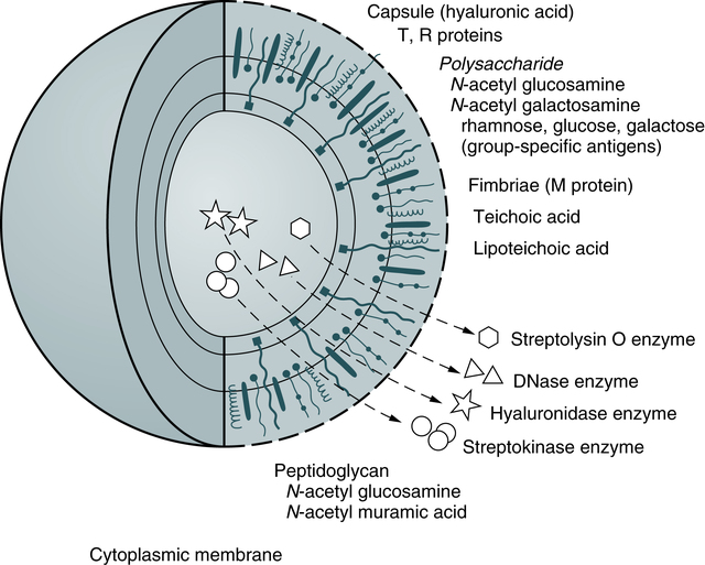
Extracellular Products

Extracellular products are important in the pathogenesis of disease and in the serologic diagnosis of streptococcal disease. Antibodies produced in response to these substances provide evidence of recent streptococcal infection. Two hemolysins, with the ability to damage human and animal erythrocytes, polymorphonuclear leukocytes (PMNs), and platelets, are produced by most group A strains, as follows:

• Streptolysin O (SLO), an oxygen-labile enzyme, binds to sterols in the red blood cell (RBC) membrane, causing stearic rearrangement. This rearrangement produces submicroscopic holes in the RBC membrane and hemoglobin diffuses from the cells. SLO is antigenic; the antibody response to it is the most frequently used serologic indicator of recent streptococcal infection.

• Streptolysin S, an oxygen-stable enzyme, is responsible for the beta (clear-appearing) hemolysis on the surface of a blood agar culture plate. Streptolysin S disrupts the selective permeability of the RBC membrane, causing osmotic lysis. It is not antigenic.

• Other substances produced by group A streptococci presumably facilitate rapid spread through subcutaneous or deeper soft tissues and include the following:

• Hyaluronidase, also called spreading factor, breaks down hyaluronic acid found in the host’s connective tissue.

• Four immunologically distinct deoxyribonucleases (DNases A, B, C, and D) degrade deoxyribonucleic acid (DNA).

• Streptokinase, an enzyme, dissolves clots by converting plasminogen to plasmin.

• Other extracellular products that can elicit an antibody response include NADase, proteinase, esterase, and amylase.

• Erythrogenic toxin is elaborated by scarlet fever–associated strains and is responsible for the characteristic rash.

Epidemiology

S. pyogenes is one of the most common and ubiquitous of human pathogens. It is found in the human respiratory tract and is always considered a potential pathogen. Upper respiratory infections caused by S. pyogenes occur most frequently in school-age children and are uncommon in children younger than 3 years. No gender or race predilection has been described.

Infection is spread by contact with large droplets produced in the upper respiratory tract. Although not as common, foodborne and milkborne epidemics do occur. Crowding enhances the spread of microorganisms.

A number of individuals, particularly school-age children, carry S. pyogenes without signs of illness. Carriers have positive cultures without serologic evidence of infection. If a person carries the organisms in the pharynx for prolonged periods after untreated infection, the number of organisms carried and their ability to produce M protein decline during carriage. This results in a progressive decline in the likelihood of spreading infection to others.

The incidence of a major complication of S. pyogenes, rheumatic fever, has decreased in the United States. It occurs primarily in the rural South and in areas of crowding and lower socioeconomic status. The incidence of rheumatic fever is 2% to 3% in epidemics and 0.1% to 1% after sporadic cases of streptococcal infection. The probability of developing rheumatic fever is age related, with younger patients more likely to develop carditis than older persons.

Rheumatic fever and resultant valvular heart disease, however, are syndromes of major importance among children in developing nations. Patients with a history of rheumatic heart disease resulting from rheumatic fever are at a significantly increased risk of developing cardiac malfunction and endocarditis later. The risk of recurrent rheumatic fever depends on factors such as the age of the patient at previous recurrences, length of time since the last recurrence, and presence of carditis. In addition, patients who develop streptococcal glomerulonephritis are at risk of later development of renal failure.

Signs and Symptoms

S. pyogenes causes a wide variety of infections, most often acute pharyngitis (strep throat) and upper respiratory infection, as well as impetigo (pyoderma). Other manifestations of infection with S. pyogenes include sinusitis, otitis, peritonsillar and retropharyngeal abscess, pneumonia, scarlet fever, erysipelas, cellulitis, puerperal sepsis, and gangrene. A concern still exists that group A streptococcus may be acquiring greater virulence.

Upper Respiratory Infection

The clinical manifestations of S. pyogenes–associated upper respiratory infection are age dependent. In an infant or young child, the infection is characterized by an insidious onset of rhinorrhea, coughing, fever, vomiting, and anorexia. Cervical adenopathy may also be present. Rhinorrhea is sometimes purulent. This syndrome is called streptococcosis.

The classic syndrome of streptococcal pharyngitis is seen in children older than 3 years. It begins with a sudden onset of sore throat and fever, which rapidly progress in severity. Pharyngeal erythema with purulent tonsillar exudate and petechiae may be observed on the palate, posterior pharynx, and tonsils. Younger children may have abdominal pain, nausea, and vomiting. Most patients, however, do not manifest the classic syndrome. It is more common for a child with S. pyogenes pharyngitis to have a fever, mild sore throat, and pharyngeal erythema without exudate.

Viral pharyngitis can produce many of the same symptoms and cannot be reliably differentiated from streptococcal pharyngitis on the basis of clinical examination alone.

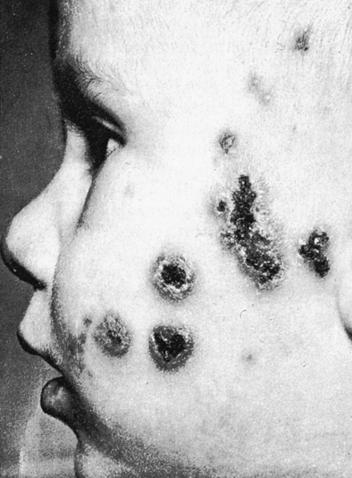
Impetigo and Cellulitis

Impetigo is a skin infection that begins as a papule (Fig. 17-2). The lesion may itch and will eventually crust over and heal. Cellulitis caused by subcutaneous infection with group A streptococci is associated with a warm, red, tender area that may be mildly swollen. Erysipelas, a distinct cellulitis syndrome, usually involves the face and may be associated with pharyngitis. This syndrome is characterized by toxicity and a high fever. If left untreated, erysipelas can be fatal.

Complications of Streptococcus pyogenes Infection

Not all infections with S. pyogenes lead to complications. Acute rheumatic fever, for example, occurs only after upper respiratory tract infection. In contrast, glomerulonephritis occurs after pharyngitis or skin infections (pyoderma). Acute rheumatic fever and poststreptococcal glomerulonephritis are considered nonsuppurative because the organs themselves are not directly infected and a purulent inflammatory response is not present in affected organs (e.g., heart, joints, blood, kidneys).

The pathogenesis of this disease process has not been fully described, but an autoimmune phenomenon may be operational. It is believed that cross-reactive antibodies, originally directed against streptococcal cell membranes, bind to myosin in human heart muscle cells. Other cross-reactive antibodies bind to components of the glomerular basement membrane and form immune complexes at the affected site. These antigen-antibody complexes attract reactive host cells and enzymes that ultimately cause the cellular damage.

All M serotypes that infect the throat appear to be capable of causing rheumatic fever. Researchers have identified a few serotypes, however, that cause a much lower proportion of rheumatic fever cases than would be expected from their frequency as a cause of pharyngitis. The incidence of rheumatic fever is directly proportional to the strength of the antibody response to SLO. The prognosis of rheumatic fever is good when carditis is absent during the initial infection.

Glomerulonephritis may follow an infection of the skin or respiratory tract with one of a limited number of nephritogenic M serotypes. These serotypes are defined by antisera against the M protein, which is also associated with virulence. Why these serotypes cause glomerulonephritis is unknown.

Immunologic Manifestations

S. pyogenes is an example of a pathogen that induces the production of several different antibodies. This coccus contains antigenic structural components and produces antigenic enzymes, each of which may elicit a specific antibody response from the infected host. In the course of an infection, the extracellular products act as antigens to which the body responds by producing specific antibodies (indications of infection).

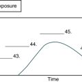
Most infected patients demonstrate increased concentration of antibody against SLO. The concentration of antibody (titer) begins to rise about 7 days after the onset of infection and reaches a maximum after 4 to 6 weeks. A rise in titer of 50 Todd units in 1 to 2 weeks is of greater diagnostic significance than a single titer.

An elevated titer indicates a relatively recent infection. Peak titers are seen at the time of acute polyarthritis of acute rheumatic fever, but these titers are no longer at their peak during the carditis of acute rheumatic fever. A patient may demonstrate an elevated antibody titer for up to 1 year after infection; therefore, the time of infection is not precisely determined by this technique. Low titers of antistreptolysin O (ASO) can be exhibited by apparently healthy persons because of the frequency of subclinical streptococcal infections, but persistently low titers rule out S. pyogenes infection.

Of the patients with S. pyogenes–related acute glomerulonephritis, 50% display a normal ASO titer but demonstrate an elevated titer to one of the other streptococcal substances (e.g., DNAse and NADase). Anti–DNase B (ADN-B) antibody appears to be the most reliable measure of recent S. pyogenes skin infection. Titers of ADN-B are elevated in more than two thirds of patients with recent streptococcal impetigo. Anti-NADase antibodies are a particularly good marker in patients who develop nephritis after pharyngitis.

Diagnostic Evaluation

In addition to throat cultures in patients with pharyngitis, antibodies to bacterial toxins and other extracellular products that display measurable activity can be tested. ASO and ADN-B are the standard serologic tests. The ability of a patient’s serum to neutralize the erythrocyte-lysing capability of SLO (ASO procedure) has been used for many years as a method for detecting previous streptococcal infection. After an infection such as pharyngitis with SLO-producing strains, most patients show a high titer of the antibody ASO. The use of rapid testing (see ASO latex procedure, Chapter 12, and Chapter 14) has replaced the use of the classic ASO procedure archived on the EVOLVE website (and on www.mlturgeon.com).

Streptococci produce the enzyme DNase B. The ADN-B neutralization test prevents the activity of this enzyme and demonstrates recent or previous S. pyogenes infection. Antistreptokinase and antihyaluronidase titers (AHTs) have also been used to diagnose streptococcal infection retrospectively.

Serologic testing should compare acute and convalescent sera collected 3 weeks apart. The ASO level becomes elevated in acute or convalescent paired specimens in 80% to 85% of patients with acute rheumatic fever. ADN-B and AHT levels are elevated in the remaining 15% to 20% of patients. In many cases, no acute serum specimen is available; therefore, the antibody titer of the convalescent serum specimen is compared with a reference range value. Reference ranges vary with age, season, and geographic area. False-positive ASO results may be demonstrated because of the presence of beta-lipoprotein, contamination of the serum specimen by bacterial growth products, or oxidation of ASO. These errors are not encountered with the ADN-B procedure, which is the serologic test of choice for acute rheumatic fever and acute glomerulonephritis after S. pyogenes infection.

Streptococcal Toxic Shock Syndrome

Streptococcal toxic shock syndrome (STSS) is caused by a highly invasive group A streptococcal infection and is associated with shock and organ failure.

Immunologic Mechanisms

Pyrogenic exotoxins cause fever in human beings and animals and also help induce shock by lowering the threshold to exogenous endotoxin. Streptococcal pyrogenic exotoxins A and B induce human mononuclear cells to synthesize not only tumor necrosis factor-α (TNF-α) but also interleukin-1 beta (IL-1β) and interleukin-6 (IL-6), suggesting that TNF could mediate the fever, shock, and tissue injury observed in patients with STSS.

M protein contributes to invasiveness through its ability to impede phagocytosis of streptococci by human PMNs.

Superantigens are capable of binding to alpha and beta T cell receptors (TCRs) and major histocompatibility complex (MHC) class II molecules. Superantigens can directly activate 1% to 2% of T cells and create high levels of cytokines in the blood. These high levels can produce shocklike symptoms.

Cytokine production by less exotic mechanisms also likely contributes to the genesis of shock and organ failure. Exotoxins such as SLO are also potent inducers of TNF-α and IL-1β. Pyrogenic exotoxin B, a proteinase precursor, has the ability to cleave pre–IL-1β to release preformed IL-1. Finally, SLO and pyrogenic exotoxin A together have additive effects in the induction of IL-1β by human mononuclear cells. Regardless of the mechanisms, induction of cytokines in vivo is likely the cause of shock and exotoxins, cell wall components, and other substances are potent inducers of TNF and IL-1.

Signs and Symptoms

The symptoms of STSS include shock; fever; blotchy rash; and a red, swollen, and painful area of infected skin. The average incubation period for STSS is 2 to 3 days, usually after minor nonpenetrating trauma.

Pain, the most common initial symptom of STSS, is abrupt in onset and severe and usually precedes tenderness or physical findings. The pain generally involves an extremity but may also mimic peritonitis, pelvic inflammatory disease, pneumonia, acute myocardial infarction, or pericarditis.

About 20% of STSS patients have an influenza-like syndrome characterized by fever, chills, myalgia, nausea, vomiting, and diarrhea. Fever is the most common early sign, although hypothermia may be present in patients with shock.

About 80% of STSS patients have clinical signs of soft tissue infection, such as localized swelling and erythema, which in 70% of one group of patients progressed to necrotizing fasciitis or myositis and required surgical débridement, fasciotomy, or amputation. An ominous sign is the progression of soft tissue swelling to the formation of vesicles and then bullae, which appear violaceous or bluish.

Group B Streptococcal Disease

Group B Streptococcus agalactiae infections causes substantial morbidity and mortality in adults and neonates.

Epidemiology

Fatality rate ranges from 26% to 70% among men and nonpregnant women with group B streptococcus (GBS) disease. Despite substantial progress in the prevention of perinatal GBS disease since the 1990s, GBS remains the leading cause of early-onset neonatal sepsis in the United States. Universal screening at 35 to 37 weeks’ gestation for maternal GBS colonization and the use of intrapartum antibiotic prophylaxis has resulted in substantial reductions in the burden of early-onset GBS disease in newborns. Although early-onset GBS disease has become relatively uncommon in recent years, the rates of maternal GBS colonization (and therefore the risk for early-onset GBS disease in the absence of intrapartum antibiotic prophylaxis) remain unchanged since the 1970s. GBS disease remains the leading infectious cause of morbidity and mortality in newborns in the United States.

Future Directions

Because of the gravity of GBS disease, especially in those who are older and those with chronic diseases, the development of a vaccine is being pursued. Determining the incidence of adult disease and groups at greatest risk helps focus prevention efforts. Intrapartum antibiotics can prevent early-onset neonatal GBS disease but have not been widely used.

CASE STUDY

This 19-year-old woman went to the emergency department (ED) with swelling and redness of her right leg. She had fallen down while rollerblading and had a number of abrasions on the skin of her leg. She also had a body temperature of 37.8° C (100° F). The ED physician ordered a culture of her leg wound, gave her a prescription for an antibiotic, and discharged her from treatment.

The following evening, the patient collapsed onto the floor of her bedroom. Her roommate found her and called 911. On arrival, the paramedics found an unconscious female with a blood pressure of 80/40 mm Hg and pronounced redness and swelling of her right leg. She was rushed to the ED and admitted to the intensive care unit, where she was immediately placed on IV fluids and medications to raise her blood pressure.

image Antistreptolysin O (ASO) Latex Test Kit

Principle

In this test (Biotech Laboratories, Suffolk, England), the ASO reagent contains latex particles coated with streptolysin O antigen. When the reagent is mixed with serum containing ASO, the particles will agglutinate, which is interpreted as a positive sample. Detection of ASO in serum may aid in the diagnosis of streptococcal infections.

Infections promoted by acute streptococcal infection result in the production of antistreptolysin O antibodies because of the presence of the SLO antigen liberated by the bacteria.