86
Stevens–Johnson
syndrome
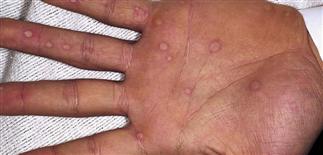
The palms, soles, dorsum of hands, and extensor surfaces are most commonly affected.
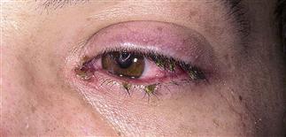
Patients with Stevens–Johnson syndrome can develop conjunctivitis. Meticulous eye care with erythromycin ointment can prevent ocular adhesions.
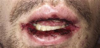
Mucosal involvement may include erythema, edema, sloughing, blistering, ulceration, and necrosis. Mouth erosions are painful and may impede adequate nutrition.
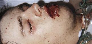
Severe bullous form. Mucosal involvement can be extensive, requiring tracheostomy.
DESCRIPTION
Severe blistering mucocutaneous syndrome, involving at least two mucous membranes.
HISTORY
• Occurs in all ages but more common in children and young adults. • Thought to be due to cytotoxic immune responses directed against keratinocytes expressing foreign infectious (Mycoplasma pneumoniae) and drug antigens (phenytoin, phenobarbital, carbamazepine, sulfonamides, aminopenicillins). • Medications started within 1 month of disease onset are more likely to cause Stevens–Johnson syndrome. • HIV infection, systemic lupus erythematosus, and malignancies treated with radiation increase the risk. • Can involve pulmonary, gastrointestinal, renal, and central nervous systems.
PHYSICAL FINDINGS
• Erythematous papules, dusky-appearing vesicles, purpura, and target lesions erupt acutely. • Lesions can be tender and burn. Oral, genital, and perianal mucosa develop bullae and erosions. • Thick hemorrhagic crusts can cover the lips. • Patients develop conjunctivitis and are at risk for corneal ulceration and uveitis. • Stevens–Johnson syndrome skin lesions are more centrally distributed on the face and trunk. Crops of lesions erupt for 10–14 days and slowly subside for the next 3–4 weeks.
TREATMENT
• Identify and treat sources of infection, withdraw suspected offending drugs, maintain fluid and nutritional requirements, provide meticulous local wound care. • Mucous membranes: frequent mouth rinses and applications of petroleum jelly (Vaseline) or Aquaphor. • Viscous lidocaine (Xylocaine) or Benadryl elixir for pain. Topical erythromycin ointment to the eyes prevents ocular adhesions. Treat eroded skin like a burn: cleanse gently, remove necrotic tissue, apply bland emollients. • Narcotics for severe pain. • Intravenous immunoglobulins in selected patients. • The use of systemic corticosteroids is controversial. Corticosteroids may be beneficial in certain cases. Sick children who have extensive cutaneous, ocular and oral lesions may respond to prednisone (20–30 mg b.i.d.) until new lesions no longer appear; it is then tapered rapidly.


