80. Stevens-Johnson Syndrome
Definition
Stevens-Johnson syndrome (SJS) is a hypersensitivity disorder mediated by immune-complex formation. It is a potentially fatal systemic disorder that is a severe type of erythema multiforme. SJS is also known as “erythema multiforme major.”
Incidence
The frequency of SJS is estimated to be about 1.2:1,000,000 to 6:1,000,000. This disorder has been reported in all racial and ethnic groups and affects both sexes about equally over all age groups.
Etiology
Stevens-Johnson syndrome is classified into four categories (see box below). Approximately half of all cases of SJS have been linked to ingestion of a medication. To date, more than 100 different medications have been linked to the development of SJS (see box on pp. 321-322). The second most common cause result from infectious agents/vectors (see box on p. 321). As many as 25% of SJS cases are deemed idiopathic, while the smallest number of SJS cases have been associated with some form of carcinoma and/or lymphoma.
Four Etiologic Categories of Stevens-Johnson Syndrome
1. Infectious
2. Drug-induced
3. Malignancy-related
4. Idiopathic
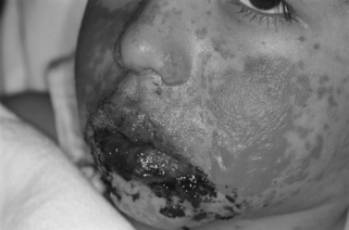 |
| Stevens-Johnson Syndrome. Severe oral and skin involvement. |
Infectious Causes of Stevens-Johnson Syndrome
• Adenoviruses
• Calmette-Guérin virus
• Deep fungal infections
• Enterobacter species
• Enteroviruses
• Herpes simplex virus
• Influenza
• Measles
• Mumps
• Mycobacterium pneumoniae
• Mycobacterium tuberculosis
• Streptococcus pneumoniae
• Syphilis
• Typhoid fever
Drugs Associated with the Development of Stevens-Johnson Syndrome
Sulfonamides
• Pyrimethamine-sulfadoxine (Fansidar)
• Sulfadiazine (Coptin)
• Sulfasalazine (Azulfidine)
• Trimethoprim-sulfamethoxazole
Anticonvulsants
• Carbamazepine
• Phenobarbital
• Phenytoin (Dilantin)
Others
• Acetaminophen
• Allopurinol (Aloprim, Zyloprim)
• Aminopenicillins
• Amithiozone
• Amoxapine (Asendin)
• Barbiturates
• Cephalosporins
• Chlormezanone (Trancopal)
• Clobazam
• Diclofenac
• Fluvoxamine (Luvox)
• Hydantoins
• Imidazole antifungals
• Indapamide (Lozol)
• Lamotrigine (Lamictal)
• Macrolides
• Mianserin
• Oxicam
• Nonsteroidal anti-inflammatory drugs (NSAIDs) (piroxicam, tenoxicam)
• Propionic NSAIDs
• Propranolol (Inderal, InnoPran XL)
• Pyrazolone derivatives (i.e., dipyrone)
• Quinolones
• Salicylates
• Sertraline (Zoloft)
• Tetracycline
• Tiapride
• Trazodone
• Valproic acid (Depakene, myproic acid)
Signs and Symptoms
• Altered level of consciousness
• Anxiety
• Arthralgia
• Coma
• Confluent erythema
• Conjunctivitis
• Corneal ulcerations
• Dehydration
• Dysuria
• Epistaxis
• Erosive vulvovaginitis
• Fever
• Headache
• Hyperventilation
• Hypotension
• Malaise
• Mild hypoxia
• Mucous membrane erythema, edema, sloughing, blisters, ulceration, and/or necrosis
• Mucous membrane lesions (e.g., oropharynx, conjunctivae, genitalia, anus, tracheobronchial tree, esophagus, bowel)
• Orthostasis
• Productive cough (thick and purulent)
• Profuse diarrhea
• Purpuric macules, papules, vesicles, or bullae
• Seizures
• Tachycardia
• Urticarial plaques
Medical Management
The most critical step is early detection of SJS followed by rapid, aggressive initiation of treatment. The patient begins treatment by discontinuing use of any medications that may be the potential causative agent. The disorder causes significant disruption of the patient’s skin barrier that closely resembles the denuded appearance of chemical or thermal burn injuries. This disorder is essentially a “burn” produced by internal mechanisms that disrupt skin and/or mucous membrane integrity—almost as if the patient is “burned” from the inside outward. The patient also frequently experiences profuse diarrhea along with significant increases in core temperature.
These symptoms—skin loss, diarrhea, and hyperthermia—may cause the patient to lose significant fluid volume. Fluid losses frequently bring about extensive shifts/losses of electrolyte concentrations. The fluid volume losses must be aggressively treated to avoid potentially lethal losses culminating in cardiovascular collapse. Fluid resuscitation requirements are generally 66% to 75% of the amount necessary to care for a burn-injured patient with a comparably sized injury. Fluid should be given via peripheral intravenous access that is as distant from the cutaneous or mucosal lesions and/or denuded areas as possible. Resuscitation via central venous access should be avoided if at all possible because of a greater potential for infection. Fluids should be warmed (if the patient is not significantly febrile) to prevent initiation of shivering, which can increase oxygen consumption by as much as 300%.
There is no single medication that can successfully treat SJS. The preponderance of treatments address symptoms. Denuded skin areas are generally treated with bactericidal creams. Occasionally, surgical intervention may be required to promote skin regeneration by application of xenographs and similar interventions.
If the tracheobronchial tree is involved, it will be manifested by hyperventilation plus mild hypoxemia. Aggressive pulmonary support is required to detect the onset of diffuse interstitial pneumonitis. With early detection of the pneumonitis, treatment can be initiated sooner to reduce the potential for, if not prevent, acute respiratory distress syndrome (ARDS).
Similar to the treatment of burn injuries, pain control is a significant problem. The skin disruption of SJS is strikingly similar to that of a deep partial-thickness thermal or chemical injury; the nerve endings are basically uncovered and are constantly in a state of hyperstimulation. Attaining adequate analgesia may require potent opioid analgesics. In addition, the patient with SJS is prone to develop stress ulcers. The patient should be treated with antacids, histamine-2 (H 2)-blockers, and/or proton pump inhibitors. The patient should be placed on deep vein thrombosis prophylaxis, which may include use of heparin.
SCORTEN Score Prognostic Factors and Mortality Rates
Variables
• Age > 40 years
• Bicarbonate < 20 mmol/L
• BUN > 10 mmol/L
• Heart rate > 120 bpm
• Initial percentage of epidermal detachment >10%
• Malignancy
• Serum glucose > 14 mmol/L
Scores and Mortality Rates
| Score | Mortality Rate |
|---|---|
| 0-1 | ≥3.2% |
| 2 | ≥12.1% |
| 3 | ≥35.3% |
| 4 | ≥58.3% |
| 5 | ≥90% |
Complications
• ARDS
• Blindness
• Burning eyes
• Conjunctival synechiae
• Corneal and conjunctival neovascularization
• Corneal scarring
• Death
• Epithelial proliferation with squamous metaplasia
• Esophageal strictures
• In-turned eyelashes
• Lacrimal duct obstruction causing epiphora
• Nephritis
• Patchwork skin
• Penile scarring
• Photophobia
• Pneumonia
• Renal tubular necrosis
• Sjögren-like sicca syndrome (see p. 316)
• Tracheobronchial shedding with respiratory failure
• Vaginal synechiae
• Visual impairment
Anesthesia Implications
The skin of the patient with SJS is extremely fragile; lesions may develop easily and just as easily rupture to produce a new skin barrier disruption. Anesthetists must take extraordinary care when positioning the patient and padding bony prominences and pressure points. The best padding for the patient with SJS is to use gelatin-filled pads that maximize the dispersal of pressure. In addition, the patient with SJS should have his or her skin protected from the shearing forces the blood pressure cuff can produce, which will exacerbate skin barrier disruption. Similarly, securing intravenous catheters, endotracheal tubes, and protecting the eyes with tape can be detrimental to the patient with SJS because removal of the tape can also remove the underlying skin. Intravenous lines may be best secured using a suture rather than tape, followed by covering the site with a sterile gauze dressing. The endotracheal tube can be secured using any of the commercially available devices. The patient’s eyes can be protected by placing lightly moistened gauze pads on the closed eyes then wrapping the head with gauze. The anesthetist should also take similar care when removing the electrocardiogram pads and should strive to minimize the shearing forces produced.
Intravenous access is a critical need for the patient with SJS for both anesthesia administration and fluid resuscitation. Central venous access is discouraged. However, if larger fluid volumes are required, it is obviously an avenue to be cautiously explored after weighing the risks versus benefits. The patient will have nutritional needs similar to those of a patient with burn injuries and may need to receive parenteral nutrition on arrival at the operating room. It is important that the parenteral nutrition be maintained throughout the duration of the anesthetic. The patient also may be receiving enteral nutritional support critical to the healing process. Although it presents the risks of a full stomach, the anesthetist should allow the supplementation to continue, possibly at a slower rate, until the patient is delivered to the operating room to be prepared for anesthesia. Induction of general anesthesia must, of necessity, follow the classic rapid sequence induction precautions.
The patient with SJS frequently has significant involvement of the oral cavity and oropharynx, and may present with major laryngeal edema. These tissues will be almost astoundingly friable. Hemorrhagic bullae form with seemingly the most simple contact; direct laryngoscopy, endotracheal intubation, oral and nasal airway insertion, placement of an esophageal stethoscope, or passing an orogastric or nasogastric tube may produce and rupture these bullae and cause immediate severe airway compromise. The use of a laryngeal mask airway is not strictly contraindicated in the patient with SJS, but its use must be very carefully considered, especially in the patient with extensive oral cavity involvement, despite the reduced use of positive pressure ventilation it can afford.
The involvement of mucous membranes in the patient with SJS can extend further down the tracheobronchial tree, resulting in development of visceral pleural blebs. These blebs are likely to rupture, producing pneumothorax. For this reason it is desirable to avoid positive pressure ventilation where possible. In cases where positive pressure ventilation is unavoidable, it is advisable to prolong the inspiratory phase with reduced peak inspiratory pressure by selecting a pressure-controlled method of ventilation available on most late-model anesthesia machines. At the same time, the lowest effect peak inspiratory pressure should be selected as well. These measures will help minimize the potential for bleb rupture. In addition, because of its rapid diffusion and expansion, nitrous oxide should not be used, especially if tracheobronchial tree involvement and/or pleural bleb formation is suspected.
Because of the patient’s high degree of pain, potent opioid analgesics may be in use with adequate or inadequate pain relief. The anesthetist should anticipate that the patient has developed tolerance to the opioid analgesics and should be prepared to administer large doses of opioids to achieve seemingly minimal results. No specific anesthesia drugs or anesthesia techniques have demonstrated clear superiority for the patient with SJS. Infiltration of local anesthetic must be undertaken cautiously, with due consideration for the condition of the patient’s skin as well as the increased potential for infection. Drugs that have been implicated in the development of SJS, such as barbiturates, should be avoided. Propofol has been used safely to anesthetize patients with SJS, as has ketamine. Both can be used as the sole anesthetic agent if necessary. The anesthetist must also consider the route of elimination for anesthesia-related drugs when planning the anesthesia care for the patient with SJS. In particular, the anesthetist should avoid potentially nephrotoxic drugs and those eliminated via the kidneys when renal involvement has been demonstrated.