128
Squamous cell carcinoma
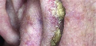
Squamous cell carcinoma is commonly found on the ears. The thick smooth mass had been present for over 1 year. Dense scale has formed on the surface.
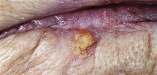
A hard mass was present for 2 years. Erosion and bleeding prompted a visit to the physician.
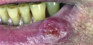
This lower lip tumor appears to be a superficial actinic keratosis. Palpation revealed a large firm mass in the dermis. Biopsy showed squamous cell carcinoma.
Retention of dense scale on a hard mass suggests the diagnosis of squamous cell carcinoma.
DESCRIPTION
Invasive, primary cutaneous malignancy arising from keratinocytes of skin and mucosal surfaces. Most commonly on the sun-damaged area of the head, neck, hands of elderly. Can occur at any location. Lesions may develop from precursor actinic keratoses or de novo. Second most common form of skin cancer; 20% of all primary cutaneous malignancies. Lifetime risk estimated to be 5–15%; > 200 000 new cases of primary squamous cell carcinoma (SCC) in USA each year, 2500 deaths annually.
HISTORY
• Occur on sun-exposed skin from years of accumulated actinic damage. • In men, 90% of cutaneous SCCs—nearly 80% in women—occur on head, neck, hands. SCC on legs more often in women. • White people with fair skin at greatest risk. • Ultraviolet radiation (sunlight) is the primary cause of most SCCs. Other factors include arsenic, tobacco, chemicals, chronic inflammation, chronic infections, chronic immunosuppression, burn scars, human papillomavirus infection. • Incidence doubles with each 8–10° decline in latitude. • Ultimately, tumors metastasize, via the lymphatics, to other organs.
PHYSICAL FINDINGS
• Typically occur on sun-exposed areas. Found within a background of sun-damaged skin with atrophy, telangiectasia, blotchy hyperpigmentation. • Early invasive SCC may have appearance of hypertrophic actinic keratosis. Red, poorly defined base and adherent, yellow-white cutaneous horn. • Untreated lesion becomes larger, more raised, developing into a firm red nodule with necrotic crusted center. • May arise de novo, appearing as sharply defined, smooth, dull-red, firm, dome-shaped nodule with crusted center. Removal of crust reveals central cavity filled with necrotic keratin debris. • Keratoacanthoma now considered a variant of invasive SCC. Most consider keratoacanthoma a low-grade form of SCC. • Diagnosis based on histology. • Metastases usually to regional lymph nodes. Detected within 2–3 years. Palpable regional lymph nodes suggest metastatic disease.
TREATMENT
• Excellent long-term prognosis for adequately treated SCC. • Increased risk of developing additional primary skin malignancies. • Metastatic rate of SCC arising on sun-exposed skin: 2–6%. • Patients on immunosuppressive therapy after organ transplantation at higher risk for all cutaneous malignancies, especially SCC, 5–10 years after transplantation. • Treatment of primary SCC involves wide local excision with histologic confirmation of margins. Mohs micrographic surgery may be useful for specific sites, such as the central face, where tissue sparing important. • Palpation of regional lymph nodes mandatory for all patients. • Lymph node biopsy indicated for suspected nodal disease. • May consider radiation therapy when surgical resection not feasible. • Careful follow-up at regular intervals recommended for all patients.


