CHAPTER 9 SPLENIC PATHOLOGY
SPENUNCULUS / ACCESSORY SPLEENS (Fig 9.1)
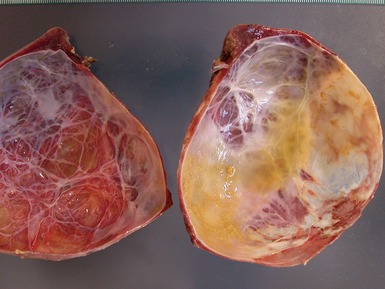
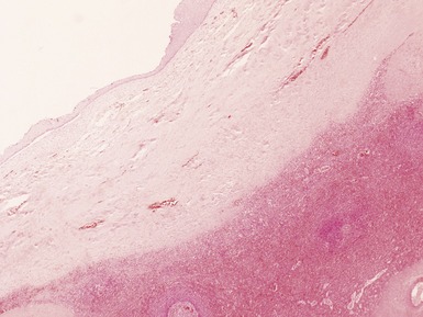
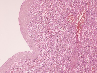
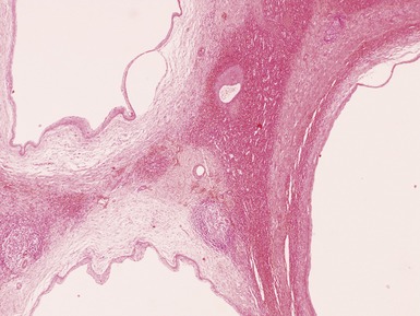
SPLENIC ‘CYSTS’
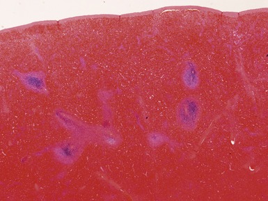
SPLENECTOMY FOR HYPERSPLENISM
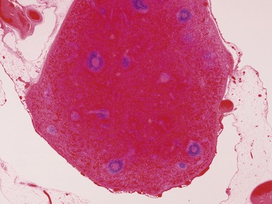
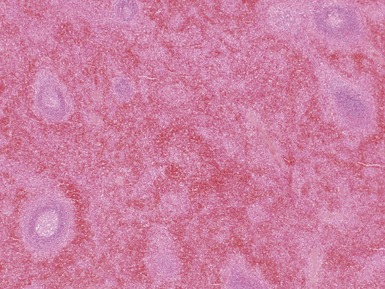
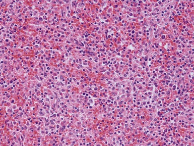
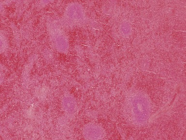
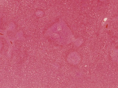
IMMUNE THROMBOCYTOPENIC PURPURA (ITP) (Figs 9.6, 9.7)
OTHER MISCELLANEOUS SPLENIC DISEASE
INVOLVEMENT BY MALIGNANCY
Chang CS, Li CY, Cha SS. Chronic idiopathic thrombocytopenic purpura. Splenic pathologic features and their clinical correlation. Arch Pathol Lab Med. 1993;117:981-985.
Chang CS, Li CY, Liang YH, Cha SS. Clinical features and splenic pathologic changes in patients with autoimmune hemolytic anemia and congenital hemolytic anemia. Mayo Clin Proc. 1993;68:757-762.
Czauderna P, Vajda P, Schaarschmidt K, et al. Nonparasitic splenic cysts in children: a multicentric study. Eur J Pediatr Surg. 2006;16:415-419.
Hayes MM, Jacobs P, Wood L, Dent DM. Splenic pathology in immune thrombocytopenia. J Clin Pathol. 1985;38:985-988.
Klatt EC, Meyer PR. Pathology of the spleen in the acquired immunodeficiency syndrome. Arch Pathol Lab Med. 1987;111:1050-1053.
Vermi W, Blanzuoli L, Kraus MD, et al. The spleen in the Wiskott–Aldrich syndrome: histopathologic abnormalities of the white pulp correlate with the clinical phenotype of the disease. Am J Surg Pathol. 1999;23:182-191.


