Low-attenuation complex fluid collection ± air-fluid levels


 May extend to subcapsular location and may rarely cause splenic rupture with generalized peritonitis

• Echinococcal (hydatid) cyst on CECT


PATHOLOGY
• Multiple different causes for splenic pyogenic abscesses


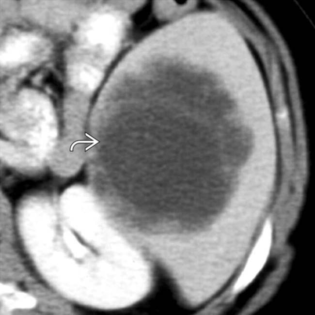
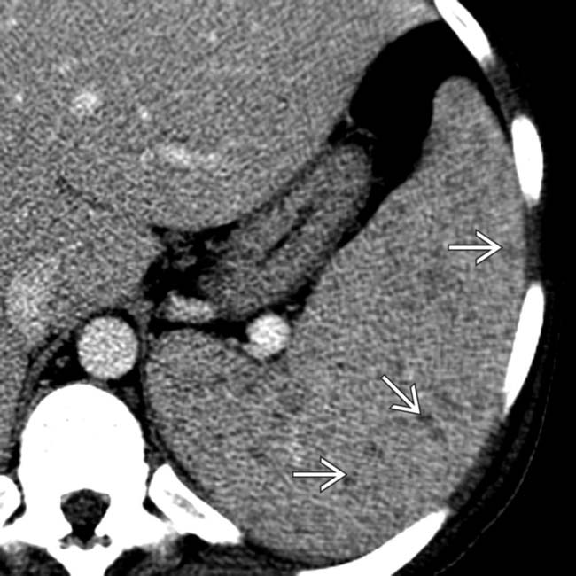
 within the spleen that proved to be a pyogenic abscess. Such large abscesses are unusual in the spleen, especially in the absence of prior splenic infarction.
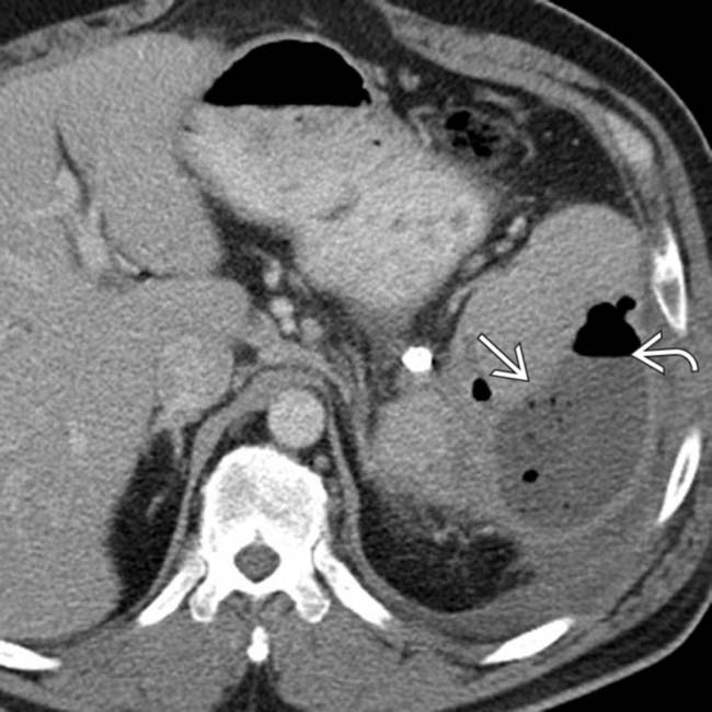
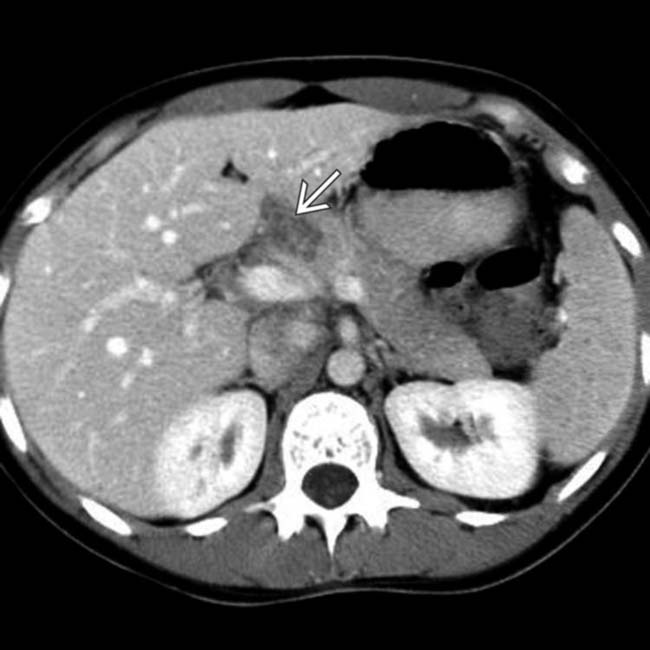
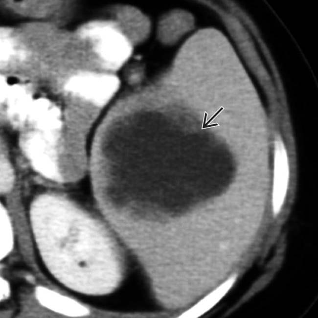
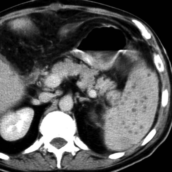
 demonstrates a rim-enhancing fluid collection with an air-fluid level
  , consistent with a splenic abscess. The patient was treated with percutaneous drainage and antibiotics.
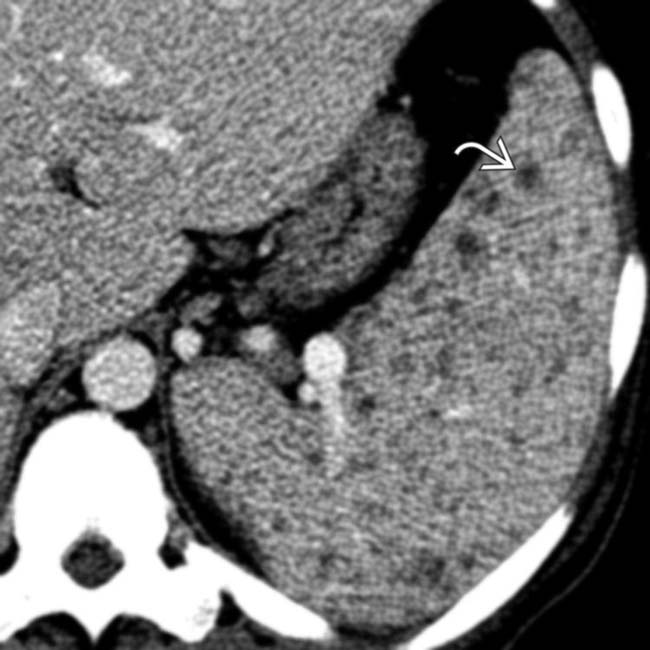
 throughout the spleen. The patient deteriorated rapidly, and multiple tuberculous abscesses were identified at autopsy.
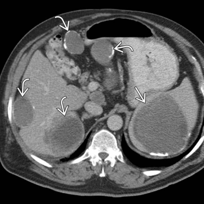
 with additional similar lesions in the liver and peritoneal cavity
  , many of which have calcified walls. This patient had a prior rupture of a hepatic hydatid cyst with diffuse spread throughout the abdomen.
IMAGING
General Features


CT Findings
• Pyogenic abscess


• Echinococcal (hydatid) cyst
 Complex cyst with multiple low density “daughter” cysts and thick, enhancing wall composed of fibrous tissue (“cyst within a cyst” appearance)
 Serpiginous, linear hypodense bands within cyst due to collapsed parasitic membranes (water lily sign)
Complex cyst with multiple low density “daughter” cysts and thick, enhancing wall composed of fibrous tissue (“cyst within a cyst” appearance) Serpiginous, linear hypodense bands within cyst due to collapsed parasitic membranes (water lily sign)


DIFFERENTIAL DIAGNOSIS
Splenic Infarct


PATHOLOGY
General Features


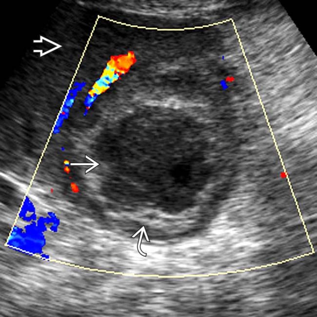
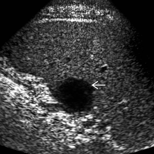
 and a thick irregular wall
  . The adjacent splenic parenchyma
  appears normal. Note the typical avascular nature of the abscess.
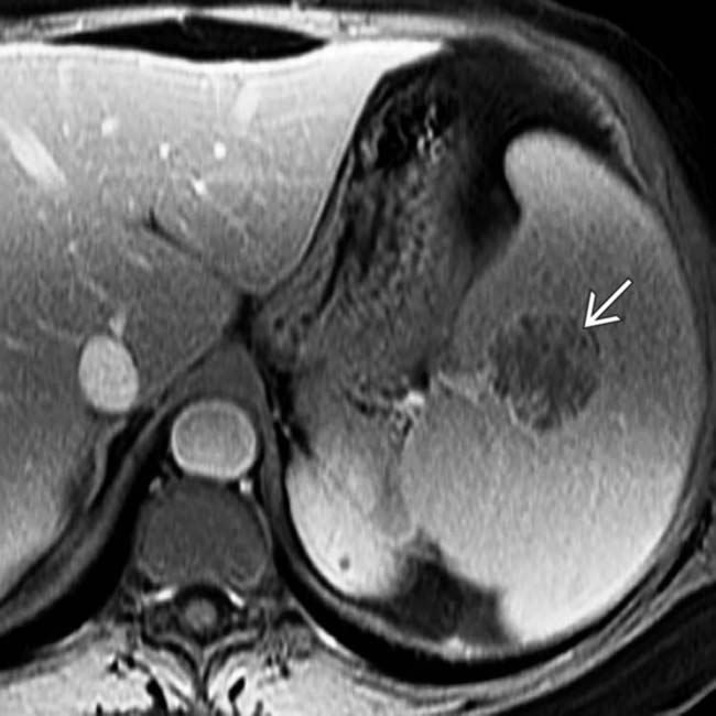
 . Blood cultures identified Staphylococcus and the patient recovered with antibiotics.
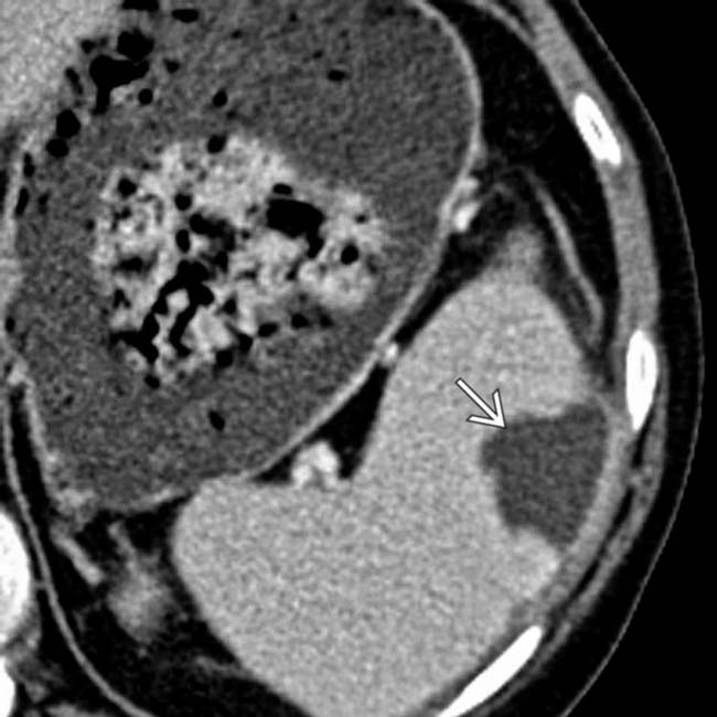
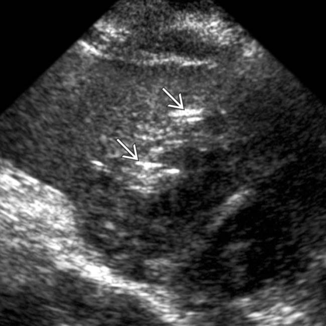
 in the periphery of the spleen’s midportion; findings that are consistent with an abscess.
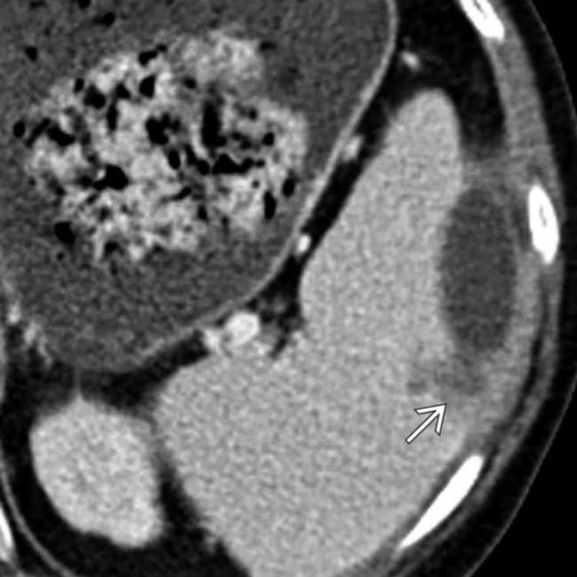
 , suggesting a possible abscess rupture. An echocardiogram showed multiple vegetations in both the aortic and mitral valves, suggesting that this abscess is due to underlying endocarditis.
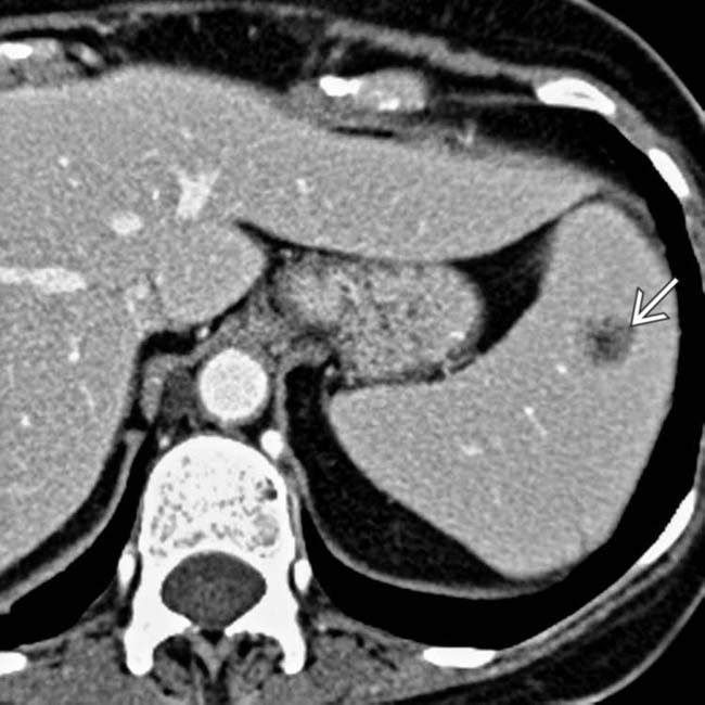
 .
.
 as well. The patient underwent an echocardiogram, which additionally revealed aortic valve vegetations from endocarditis (not shown).
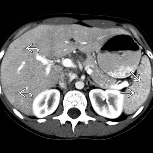
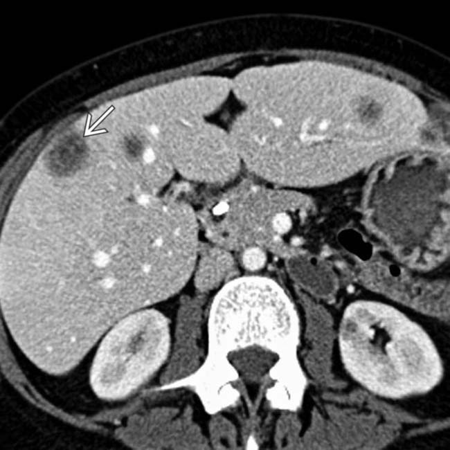
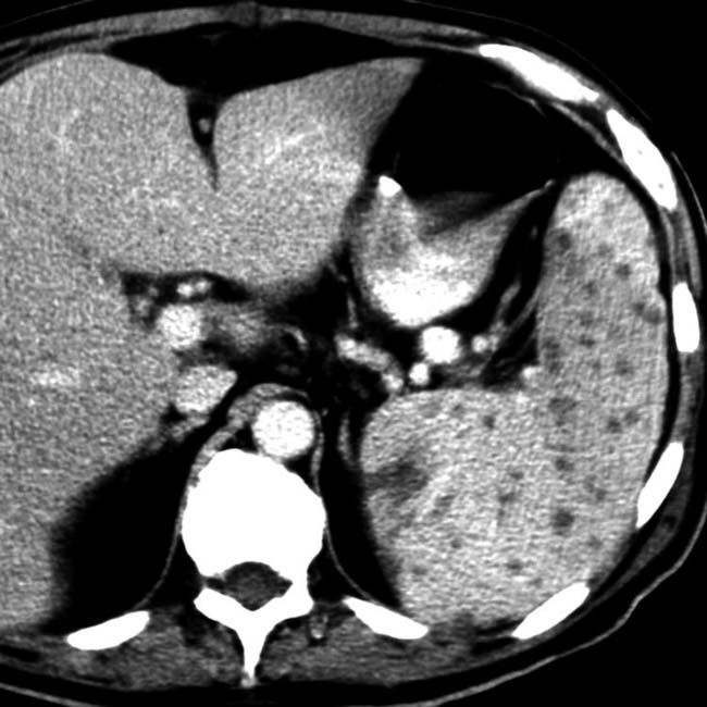
 and fewer, similar lesions in the liver
  .
.
 . Liver biopsy and blood cultures confirmed a systemic infection with histoplasmosis.
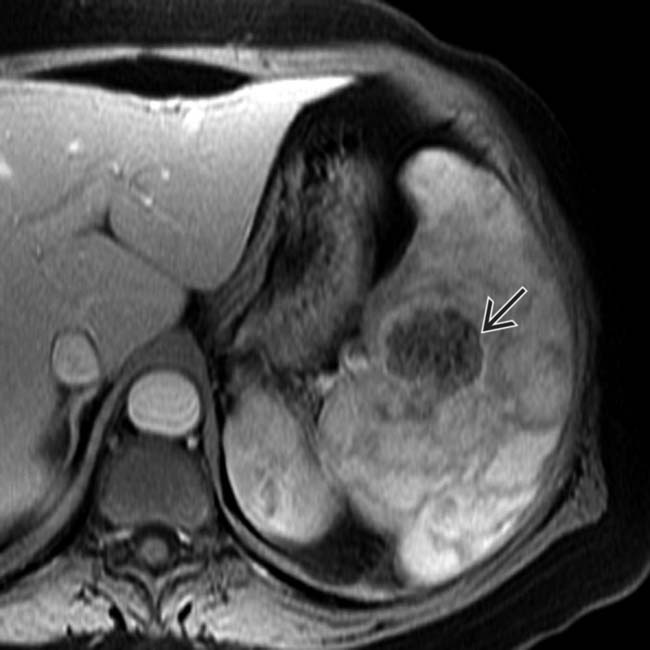
 .
.
 .
.
 .
.
 .
.
 .
.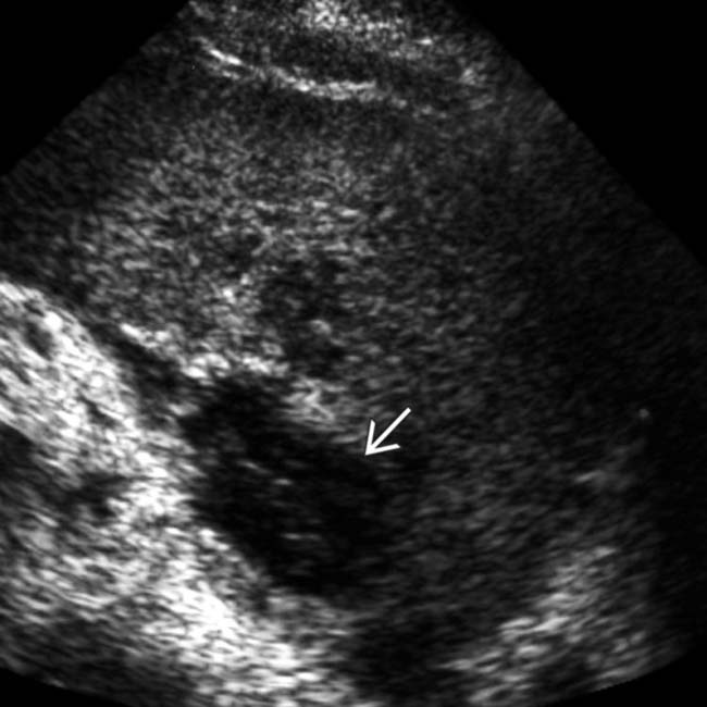
 with little distal acoustic enhancement.
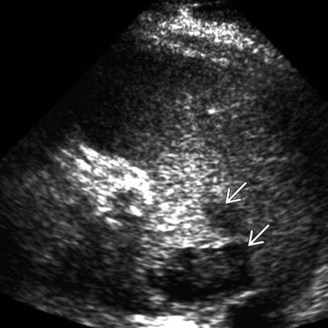
 .
.
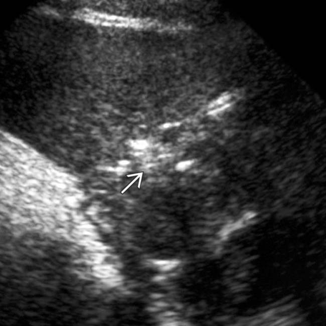
 in the midportion of the spleen, findings consistent with an abscess.
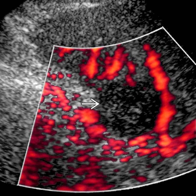
 , which excludes a solid lesion such as lymphoma or metastasis. Blood cultures were positive for anaerobic Streptococcus, and the lesion resolved with antibiotic therapy.

