148
Spider angioma (nevus araneus)
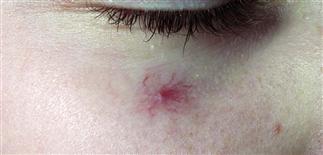
The lower eyelid is a common location for these tortuous angiomas. The face, hands, and fingers are common locations in children.
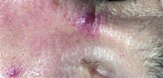
Spider angiomas are dilatations of a pre-existing blood vessel; notice the tortuous small blood vessels leading into the dorsal aspect of the nose.
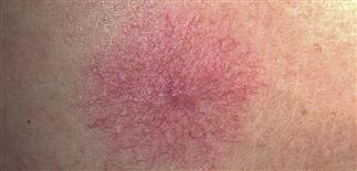
Spider angiomas blanch with pressure. Multiple spider angiomas can be seen on sun-exposed surfaces, with liver disease, and with pregnancy.
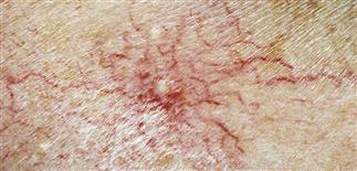
Spider angioma with long vessels leading to the center. The vessels were destroyed for cosmetic purposes with careful, conservative electrocautery.
DESCRIPTION
Asymptomatic blanchable pink papule due to a central dilated arteriole and very fine radial branches.
HISTORY
• Found in 10–15% of normal adults and children. • Seen with increased frequency in pregnancy and chronic liver disease (states of relative estrogen excess). Lesions arising during pregnancy tend to resolve after the birth. Those found in patients with liver disease are persistent. • Spider angiomas are due to dilatation of a previously existing vessel rather than a neoplasm. • Spider angiomas arising during pregnancy and those occurring in children tend to disappear spontaneously over a period of 3–4 years.
PHYSICAL FINDINGS
• A central, slightly raised, bright-red vascular papule from which fine blood vessels radiate. • Firm pressure easily blanches the radiating vessels, whereas the central papule is less easily blanched. Pulsation of the central papule with this technique confirms the arteriolar nature of the papule. • Most commonly seen on face, neck, upper trunk, upper arms, hands, fingers.
TREATMENT
• Most resolve spontaneously and do not require treatment. • For persistent, cosmetically bothersome lesions, pulsed dye laser or electrocautery are effective. • There is a slight risk of dyspigmentation and scarring, and lesions may recur.


