Chapter 12 Specific treatment techniques
Introduction
Physiotherapists working in neurological rehabilitation employ a large variety of techniques. When examining treatment approaches from different philosophical backgrounds, it is apparent that similar techniques may be being utilized (see Ch. 11). A technique can be defined as a ‘method or skill used for a particular task’ (Collins English Dictionary). With this definition in mind it is important to consider to what purpose different techniques are employed. It is also important that the technique is appropriate in order to help meet the treatment goals.
This chapter illustrates the diversity of the techniques used by physiotherapists. It is clear that there is a wealth of research supporting the use of some techniques and a lack of a clear evidence base to justify the use of others. Many techniques continue to rely on anecdotal evidence to support their use. In this chapter a variety of techniques are reviewed and the evidence available to support their use is considered. Physiotherapists are challenged to provide best practice based on evidence. However, such evidence may be unavailable or incomplete (Jones et al., 2006) and this is reflected in the field of specific treatment techniques used in neurological physiotherapy.
Facilitation
Many of the techniques used in neurological rehabilitation are applied to facilitate and enhance muscle activity, and thus help achieve improved control of movement. It is also proposed that these interventions are chosen to facilitate neuroplasticity (Umphred et al., 2007a). Many of the specific techniques used for facilitation have their origins in the work of Margaret Rood. For a comprehensive examination of the Rood approach and the most recent interpretation of its relevance, the reader is directed to Baily Metcalfe and Lawes (1998) and Schultz-Krohn et al. (2006). Some of those most commonly used techniques are outlined below.
Brushing
In the 1950s Rood proposed that fast brushing, using a battery-operated brush, of the skin overlying a muscle could be used to facilitate a muscle contraction. Brushing has been used widely by physiotherapists, applied either using an electrically operated brush or manually using a bottle brush, but there is little indication given about the required rate or duration of the brushing, or pressure to be applied. It would make sense that the skin being brushed and the muscle being facilitated should be supplied by the same spinal segment. Although Garland and Hayes (1987) observed an effect of brusing in hemiplegic subjects with foot drop, there is little evidence to support its effectiveness. In subjects who received a combination of brushing preceded by voluntary contraction of the tibialis anterior, a significant change in electromyographic (EMG) activity was seen both immediately and 30 minutes after stimulation.
Brushing may be a powerful method of facilitation but it is clearly not well researched in terms of its continued effects, particularly as much of the work has been carried out on subjects with no neurological impairments. It is worth noting that caution in its use has been advised (Farber, 1982) and that Schultz-Krohn et al. (2006) consider it beyond the scope of entry-level practice.
Ice – brief
In order to facilitate a motor response, an ice cube is quickly swept over the chosen muscle belly (Umphred et al., 2007a). Following each swipe the iced area is blotted with a towel. After three swipes the patient is asked to produce an active muscle contraction. If ice is being used to facilitate lip closure and encourage feeding and sucking, an ice lolly can be placed in the mouth with pressure on the tongue (Farber, 1982).
When using ice as a stimulating technique it is important to remember that it can be a potent stimulus and results can be unpredictable. Putting ice on the face above the level of the lips and to the midline of the trunk should be avoided, as it has been reported that undesirable behavioural and autonomic responses may be provoked (Umphred et al., 2007a; Schultz-Krohn et al., 2006).
Retraining of sensory function has received little attention in the physiotherapy literature. However, the use of repeated ice water immersions of the affected hand in chronic stroke patients was investigated by Bohls and McIntyre (2005) in a small scale study. Although wrist position sense was not improved by the intervention it was suggested that a positive effect may be found in relation to the sensation of light touch and temperature discrimination.
Tapping
Tapping is the use of a light force applied manually over a tendon or muscle belly to facilitate a voluntary contraction. Tapping over a tendon would usually be used to assess reflex activity. A normal response would be a brisk muscle contraction. It is not therefore recommended that tendon tapping be used in a treatment situation, as the response is a crude muscle contraction and will be of little use to help a patient produce a graded, functional movement (Umphred et al., 2007a).
Rood recommended three to five taps over the belly of the muscle being facilitated. In addition, tapping can be applied to a muscle that has been stretched by the effect of gravity. Once the muscle responds to the stretch produced by gravity the therapist taps the muscle, using the hand, facilitating further activity (O’Sullivan, 2007). For example, with a patient who is standing, weight-bearing through both legs, if one knee gives way gravity will stretch the quadriceps muscle group. The therapist can then tap the muscle, facilitating a return to full knee extension.
Sweep tapping is a light touch sweeping movement applied by the back of the therapist’s fingers over the dermatomal area innervating the muscles the patient is required to contract (Umphred et al., 2007a). Davies (2000) described the use of sweep tapping to provide an excitatory stimulus to activate the finger extensors in hemiplegia. This is applied by providing support to the affected upper limb with one hand, while the other hand sweeps firmly and briskly over the extensors of the wrist and fingers; the sweep commences just below the elbow and continues over the dorsum of the hand and fingers. In common with other tapping techniques, an active response is requested from the patient following its application.
Joint compression
Receptors in joints and muscles are involved with the awareness of joint position and movement. Compression of a joint stimulates these receptors and can produce both inhibitory and facilitatory effects. Joint compression (approximation) is achieved either by normal body weight (or less) being applied through the longitudinal axis of the bone (light compression) or as heavy joint compression, where the approximation is greater than that produced by body weight (Schultz-Krohn et al., 2006). Heavy joint compression is thought to facilitate cocontraction at the joint undergoing compression, whereas light joint compression is reported to produce an inhibitory (relaxing) effect on spastic muscles around joints (Schultz-Krohn et al., 2006).
Bone pounding or jamming is used to inhibit plantarflexion and facilitate cocontraction around the ankle. It can be applied with the patient sitting, by pounding the heel on the floor whilst supporting the knee. Alternatively, with the patient lying prone over a pillow with some degree of flexion at the hips and knees, force can be applied to the heel by the therapist using the ulnar side of a clenched fist (Umphred et al., 2007a).
Other techniques that use joint compression include weight-bearing through a hemiplegic arm to facilitate cocontraction and activation of the muscles around the shoulder joint (Davies, 2000). Weight belts and weighted wrist or ankle cuffs have also been used to increase joint compression. Joint compression can be applied to many joints using a variety of positions or patterns of movement. For example, using four-point kneeling as a starting position, joint compression can be applied to the shoulders and/or hips. Ideally, joint compression should be applied in a functional position, but if this is not possible treatment should quickly progress to using the joint in a functional manner. Joint compression is also a procedure used in PNF and is considered in this context below.
Several authors described joint compression, either in terms of normal weight approximation or by other means, but only anecdotal evidence is given to support its use (Davies, 2000; Farber, 1982; Schultz-Krohn et al., 2006; Umphred et al. 2007a).
Vibration
Muscle vibration
Therapeutic vibration is a directly applied stimulus of high frequency (100–300 Hz) and low amplitude, which stretches the muscle spindle and activates type 1a afferent fibres. Vibration is generally applied directly to the chosen muscle or its tendon. Bishop (1974) identified three motor effects achievable by vibrating a muscle: (1) a sustained contraction of the vibrated muscle (via the tonic vibration reflex); (2) the depression of the motoneurones innervating the antagonistic muscles (reciprocal inhibition or antagonist inhibition); and (3) suppression of the monosynaptic stretch reflexes of the vibrated muscle (during the period of vibration). There appears to be disagreement, however, as to whether vibration has a sustained effect on muscle contractility (Umphred et al., 2007a) and thus any long-term benefit.
It would appear that vibration has potential clinical applications via agonist facilitation or antagonist inhibition. Bishop (1974) identified four factors that influenced the strength of the tonic vibration reflex (TVR):
Application of the vibrator to the belly of a stretched muscle or over the tendon allows easy facilitation of the TVR. It appears that the tonic neck reflexes and body-righting reflexes (Rothwell, 1994; Shepherd, 1994) interact with the TVR; so, treatment in the supine position results in an improved extensor TVR, and that in the prone position results in increased flexor TVR. Finally, increasing the amplitude of the vibration increases the stretch on the muscle but, more significantly, the TVR is greater as the frequency of the vibratory stimulus increases.
Another quite different investigation involving vibration was made by Lovgreen et al. (1993), who studied the effects of muscle vibration on the voluntary movements of patients with cerebellar dysmetria. Part of the study was to consider whether vibration could improve movement accuracy and reduce hypermetria. They found that antagonist vibration reduced the amplitude of patients’ movements and suggested that vibration had potential for use in both hyper- and hypometria, although the feasibility of its application would require careful thought.
Vibration has the potential to be a potent treatment technique but there are various precautions that must be considered when using it. Key points to remember include: vibration will generate heat at its point of application and there is potential to cause damage to the skin, particularly at high amplitudes (Farber, 1982). Vibration to augment a muscle contraction should not be applied with cutanteous pressure, which is known to cause inhibition as the two oposing effects could negate each other (Umphred et al., 2007a).
Umphred et al. (2007a) recommend that vibrators registering 100–125 Hz be used and noted that most battery-operated hand-held vibrators register only 50–90 Hz. There is a wide range of commercially available vibrators, so the available frequency range should always be checked prior to purchase.
The use of more generalized vibration has also been considered. Its current use as part of exercise training formed the basis of a study by Jackson et al. (2008) examining its effect on lower limb performance in patients with multiple sclerosis, although results were inconclusive.
Whole body vibration
Whole body vibration is a relatively new modality in neurological rehabilitation, which involves the patient standing on a vibrating platform (Wunderer et al., 2008). The effect of vibration compared to conventional physiotherapy to improve balance and gait in Parkinson’s disease was explored by Ebersbach et al. (2008). However, improvements were demonstrated in both groups, so its superior efficacy in comparison to a standard approach to rehabilitation was not established. Another study exploring its use in Parkinson’s disease (Arias et al., 2009) concluded that any effect from whole body vibration in Parkinson’s disease was solely due to a placebo effect. A systematic review by Wunderer et al. (2008) concluded that whole body vibration appeared to produce similar gains to traditional exercise and resistance training but also limited fatigue, so its use could therefore be considered beneficial in neurological patients.
Vestibular stimulation
Any static position or movement will have an effect on the vestibular system, so many interventions will result in vestibular stimulation in some way or other. However, specific vestibular stimulation has not been widely used in neurological physiotherapy and was, until recently, mainly described in relation to a multisensory approach to neurological rehabilitation in paediatrics. Advocates of its use are anxious to remind others that vestibular stimulation is a powerful form of stimulation that should be used with care. Umphred et al. (2007a, p. 231) stated that it is important to remember that ‘the rate of vestibular stimulation determines the effects. A constant, slow, repetitive rocking pattern, irrespective of plan or direction, generally causes inhibition of total-body responses, whereas a fast spin or fast linear movement tends to heighten both alertness and the motor responses.’
The management of vestibular dysfunction has evolved from increasing research to become recognized as a specialist area within physiotherapy. Chapter 13 explains that patients with a primary problem of vestibular dysfunction require vestibular rehabilitation, which involves specific assessment and treatment techniques.
Facilitation of movement
Facilitated movements do not require the patient to activate the nervous system to produce the required movement. This lack of self-initiation of movement has been criticized for not providing a basis for the learning of movement. However, it could be argued that once movement can be initiated in patients the possibility of the production of an active response then exists with the potential for learning of functional movements (Baily Metcalfe & Lawes, 1998). However, it must be remembered that eventually the patient must become independent of the physiotherapist in order to produce the movements required for functional indpendence.
Normalization of tone and the maintenance of soft-tissue length
The Bobath approach is widely used as a treatment approach in neurological physiotherapy (Lennon, 2003; Sackley & Lincoln, 1996) and the control and normalization of tone clearly contribute to the theoretical assumptions of the Bobath concept of stroke rehabilitation (Raine 2007; also see Ch. 11). An awareness of the potential for changes in the musculoskeletal system and the subsequent loss of range of movement associated with neurological dysfunction (Ch. 14) is essential for effective management of patients with neurological disorders.
Passive stretching – slow
Slow stretch is applied to a muscle or joint such that a stretch reflex is not elicited and the effect is, therefore, inhibitory in terms of the neural response. The effect of prolonged, slow stretching on muscle is not entirely clear, although it certainly varies depending upon the time for which the stretch is maintained. It appears to have an influence on both the neural components of muscle, via the Golgi tendon organs and muscle spindles (O’Sullivan, 2007), and the structural components in the long term, via the number and length of sarcomeres (Hale et al., 1995).
Changes in muscle length
The presence of increased tone, possibly combined with paresis and/or weakness, can ultimately lead to joint contracture and changes in muscle length (see Ch. 14). Slow, prolonged stretching is therefore applied to maintain or prevent loss of range of movement (ROM). It has been demonstrated in animal studies that if a muscle is immobilized in a shortened position, sarcomeres will be lost and, conversely, a muscle immobilized in a lengthened position will add on sarcomeres (Goldspink & Williams, 1990). A shortened immobilized muscle will also show an increase in stiffness related to an increase in connective tissue within the muscle (Williams et al., 1988). However, it has been demonstrated in mice that a stretch of 30 minutes daily will prevent the loss of sarcomeres and changes in the connective tissue of an immobilized muscle (Williams, 1990). The timescale relating to changes in the mouse may not be relevant to humans.
Manual stretching
A prolonged muscle stretch can be applied manually, using the effect of gravity and body weight, or mechanically (by machine or splint). When applied, the stretch should provide sufficient force to overcome the hypertonicity and passively lengthen the muscle. When contractures are already present, it is doubtful whether the use of manual stretching alone will be sufficient to provide a sustained improvement in the ROM, if any was achieved. A systematic review exploring the effects of stretching in spasticity by Bovend’Eerdt et al. (2008) found some positive evidence to support the immediate effects of one stretching session, but the long-term effects were unclear. Overall the heterogeneic nature of the studies made a meta-analysis unfeasible and they concluded that the available evidence related to the clinical benefits of stretching and spasticity was inconclusive.
Splinting
Low-force stretching of long duration can be provided by splinting. The clinical practice guidelines on splinting adults with neurological dysfunction (Association of Chartered Physiotherapists Interested in Neurology (ACPIN), 1998) identified a paucity of research in the area, making it almost impossible to adopt an evidence-based approach to the use of splints. Over a decade later, little has changed.
Dynamic Lycra splints have been used as part of the management of patients with hemiplegia (Gracies et al., 2000). Lycra splints are custom-made, individually designed garments – it is claimed that Lycra splinting is effective in managing posture, and motor and sensory changes following a stroke. The study by Gracies et al. (2000) investigated acceptability and effects on swelling, resting posture, spasticity, active ROM and passive ROM of an upper-limb Lycra garment when worn for 3 hours by patients with a hemiplegia. The findings from this small-scale study, using a convenience sample, indicate some support for the use of these garments in reducing spasticity and swelling. Overall the use of lycra splits or orthoses in adult patients with neurological dysfunction is not widespread. In contrast there is increasing use of lycra splinting in children with cerebral palsy, although a review by Attard and Rithalia (2004) identified a lack of scientific research to support their use.
Different types of splinting and the rationale for use are discussed in Chapter 14, with further details being provided by Edwards and Charlton (2002). Examples of splints used for peripheral nerve injuries are illustrated in photographs in Chapter 9.
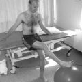
Weight-bearing
Several studies report the use of weight-bearing to reduce contractures in joints of the lower limb (Bohannon, 1993; Richardson, 1991). These reports illustrate the effectiveness of using a tilt table to achieve a sustainable position in which a prolonged stretch is applied. The angle of table tilt needs to be considered when standing patients with knee joint contractures, as the supporting straps bear more of the body weight than when the knees are extended (Morgan et al., 2003). Force exerted at the supporting straps is greater the higher the degree of flexion, and is more pronounced with greater body weight, but can be modified by reducing table incline, thus reducing the pressure on underlying tissues. However, a study by Ben et al. (2005) challenged some of the assumptions about the benefits of standing where small changes to ankle mobility and little or none to mineral density were found during a single blinded randomized trial examining the effects of a 12-week standing programme in patients with recent spinal cord injuries. Illustrated examples of equipment to assist standing can be seen in Chapters 4 and 14.
Serial casting
Serial plaster casting is another technique used to prevent or reduce contractures (O’Sullivan 2007), which may be most effective when the contractures result from spasticity. Serial casting methods were described and illustrated by Edwards and Charlton (2002) and a comprehensive overview of the practicalities of casting the lower limb in neurology was provided by Young and Nicklin (2000).
The use of a soft splint has been shown to be effective in the acute management of elbow hypertonicity (Wallen & Mackay, 1995). This splint has certain advantages over casting in that it is more dynamic in nature, less likely to cause unwanted pressure and provides neutral warmth (Wallen & O’Flaherty, 1991). However, it is also easily removed and thus a level of compliance is necessary!
Moseley (1997) also demonstrated the effectiveness of serial casting and stretching on regaining ROM in the ankle due to established shortening of the calf muscles. Jones (1999) undertook a series of single-system studies to examine the efficacy of lower-extremity serial casts on gait in four adults with hemiparetic gait patterns. There was an improvement in walking speed and a reduction in the level of assistance required during walking following the intervention. Singer et al. (2004) reported a descriptive study of the non-surgical management of ankle contracture in patients with acquired brain injury. Serial casting was used when the contracture appeared to be worsening, despite standard physiotherapy. In some cases of serial casting this also included an injection of botulinum toxin type A.
When spasticity is present, physiotherapists are often reluctant to use splints or other externally applied devices for stretching as, despite the lack of supporting evidence, it is thought that splinting can lead to an increase in muscle tone. However, it has been demonstrated that inhibitory splinting can reduce contractures without causing detrimental effects to muscle tone (Mills, 1986). Indeed, the ACPIN guidelines (1998) recommend that patients suitable for splinting are those who may have, or be at risk of, contractures as a result of significant increases in muscle tone or immobility.
Duration of stretch to reduce spasticity
Although it has been shown that prolonged stretching can reduce spasticity, the time needed is not clear. Hale et al. (1995) found that the most beneficial duration of stretch applied to reduce spasticity was 10 minutes. This study used a variety of methods to assess the level of spasticity, including both subjective and objective measures. The results illustrated the difficulties that arise when measuring spasticity (see Ch. 14), and that perhaps the concurrent problems of length-associated changes in muscle required greater consideration.
Duration of stretch to prevent contracture
Tardieu et al. (1988) investigated how long it was necessary to stretch the soleus muscle each day to prevent contracture in children with cerebral palsy and concluded that it must be stretched for 6 hours a day.
Some work has been done to evaluate the effect of stretching, mainly on normal subjects (Harvey et al., 2002). However, it is clear that further work is still required to establish the appropriate stretching techniques and the duration required to produce the desired effect in different situations.
Positioning
Positioning is used widely by physiotherapists to prevent the development of contractures (Fraser, 2009) and to discourage unwanted reflex activity (Carr & Kenney, 1992; Pope, 2002). A survey of practice of positioning for stroke patients identified that one of the most common aims of physiotherapists advocating its use was to modulate muscle tone and prevent damage to affected limbs (Chatterton et al., 2001). A study by Ada et al. (2005a) of stretching in patients with a recent stroke, in which the affected shoulders were positioned in 90° of flexion with the maximum external rotation tolerable for two 30 minute sessions a day, 5 days a week, for 4 weeks, found significantly reduced incidence of contracture when compared with a control group. This contrasted with Dean et al. (2000) who failed to demonstrate a significant effect of prolonged positioning of the shoulder, applied daily for 6 weeks, in patients undergoing a multidisciplinary rehabilitation programme. Turton et al. (2005), although supporting the principle that early treatment to prevent the loss of range of movement in the affected limb post stroke is essential, also raised the issue of patient compliance, which must be considered in any intervention requiring patients to adopt a sustained position. However, a recent meta-analysis of positioning to prevent loss of range of movement in the shoulder post stroke did not support its use (Borisova & Bohannon, 2009).
Specific positions are often adopted to achieve a slow maintained stretch on a particular muscle and the thinking behind this has already been explored. Bromley (2006) gave detailed guidelines for the positioning of patients following spinal cord injury and described its importance for: correct alignment of fractures, prevention of contractures, prevention of pressure sores and inhibiting the onset of severe spasticity.
Indeed, historically many of the positions advocated by physiotherapists relate to the desire to avoid the development of spastic patterns of movement (Bobath, 1990). Positions are chosen to minimize the influence of the primitive reflexes. The three reflexes, which are normally under cortical control and whose release can be influenced by careful choice and use of positions, are: (1) the symmetrical tonic neck reflex; (2) the asymmetrical tonic neck reflex; and (3) the labyrinthine reflex (Carr & Kenney, 1992).
Davies (2000) gave fairly detailed descriptions of desirable positions that should be used following stroke, urging the avoidance of supine lying, as in this position the influences of the tonic neck and labyrinthine reflexes are great and this could result in an overall increase in extensor activity throughout the body. Fraser (2009) advocates the use of good seating and positioning to facilitate appropriate alignment and stability of the trunk and limbs, thus avoiding compensatory responses to prevent falling.
Careful positioning to limit musculoskeletal changes is essential, but it appears that there is a lack of consensus about the precise positions necessary to limit the onset of spasticity and unwanted patterns of movement, particularly after stroke (Carr & Kenney, 1992; Chatterton et al., 2001). Certainly, Bobath (1990) identified a need to be more dynamic and advocated the use of reflex-inhibiting patterns of movement, rather than static postures, to inhibit abnormal postural reactions and facilitate automatic and voluntary movements.
These concepts are discussed in Chapter 11. Positioning is also discussed in Chapters 4 and 14, where illustrations show various types of equipment used for posture and seating.
Pressure
Pressure is used by physiotherapists both to facilitate and inhibit a response in muscle, more especially in muscle tone (O’Sullivan, 2007). This pressure can be applied in a variety of ways, including the use of air-filled splints (Johnstone, 1995), tone-inhibiting casts (Zachazewski et al., 1982) or manually (Umphred et al., 2007a). Pressure can be applied directly over a tendon (Leone & Kukulka, 1988) or over the muscle itself (Robichaud et al., 1992). The pressure can be sustained or intermittent, and variable in terms of the degree applied.
Most of the research investigating the effects of a variety of pressure conditions has measured motoneurone excitability, via change in the Hoffman reflex (H reflex). Studies have suggested that the characteristic appearance of the H reflex reflects spinal motor function and, therefore, it can be used to evaluate the effects of therapeutic interventions that aim to reduce motoneurone excitability (Suzuki et al., 1995). It is important to remember, however, the problems of quantifying that part of muscle tone that occurs as a direct result of reflex activity.
Leone and Kukulka (1988) investigated the effects of Achilles tendon pressure on the H reflex in stroke patients. The assumption was made that any change in motoneurone excitability would be reflected in an associated alteration in tone as, again, no direct measurement of tone was made. Pressure was applied both continuously and intermittently, and under both conditions depression of the H reflex occurred. Intermittent pressure, however, was significantly more effective than continuous. Further investigation revealed that increasing the amount of pressure had no greater effect, and the effect of the pressure was sustained only during its actual application. No carryover effect was observed, but it is suggested that tendon pressure could be used therapeutically, e.g. when a short-term reduction in tone would allow achievement of an improved patient position in bed.
The strongest proponent of the use of pressure during treatment was Johnstone (1995), who advocated the use of constant pressure provided by orally inflated splints and intermittent pressure produced by a machine. The uses of the splint are to: reduce the therapist’s need for extra hands; provide stability to the limb; divert associated reactions; allow early weight-bearing through the affected limb; and increase sensory input (Johnstone, 1995). It was claimed that when the antigravity muscles of the upper limb are held in a position of sustained stretch using the air splints, tonic and phasic wrist flexor EMG activity is reduced (Johnstone, 1995).
Robichaud et al. (1992) supported the use of air-splint pressure to reduce motoneurone excitability of the soleus muscle when circumferential pressure was applied around the lower leg. As in the tendon pressure study, the reduction was not sustained once the pressure had been released. Conversely, an increase in motoneurone excitability following the application of muscle pressure has been reported (Kukulka et al., 1987). This may reflect the different methods employed to apply pressure, which can include tapping and massage (Umphred et al., 2007a).
It is clear that the application of pressure has many potential effects, some of which are still not understood. Externally applied pressure over muscle or tendon must also cause a disturbance in the cutaneous mechanoreceptors. Because of the wealth of afferent activity caused by pressure, its application poses many questions yet to be answered. Pressure is postulated to be one of the mechanisms supporting the therapeutic use of lycra body suits (Attard & Rithalia, 2004). The efficacy of using pressure as a technique has largely been supported by observations of therapists, but outcome studies are now required (Umphred et al., 2007a).
Neutral warmth
When considering exteroceptive input techniques, Umphred et al. (2007a) identified an additional use for air splints – that of the provision of neutral warmth. Johnstone (1995) also advocated their use to provide sensory stimulation of soft tissues, causing inhibition of the area under which the neutral warmth is applied. Alternative techniques used for achieving neutral warmth are tepid baths, whole-body wrapping and wrapping of isolated body parts (O’Sullivan, 2007). The required range of temperatures that should be utilized for this technique is 35–37°C (Farber, 1982).
There appears to be little research to support the use of this concept of neutral warmth. Baily Metcalf and Lawes (1998) suggested that the inhibition seen is due to inhibition of tonic muscles via the stimulation of low-threshold mechanoreceptors through light touch. One study looked specifically at the effect of a wrapping technique on a passive ROM in a spastic upper extremity (Twist, 1985). Wrapping (elastic wrap bandages and gloves) was applied to spastic upper limbs for 3 hours, three times a week on alternate days over a period of 2–4 weeks. Results showed statistically significant increases in passive ROM, with subjective reports of reduced pain. Although this study contained several shortcomings (small subject numbers and lack of control), it did indicate an effect.
Ice – prolonged
Prolonged use of ice reduces afferent and efferent neurotransmission. To be effective in reducing spasticity, the muscle spindles must themselves be cooled. The ice must be applied until there is no longer an excessive reflex response to stretching (Lehmann & De Lateur, 1990). It is considered that a reduction of spasticity lasting 1–2 hours can be achieved, such that stretching or active exercises can be applied to greater effect.
The most common form of application of ice to reduce spasticity is local immersion; this is particularly effective for reducing flexor spasticity in the hand. A mixture of tap water and flaked ice is used, in the ratio of one-third water to two-thirds ice. Davies (2000) advocated that the hand is immersed three times for 3 seconds, with only a few seconds between immersions. The therapist should hold the patient’s hand in the ice–water mixture. This procedure can result in a dramatic reduction in spasticity.
General immersion, where the patient sits in a bath of cold water, has been used to reduce spasticity. Patients can tolerate water temperatures of 20–22°C for 10–15 minutes (Lee et al., 1978). Neither local nor general cooling has been found to have any long-term effect on spasticity, so any short-term reduction achieved must be fully exploited.
When using ice it is important to remember that the patient must be receptive to its use. If ice causes the patient distress and anxiety, the inhibitory effect may be blocked (Farber, 1982). A sensory assessment of the patient should be carried out before using ice and the presence of sensory deficits is a contraindication to its use (Umphred et al., 2007a).
Vibration
Vibration can also be used to produce inhibitory effects. In an effort to support the efficacy of its use to treat patients with disorders of muscle tone, Ageranioti and Hayes (1990) investigated the effects of vibration on hypertonia and hyperreflexia in the wrist joints of patients with spastic hemiplegia. They found that immediately after vibration, hypertonia and hyperreflexia were significantly reduced and concluded that in patients with spastic hemiplegia vibration gave short-term symptomatic relief. However, they also acknowledged that, despite using a relatively homogeneous group of subjects, there were many different patterns of hyperreflexia and this could possibly explain previous anecdotal reports where vibration was of no benefit in apparently similar cases.
Vibration has also been used at low frequencies (60–90 Hz) to normalize or reduce sensitivity in the skin (Farber, 1982; Umphred et al., 2007a). Hochreiter et al. (1983) found that in the ‘normal’ hand, vibration increased the tactile threshold, with the effect lasting for at least 10 minutes. There appears to be a lack of clinically applied studies in this area.
Certain precautions need to be considered when applying vibration (Farber, 1982), and these are outlined above in the section on facilitation.
Massage
Massage was a core element of physiotherapy in the UK and has been described as one of the ‘roots of our profession’ (Murphy, 1993). How widely massage is used or should be used is the subject of much debate that will not be explored here. For an extensive overview of massage, its application and effects, the reader is directed to Holey and Cook (2003) or Hollis and Jones (2009).
Massage has two main physical effects – mechanical and physiological. The inhibitory effects of massage are of particular interest to the physiotherapist working in neurology when the aim is to achieve a reduction in muscle tone or muscle spasm. Slow stroking applied to patients with multiple sclerosis has been found to achieve a significant reduction in the amplitude of the H reflex (a measure of motoneurone excitability). The stroking was of light pressure and applied over the posterior primary rami (Brouwer & Sousa de Andrade, 1995).
Studies on neurologically healthy subjects have found similar results. Goldberg et al. (1992) found that deep massage produced a greater inhibitory response than light massage when applied to the leg. Sullivan et al. (1991) indicated, by their results, a specificity of the effect of massage on the muscle group being massaged. This was contrary to their expectations that the inhibitory effects of massage would extend beyond the muscle being massaged.
Exercise and movement
This section includes well-established treatments and those that are emerging in neurological rehabilitation (also see Ch. 18). A major area that is not included is gait re-education, which is a vast field and the reader is referred to the chapters in this book on the different neurological conditions, as well as to Kisner and Colby (2007), Whittle (2007) and Kerrigan and Sheffler (1995).
Hydrotherapy
It must be remembered, when hydrotherapy is incorporated into a rehabilitation programme, that the effects of gravity are altered when in water. Many of the problems associated with neurological dysfunction arise from an individual’s inability to respond normally to the effect of gravity and, therefore, hydrotherapy is unlikely to be the sole method of treatment. However, water is an environment that permits a freedom of movement seldom achieved elsewhere. Water is also quite unique in being able to take over some of the physiotherapist’s work (Gray, 1997), particularly in terms of supporting the patient.
Muscle stretching, reducing contractures, re-education of motor patterns, re-education of balance and equilibrium reactions, gait retraining and breathing exercises are all areas covered by Gray (1997) and Bennie (1997), along with details of examples of suitable procedures used in hydrotherapy for neurological rehabilitation. Bad Ragaz techniques, where the buoyancy of the water is used to provide support rather than resistance to the patient, are covered by Davis and Harrison (1988). This approach has been advocated to achieve improvement in stability and motor control in neurological rehabilitation (Cameron, 2009).
As with any technique, careful assessment of the patient before and after treatment will allow the physiotherapist to monitor the effect of hydrotherapy. There are anecdotal reports of increased tone following exercise in hot water, but there is little evidence to substantiate this claim. The anxiety experienced by a patient being treated in water should be minimized by the reassurance provided by careful teaching skills (Reid Campion, 1997).
Swimming can form an integral part of hydrotherapy. The Halliwick method of swimming for the disabled (Lambeck et al., 2004; Reid Campion, 1997) is suitable for nearly any degree of disability at any age. A demonstrable improvement in postural balance and knee flexor strength was found by Noh et al. (2008) in stroke survivors undertaking an Ai Chi and Halliwick based exercise programme in water. Water-based exercise for improving cardiovascular fitness in people with chronic stroke was investigated by Chu et al. (2004) who demonstrated significant improvements in fitness and mobility. Both of these small-scale randomized trials of relatively short duration identify effects that warrant further investigation. Despite the widespread use of hydrotherapy with children with neurological impairments, a lack of evidence-based research was identified by Getz et al. (2006).
For further details of the principles, applications and techniques of hydrotherapy, the reader is directed to Schrepfer (2007), Cole and Becker (2004), Hecox and Leinanger (2006) and Reid Campion (1997).
Gymnastic balls
A comprehensive description of the use of the gymnastic ball can be found in Carriere (1998, 1999) and Davies (1990), and an example of use is illustrated in Chapter 4 (Figure 4.15). A wide range of exercises using the gymnastic ball can be found in Kisner and Colby (2007).
Proprioceptive neuromuscular facilitation
Proprioceptive neuromuscular facilitation (PNF) was developed as a therapeutic approach over 40 years ago. It is a very labour-intensive method of treatment, in which the physiotherapist facilitates the achievement of specific movement patterns by the patient with particular use of the therapist’s hands. Some of the basic procedures and techniques that are utilized will be considered here in relation to their use in neurological rehabilitation. For a complete overview of PNF the reader is referred to Voss et al. (1985), Kisner and Colby (2007) and Adler et al. (2008); combined, these texts give an extensive theoretical and practical review of the thoughts of some of the proponents of PNF.
Ten basic procedures for facilitation have been identified by Adler et al. (2008):
These two procedures of resistance and the resulting irradiation and reinforcement are possibly two of the reasons why PNF is no longer used extensively for neurological rehabilitation in the UK. The use of resistance does not fit comfortably with the other neurophysiological approaches, such as the Bobath approach. This, combined with the diagonal and spiral patterns of movement in the three anatomical planes, makes its relevance to normal movement difficult to comprehend. It is interesting to note, however, that Adler et al. (2008) felt that the patterns are not essential for the application of PNF and it is possible to use only the philosophy and appropriate procedures.
Adler et al. (2008) grouped the techniques so that those with similar functions or actions were together. They gave detailed descriptions and examples of the techniques and indications for their use.
Although the core PNF texts previously cited gave examples of its use with neurological dysfunction, PNF is certainly not in common use in neurology gymnasia in the UK. One study investigated its effect on the gait of patients with hemiplegia of long and short duration and found its cumulative effects were more beneficial than the immediate effects (Wang, 1994). However, as no control groups were used, the possible inferences from this study are limited. An earlier study by Dickstein et al. (1986) compared three exercise therapy approaches including PNF and found that no substantial advantages could be attributed to any of the three therapeutic approaches used.
It has been identified that some of the underlying assumptions of the procedures and techniques used in PNF are now out of date (Morris & Sharpe, 1993), but there still appears to be a vast potential for research involving its use, especially in modified forms. An attempt has been made to explore the rationale behind the PNF relaxation techniques by studying postcontraction depression of the H reflex (Moore & Kukulka, 1991). The techniques did produce a strong but brief neuromuscular inhibition; however the results of this study, performed on subjects with no neurological dysfunction, cannot be directly applied to patients.
More recent publications are available via the International PNF Association (http://www.ipnfa.org).
Cardiovascular exercise, strength training and exercise on prescription
The use of exercise to increase muscle strength in neurological rehabilitation is controversial and many physiotherapists have believed that muscle strength is not appropriate for treatment or measurement. However, there is increasing evidence that use of exercise and strength training, including the use of treadmills and static bikes, is beneficial in the management of patients with neurological dysfunction. Refshauge et al. (2005) provide evidence of the importance of exercise and training for individuals with chronic disability. Chapter 18 reviews the emerging literature, which reveals the benefits of exercise without the adverse effects traditionally feared by physiotherapists, such as increasing muscle tone in patients with spasticity.
There are currently no UK guidelines for exercise prescriptions for people with chronic neurological conditions (Glynn & Fiddler, 2009) so any recommendations are usually based on those for cardiovascular training. The guidelines produced by the American College of Sports Medicine are widely recognized as a resource for those planning to undertake training with special populations (ACSM, 2009).
Treadmill training
Treadmill training has been used across a range of different neurological patient groups to improve locomotion (see Ch. 18). Treadmill training is based upon the principle of task-specific repetitive training in that to learn to walk or improve walking practice is essential. Partial bodyweight-supported treadmill training can be used with non-ambulatory patients following a stroke to enable them to practice the complex requirements of the gait cycle (Hesse, 2008). Treadmill training has also been investigated in chronic stroke where it was found to be more effective in improving walking speed than resisted leg cycling alone (Sullivan et al., 2007). A recent review by Damiano and Dejong (2009) of the effectiveness of treadmill training in paediatric rehabilitation concluded that its efficacy had been demonstrated in children with Down’s syndrome, but although positive effects had been noted with other patient groups, including cerebral palsy, further research was required to support its use. This conclusion is supported in another review specific to children with cerebral palsy (Mutlu et al., 2009).
It should be acknowledged that the benefits of treadmill may extend beyond the improvement of walking ability. For some patient groups, for example spinal cord injured patients, it has been argued that the physiological and psychological benefits may justify its use even where it has not been shown to be superior to conventional physiotherapy in improving locomotor ability (Hicks & Martin Ginis, 2008). The potential contribution of treadmill training in relation to cardiorespiratory fitness should also be considered (Kilbreath & Davis, 2005).
Pilates-based rehabilitation
Pilates exercise has evolved from its use with elite dancers into different areas of rehabilitation (Anderson & Spector, 2000). Pilates-based rehabilitation was introduced from the USA in the 1990s and interest amongst physiotherapists is growing. For a summary of the principles behind its use and possible clinical applications the reader is directed to Anderson and Butler (2007). Despite the proliferation of Pilates courses for physiotherapists there is almost no research into its efficacy as part of a rehabilitation programme for patients with neurolocial dysfunction. A recent article by King and Horak (2009) included Pilates as part of a sensorimotor programme for patients with Parkinson’s disease. Although a rationale for such an approach was presented it has not yet been tested.
As acknowledged in the above reviews, research is needed to provide evidence of its effectiveness. However, it has been claimed that one of the defining characteristics of Pilates-inspired exercises is to enhance core, shoulder girdle and limb control (Lange et al., 2000) and this philosophy in many cases has a good fit with the aims of physiotherapy. Pilates classes in sports gyms are also very popular but people with neurological conditions would be advised to seek specialist supervision from a physiotherapist trained in Pilates, at least initially.
Tai chi
Tai chi is increasingly being incorporated into the range of interventions recommended or used by physiotherapists. It is used across the world as a form of exercise for health and fitness (Wang et al., 2004) despite little evidence of its benefits. A systematic review by Wang et al. (2004) explored the physical and psychological effects of tai chi on various chronic conditions, concluding that due to the lack of rigour in the majority of the studies considered it was difficult to make recommendations to support its use despite the apparent benefits. However, it appeared safe and was useful in relation to balance control, flexibility and cardiovascular fitness. A more recent review by Harling and Simpson (2008) concluded that there was strong evidence to support the use of tai chi in reducing the fear of falling in older adults, although the evidence to support its effectiveness in actually reducing the incidence of falling was weak. Given the anxiety in relation to falling experienced by many people with neurological conditions, it would seem reasonable therefore to consider its use within such populations. There is clearly a need for additional research and this view was supported by Lee et al. (2008) who reviewed its use in relation to Parkinson’s disease and found insufficent evidence to support its use.
Constraint-induced (forced-use) therapy
The principle of forced use to overcome the effects of ‘learned non-use’ is the basis for the increasing use of constraint therapy (Morris & Taub, 2001). It is used particularly in relation to rehabilitation of the upper limb in adults following a stroke and children with cerebral palsy and is discussed in Chapter 18. A systematic review (Hakkennes & Keating, 2005) of constraint-induced (forced-use) therapy (CIMT) following stroke concluded that it may improve upper limb function for some patients when compared to alternative or no treatment. This is reflected in the National Clinical Guidelines for Stroke (Intercollegiate Working Party for Stroke, 2008), which highlight the great commitment required from the patient when using CIMT and the need to use specific selection criteria and full patient agreement. There is considerably more research exploring its use in adults with stroke than children with cerebral palsy. However, a review by Hoare et al. (2007) also found positive trends supporting its use with children.
For a historical perspective and overview of the use of CIMT the reader is directed to Wolf (2007) who explores its theoretical underpinnings, the rationale behind its use and the associated strengths, uncertainties and limitations.
Robotics
The use of robotics within neurorehabilitation provides an alternative method of enhancing both upper and lower limb function in patients with movement disorders (Umphred et al., 2007a). It is another form of an augmented intervention along with bodyweight-supported treadmill training and CIMT, and has the most potential to exploit the increasing use of technology in rehabilitation (Umphred et al., 2007a). As the most recent of these interventions, it has the most limited evidence base for its use. The majority of support exists where the focus is on rehabilitation of the upper limb and it has been suggested that it could complement conventional therapy (Masiero et al., 2007). A review by Kwakkel et al. (2008) highlighted the challenges of making sense of the existing research due to the heterogeneity of the studies available. However, it appeared that a significant improvement in upper limb motor function after stroke could be identified with the use of upper limb robotics.
Cueing
Cueing can be thought of as a way of prompting a response. This prompt can be given in many different ways, including a verbal command, a noise, touch or visual stimulus. Its use has been explored extensively in Parkinson’s disease, where it is used to circumvent the dopamine deficits associated with the disease (Rubinstein et al., 2002; also see Ch. 6). External cueing has been defined as, ‘applying temporal (rhythmical) or spatial stimuli associated with the initiation and ongoing facilitation of motor activity.’ (Lim et al., 2005). However, the evidence base is limited and in a systematic review by Lim et al. (2005) only one high-quality randomized controlled trial was identified exploring the effects of auditory rhythmical cueing. This study suggested that the walking speed of patients with Parkinson’s disease could be positively influenced by such cueing, but it is unclear whether the effects would be translated into the real world. Insufficient evidence was found to support the use of visual or somatosensory cueing. In another study exploring bilateral arm training in chronic stroke, rhythmic auditory cueing appeared to induce significant changes identified by functional magnetic resonance imaging, although no significant functional outcomes were found (Luft et al., 2004).
More recently the RESCUE project has published a CD-ROM which provides guidelines for therapists using cueing to improve mobility in Parkinson’s disease (http://www.rescueproject.org/).
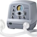
Electrical stimulation techniques
Transcutaneous electrical nerve stimulation
Pain relief
The management of pain in neurological rehabilitation would possibly not be identified as a key area for the physiotherapist working in neurology. However, the physiotherpist may have a significant role in the management of a patient’s pain. Chapter 16 reviews the specific management of pain in neurological rehabilitation.
TENS has been used specifically in the management of hemiplegic shoulder pain (Leandri et al., 1990). However a systematic review by Price and Pandyan (2000) concluded that it was not possible to confirm or refute the use of electrical stimulation for shoulder pain post stroke.
Details about TENS and its application in pain relief can be found in Kitchen (2002) and Robertson et al. (2006), which also provide a comprehensive overview of electrotherapy and its principles and practice.
Management of spasticity using transcutaneous electrical nerve stimulation
An alternative use of TENS has been in the treatment of spasticity. Studies investigating its effects on spasticity have had mixed results. Goulet et al. (1994) postulated that TENS would have an inhibitory effect on the amplitude of the soleus H reflex. They failed to demonstrate any consistent effects and no significant treatment effects were found following stimulation (at 50 or 99 Hz) on a mixed or sensory nerve. These results could reflect the difficulties of obtaining consistent H-reflex amplitudes in normal subjects and in those with neurological dysfunction.
Seib et al. (1994) used the spasticity measurement scale, in which neurophysiological and biomechanical responses are evaluated, to investigate the effect of cutaneous ES (over the tibialis anterior muscle) on spasticity of the gastrocnemius–soleus–achilles tendon unit. Using two groups of subjects, one with traumatic brain injuries and the other with spinal cord injuries; a significant reduction in spasticity was found which lasted for 6 hours or more following the stimulation. Based on these results the authors proposed that TENS could be of use for decreasing spasticity prior to other physiotherapeutic interventions such as stretching. Sonde et al. (2000) evaluated whether high-frequency TENS on a specific acupuncture point would influence the level of spasticity in the paretic leg after stroke. They found a reduction in spasticity, which was sustained in some patients for 2 weeks after treatment. Information about acupuncture points used with TENS is available in Fox and Sharp (2007).
More recently the effects of TENS and exercises on the sensorimotor and functional recovery of the upper limb in acute stroke were investigated by Yozbatiran et al. (2006). It was concluded that additonal stimulation of the hand and fingers improved sensorimotor outcome immediately after the intervention, although it was acknowledged that the implications of this finding in relation to functional performance were not investigated.
Electrical stimulation of muscle
Maintaining ROM is often an important goal in neurological dysfunction. If patients are unable to maintain range by moving a joint themselves, or having it moved passively, neuromuscular ES may be used to provide assistance or as a substitute. It can provide a consistent controlled treatment that the patient can apply and use at home (Baker, 1991).
The effects of ES on shoulder subluxation, functional recovery of the upper limb and shoulder pain in stroke patients have been studied (Faghri et al., 1994). Using radiographs to assess the degree of subluxation, a significant reduction in the amount of displacement was achieved in the experimental group who received ES to supraspinatus and posterior deltoid muscles. A larger study by Chantraine et al. (1999) also supported the early use of ES in order to reduce the degree of shoulder subluxation poststroke.
A review by De Kroon et al. (2002) of therapeutic electrical stimulation on the functional abilities of the upper limb post stroke was unable to draw specific conclusions due to limitations within the studies reviewed. However the positive effect noted on motor control did support the need for further research in this area.
Functional electrical stimulation
Functional electrical stimulation (FES) is the term used when the aim of treatment is to enhance or produce a functional movement (McDonough & Kitchen, 2002).
When used as an orthotic substitute, ES can possibly be considered to be truly functional. However, opinions vary as to the efficacy of its use in this area. Petrofsky (1988) identified that FES can be used, often in conjunction with lightweight braces, to provide a method of independent ambulation, but that walking in this way is only part of a comprehensive physical training programme. Melis et al. (1995) concluded that the use of ambulatory assistive devices and FES could help patients with spinal cord injuries to regain independent locomotion and improve their quality of life. Much of the literature available about FES of the lower limbs focuses on its use in spinal cord injury. Whalley Hammell (1995) considered the financial implications of FES which, despite two decades of research, still cannot produce a functional level of walking. However, research has continued and Kim et al. (2004) concluded that a combination of a hinged ankle-foot orthosis and FES provided greater benefits in overall gait function that either device used alone in patients with incomplete spinal cord injury. Another use of FES in this patient group is in the upper limb to improve hand function, but it appears that the fine control required here is as difficult to reproduce as the combination of balance and movement required in walking (Baker, 1991).
The use of FES as part of a rehabilitation programme must be accompanied by an accurate explanation to the patient, including setting achievable goals so that the patient’s expectations are realistic. It should be noted that the National Clinical Guidelines for Stroke (Intercollegiate Working Party for Stroke, 2008) do not recommend the routine use of FES after stroke. This recommendation is based on the work of Handy et al. (2003) and De Kroon et al. (2002). However, a review by Roche et al. (2009) concluded that FES could have a positive orthotic effect for gait speed and physiological cost index in chronic post stroke patients, although the therapeutic effect (presence of the effect with the FES device removed) was less evident so it appears to be an area with potential for more research.
Electrical stimulation for reducing spasticity
Establishing the effect of ES on spasticity has been hindered by the difficulties of quantifying spasticity. Vang et al. (1995) used a single-case-study design to investigate the effect of ES on a patient experiencing problems with upper limb function due to spasticity, secondary to cerebral palsy. Using a test of hand function to evaluate the level of spasticity, ES resulted in a measurable reduction in spasticity. However, a systematic review of the literature relating to the use of ES for preventing and treating poststroke shoulder pain concluded that there was no significant effect on upper limb spasticity (Price & Pandyan, 2000).
Considerations when using electrical stimulation
It is important to be aware of the safety aspects of using ES and the adverse effects it may have on abnormal neuromuscular systems, as much of the research has so far been conducted on normal muscle. Stokes and Cooper (1989) considered the problems of fatigue when stimulating muscles, the physiological effects of ES and the potential dangers when ES is used indiscriminately for therapeutic stimulation. Indeed, initial studies using stimulation to allow paraplegic and quadriplegic subjects to stand and walk short distances found that fatigue limited the distance walked and excessive stress was placed on the cardiorespiratory system and the legs (Petrofsky, 1988). These problems have been partly overcome by the combined use of bracing and FES, and preparation of the muscle for FES by low-frequency conditioning stimulation to improve endurance.
Increased resistance to fatigue in response to conditioning stimulation is achieved by biochemical and physiological adaptations in the muscle (Pette, 1986). Furthermore, the frequency patterns used during conditioning stimulation are important, as a single low frequency can cause muscle weakness, but intermittent bursts of high frequency can maintain strength and still improve endurance (Rutherford & Jones, 1988). When ES is used to strengthen muscle, a stimulation pattern similar to the normal motor unit firing pattern has been shown to be more effective than uniform frequency or random frequencies (Oldham et al., 1995). This finding was in patients with rheumatoid arthritis and hand muscle weakness. Stimulation patterns have also been studied in the quadriceps in patients with patellofemoral pain (Callaghan et al., 2001). Whilst this approach appears promising, these studies involved conditions in which muscles and nerves were not diseased, so further research is required in patients with muscular and neurological disorders.
It also appears that further studies are necessary to monitor the effects of ES in specific neuromuscular disorders. Studies such as those which examined the effects of ES on patients with progressive muscular dystrophy (Zupan & Gregoric, 1995) and other neurological disorders (Scott et al., 1986) may allow physiotherapists to make informed decisions about the usefulness of ES as part of a therapeutic programme. Research is also needed to establish appropriate stimulation parameters for the different applications of ES. The reader is directed to McDonough and Kitchen (2002) and Fox and Sharp (2007) for specific guidance to the use of ES.
Other techniques
In this section, various unrelated techniques are discussed. Two treatments that were mainly used in orthopaedics before being applied to neurology are acupuncture and neurodynamics (see below). Another orthopaedic treatment not discussed here is the correction of muscle imbalance (Sharmann, 2002).
Biofeedback
Biofeedback has been used widely in physiotherapy; a detailed description of its use in neurology can be found in Umphred et al. (2007a) and Robertson et al. (2006). It has been defined as ‘procedures whereby information about an aspect of bodily functioning is fed back by some visual or auditory signal’ (Caudrey & Seeger, 1981). External or augmented feedback can be used to provide patients with knowledge of results or knowledge of performance with the goal of improving motor control (Dutton, 2007). Biofeedback therapy seeks to allow subjects to gain conscious control over a voluntary but latent activity (Glanz et al., 1995).
The most commonly used form of biofeedback in neurological rehabilitation using technology is EMG using surface electrodes. Most EMG feedback equipment will provide both auditory and visual feedback to the patient and therapist. For the purposes of providing feedback, changes in the EMG signal can be taken to indicate changes in muscle activity. This does not provide a measure of changes in force, since EMG and force are known to dissociate when muscle fatigues, as shown by the classic experiment of Edwards and Lippold in 1956. EMG biofeedback therefore reflects muscular effort and not force.
Force can be reflected more accurately by recording the mechanical activity of muscle, using the technique of mechanomyography or MMG (Orizio, 1993; Stokes & Blythe, 2001). A small recording device is placed on the skin to record the vibrations (often referred to as muscle sounds) produced when a muscle contracts. This technique is currently used in research to examine the contractile properties of muscle and has been used clinically, including for biofeedback. MMG can be used to record from muscles in which force cannot be measured directly, e.g. paraspinal muscles. The potential for MMG as a clinical tool is, therefore, very promising but some technical limitations need to be overcome before it can be used in routine clinical practice.
It should be noted that the most recent National Clinical Guidelines for Stroke (Intercollegiate Working Party for Stroke, 2008) no longer recommend the use of biofeedback based on Woodford and Price (2007) and Van et al. (2005).
Caudrey and Seeger (1981) provided a clear review of biofeedback devices other than EMG, which could be used as adjuncts to conventional physiotherapy. These include posture control equipment and the head position trainer, the limb load monitor and devices for improving orofacial control.
Another technique that is becoming a useful biofeedback tool in physiotherapy is real-time ultrasound imaging of muscle (see Whittaker et al., 2007, for a review). For example, re-education of the lumbar multifidus muscle, which can be difficult to teach, was shown to be enhanced using ultrasound imaging as visual feedback (Hides et al., 1998). It is stressed that adoption of the ultrasound technique by physiotherapists requires training and knowledge of its technical aspects, and adherence to safety guidelines (see www.bmus.org).
Neurodynamics
With any form of neurological dysfunction, the normal adaptive lengthening or shortening which occurs within the nervous system may be interrupted (Shacklock, 2005). Maintaining and restoring a mobile, extensible nervous system and a knowledge of normal neurodynamics could therefore be considered an essential part of the management of the neurological patient. However, it should be noted that a review by Ellis and Hing (2008) concluded that there was a lack of evidence in terms of quality and quantity to support its use.
Orthoses
An orthosis is a device that, when correctly applied to the appropriate external surface of the body, will achieve one or more of the following (Leonard et al., 1989):
A variety of materials and designs can be used in the construction of an orthosis. The word ‘splint’ suggests an orthotic device designed for temporary use (Edelstein, 2007); examples of some splints were considered above in the section on inhibitory stretching. Ideally, most orthoses are designed, made and fitted by an orthotist.
Orthoses tend to be named in relation to the joints they surround. Foot orthoses (FOs) are applied to the foot, either inside or outside the shoe (arch supports, heel lifts). Ankle–foot orthoses (AFOs) encompass the foot and ankle, generally extending to just below the knee (see Ch. 8, Figure 8.3). Knee–ankle–foot orthoses (KAFOs) extend from foot to thigh; those extending above the hip are hip–knee–ankle–foot orthoses (HKAFOs; Edwards & Charlton, 2002).
Orthoses should help the patient meet identified functional objectives; in the case of those applied to the lower limbs, this frequently relates to walking. In order to use orthoses effectively to improve walking, it is essential to consider the normal biomechanics of walking. When using an orthosis, forces are applied to the lower limb as a series of three-point force systems (Leonard et al., 1989). It is essential that these forces are correctly applied so that the desired effect is achieved.
Of particular biomechanical interest is the ability to visualize the ground reaction forces during activities of the lower limb. In a laboratory situation it is possible, using a force plate and video vector generator, to evaluate the effects of an orthosis, using a real-time ground reaction vector. Abnormal moments or turning effects on joints can be noted; energy demand is reduced by minimizing the moments that must be resisted during walking, and this can be achieved by altering the forces applied by the orthosis. Butler and Nene (1991) illustrated this clearly in relation to the application of fixed AFOs used in the management of children with cerebral palsy.
Upper limb orthoses used in neurological dysfunction are often employed to provide a dynamic force on a joint to reduce contractures (Leonard et al., 1989), a use already outlined above. Orthoses to enhance upper limb function are illustrated in Chapter 9. Perhaps the most common orthotic device used by physiotherapists working in neurology is some form of shoulder support for patients with subluxation following stroke. Various types of shoulder support have been used; one of the most commonly used supports in the UK is based upon a method outlined by Bertha Bobath (Leddy, 1981). There is currently insufficient evidence to support the use of a supportive device in the prevention and treatment of subluxation of the shoulder after stroke (Ada et al., 2005b). In a randomized controlled trial strapping patients ‘at risk’ of developing hemiplegic shoulder pain, Griffin and Bernhardt (2006) found that therapeutic strapping limited development of hemiplegic shoulder pain, although placebo strapping also had an effect.
Acupuncture
The use of acupuncture for stroke rehabilitation was reviewed by Wu et al. (2006) who concluded that the quality of the trials was poor, so no definite conclusions could be reached. A similar judgement was reached by He et al. (2006) when considering its use in relation to Bell’s palsy. More information and an overview of acupuncture is available in Umphred et al. (2007b). Electro-acupuncture was investigated by Mukherjee et al. (2007) to ascertain whether it could be used to influence spasticity of the impaired wrist joint in chronic stroke patients. A combination of the electro-acupuncture and resisted exercise for 6 weeks was found to significantly reduce spasticity using a variety of outcome measures.
String wrapping
Flowers (1988) advocated the use of string wrapping, also referred to as compressive centripetal wrapping, to help control oedema. This is an easily applied method for reducing oedema, particularly in the swollen paralysed hand. Each digit, the thumb and hand, are wrapped from distal to proximal using string of 1–2 mm diameter. A loop is made as the wrapping commences and the wrapping is applied firmly and continuously. Once applied, the wrapping is immediately removed by pulling on the free end of the loop (Davies, 2000). The reduction of swelling allows greater facilitation of active movement. This is a treatment that can easily be applied by carers prior to, and in between, episodes of physiotherapy. It has very specific, local effects and may prove very useful when swelling is restricting functional improvement in the hand.
Case histories
Ada L., Goddard E., McCully J., Stavrinos T., Bampton J. Thirty minutes of positioning reduces the development of shouder external rotation contracture after stroke: a randomized controlled trial. Arch. Phys. Med. Rehabil.. 2005;86:230-234.
Ada L., Foongchomcheay A., Canning C.G. Supportive devices for preventing and treating subluxation of the shoulder after stroke. Cochrane Database Syst. Rev.. 1, 2005. CD003863
Adler S.J., Beckers D., Buck M. PNF in Practice. Heidelberg: Springer Medizin Verlag, 2008.
Ageranioti S., Hayes K. Effects of vibration in hypertonia and hyperreflexia in the wrist joint of patients with spastic hemiparesis. Physiother. Can.. 1990;42:24-33.
American College of Sports Medicine. ACSM guidelines for exercise testing and prescription. Philadelphia: Williams and Wilkins, 2009.
Anderson B.D., Butler M.N. The Pilates Method. In: Umphred D.A., editor. Neurological Rehabilitation. St Louis: Mosby; 2007:1145-1148.
Anderson B.D., Spector A. Introduction to Pilates-based rehabilitation. Orthopedic Physical Therapy North America. 2000;9:395-410.
Arias P., Chouza M., Vivas J., Cudeiro J. Effect of whole body vibration in Parkinson’s disease: a controlled study. Mov. Disord.. 2009;24:891-898.
Association of Chartered Physiotherapists Interested in Neurology (ACPIN). Clinical Practice Guidelines on Splinting Adults with Neurological Dysfunction. London: Chartered Society of Physiotherapy, 1998.
Attard J., Rithalia S. A review of the use of lycra pressure orthoses for children with cerebral palsy. International Journal of Therapy and Rehabilitation. 2004;11:120-126.
Baily Metcalf A., Lawes N. A modern approach of the Rood approach. Phys. Ther. Rev.. 1998;3:195-212.
Baker L. Clinical uses of neuromuscular electrical stimulation. In: Nelson R., Currier D., editors. Clinical Electrotherapy. Appleton and Lange: Connecticut; 1991:143-170.
Ben M., Harvey L., Denis S., Glinsky J., Goehl G., Chee S., et al. Does 12 weeks of regular standing prevent loss of ankle mobility and bone mineral density in people with recent spinal cord injuries? Aust. J. Physiother.. 2005;51:251-256.
Bennie A. Spinal cord injuries. In: Reid Campion M., editor. Hydrotherapy: Principles and Practice. Oxford: Butterworth-Heinemann; 1997:242-251.
Bishop B. Neurophysiology of motor responses evoked by vibratory stimulation. Phys. Ther.. 1974;54:1273-1282.
Bobath B. Adult Hemiplegia: Evaluation and Treatment. Oxford: Heinemann, 1990.
Bohannon R.W. Tilt table standing for reducing spasticity after spinal cord injury. Arch. Phys. Med. Rehabil.. 1993;74:1121-1122.
Bohls C., McIntyre A. The effect of ice stimulation on sensory loss in chronic stroke patients – a feasibility study. Physiother.. 2005;91:237-241.
Bovend’Eerdt T.J., Newman M., Barker K., Dawes H., Minelli C., Wade D.T. The effects of stretching in spasticity: a systematic review. Arch. Phys. Med. Rehabil.. 2008;89:1395-1406.
Bromley I. Tetraplegia and Paraplegia. A Guide for Physiotherapists. Churchill Livingstone: Elsevier, 2006.
Brouwer B., Sousa de Andrade V. The effects of slow stroking on spasticity in patients with multiple sclerosis: a pilot study. Physiother. Theory. Pract.. 1995;11:13-21.
Borisova Y., Bohannon R.W. Positioning to prevent or reduce shoulder range of motion impairments after stroke: a meta-analysis. Clin. Rehabil.. 2009;23:681-686.
Butler P., Nene A. The biomechanics of fixed ankle foot orthoses and their potential in the management of cerebral palsied children. Physiotherapy. 1991;77:81-88.
Callaghan M.J., Oldham J.A., Winstanley J. A comparison of two types of electrical stimulation of the quadriceps in the treatment of patellofemoral pain syndrome. A pilot study. Clin. Rehabil.. 2001;5:637-646.
Cameron M.H. Physical agents in rehabilitation. St Louis: Saunders, 2009.
Carr E.K., Kenney F.D. Positioning of the stroke patient: a review of the literature. Int. J. Nurs. Stud.. 1992;29:355-356.
Carriere B. The Swiss Ball: Theory, Basic Exercises and Clinical Applications. Berlin: Springer Verlag, 1998.
Carriere B. The ‘Swiss ball. Physiotherapy. 1999;85:552-561.
Caudrey D., Seeger B. Biofeedback devices as an adjunct to physiotherapy. Physiotherapy. 1981;67:371-376.
Chantraine A., Baribeault A., Uebelhart D., et al. Shoulder pain and dysfunction in hemiplegia. Arch. Phys. Med. Rehabil.. 1999;80:328-331.
Chatterton H.J., Pomeroy V.M., Gratton J. Positioning for stroke patients: a survey of physiotherapists’ aims and practices. Disabil. Rehabil.. 2001;23:413-421.
Chu K.S., Eng J.J., Dawson A.S., Harris J.E., Ozkaplan A., Gylfadottir S., et al. Water based exercise for cardiovasucular fitness in people with chronic stroke: a randomized controlled trial. Arch. Phys. Med. Rehabil.. 2004;85:870-874.
Cole A.J., Becker B.E. Comprehensive aquatic therapy. Philadelphia: Butterworth Heinemann, 2004.
Damiano D.L., DeJong S.L. A systematic review of the effectiveness treadmill training and body weight support in pediatric rehabilitation. J. Neurol. Phys. Ther. 2009;33:27-44.
Davies P.M. Right in the Middle. Selective Trunk Activity in the Treatment of Adult Hemiplegia. Berlin: Springer-Verlag, 1990.
Davies P.M. Steps to Follow: The Comprehensive Treatment of Patients with Hemiplegia. Berlin: Springer-Verlag, 2000.
Davis B.C., Harrison R.A. Hydrotherapy in Practice. Edinburgh: Churchill Livingstone, 1988.
Dean C.M., Mackey F.H., Katrak P. Examination of shoulder positioning after stroke: a randomized controlled pilot trial. Aust. J. Physiother.. 2000;46:35-40.
De Kroon J.R., van der Lee J.H., IJzerman M.J., Lankhorst G.J. Therapeutic electrical stimulation to improve motor control and functional abilities of the upper extremity after stroke: a systematic review. Clin. Rehabil.. 2002;16:350-360.
Dickstein R., Hocherman S., Pillar T., et al. Stroke rehabilitation three exercise therapy approaches. Phys. Ther.. 1986;66:1233-1237.
Dutton L.L. Adult nonprogressive central nervous system disorders. Cameron M.H., Monroe L.G., editors. Phys. Rehabil. 2007:405-435.
Ebersbach G., Edler D., Kaufhold O., Wissel J. Whole body vibration versus conventional physiotherapy to improve balance and gain in Parkinson’s disease. Arch. Phys. Med. Rehabil.. 2008;89:399-403.
Edelstein J.E. Orthotics. In: Cameron M.H., Monroe L.G., editors. Physical rehabilitation. St Louis: Elsevier; 2007:897-917.
Edwards S., Charlton P. Splinting and the use of orthoses in the management of patients with neurological disorders. In: Edwards S., editor. Neurological Physiotherapy: A Problem-solving Approach. Edinburgh: Churchill Livingstone; 2002:219-253.
Edwards R.G., Lippold O.C.J. The relation between force and integrated electrical activity in fatigued muscle. J. Physiol.. 1956;312:677-681.
Ellis R.F., Hing W.A. Neural mobilization: a systematic review of randomized controlled trials with analysis of therapeutic efficacy. J. Man. Manip. Ther.. 2008;16:8-22.
Faghri P.D., Rodgers M.M., Glaser R.M., et al. The effects of functional electrical stimulation on shoulder subluxation, arm function recovery and shoulder pain in hemiplegic stroke patients. Arch. Phys. Med. Rehabil.. 1994;75:73-79.
Farber S. A multisensory approach to neurorehabilitation. In: Farber S., editor. Neurorehabilitation: A Multisensory Approach. Philadelphia: WB Saunders; 1982:115-177.
Flowers K. String wrapping versus massage for reducing digital volume. Phys. Ther.. 1988;68:57-59.
Fox J., Sharp T. Practical electotherapy: a guide to safe application. Edinburgh: Churchill Livingstone, 2007.
Fraser C. Exploring partnerships in the rehabilitation setting: the 24-hour approach of the Bobath concept. In: Raine S., Meadows L., Lynch-Ellerington M., editors. Bobath concept Theory and clinical practice in neurological rehabilitation. Oxford: Wiley Blackwell; 2009:182-204.
Garland S.J., Hayes K.C. Effects of brushing on electromyographic activity and ankle dorsiflexion in hemiplegic subjects with foot drop. Physiother. Can.. 1987;39:239-247.
Getz M., Hutzler Y., Vermeer A. Effects of aquatic interventions in children with neuromotor impairments: a systematic review of the literature. Clin. Rehabil.. 2006;20:927-936.
Glanz M., Klawansky S., Stason W., et al. Biofeedback therapy in poststroke rehabilitation: a meta-analysis of the randomised controlled trials. Arch. Phys. Med. Rehabil.. 1995;76:508-515.
Glynn A., Fiddler H. The physiotherapist’s pocket guide to exercise assessment, prescription and training. Edinburgh: Churchill Livingstone, 2009.
Goldberg J., Sullivan S.J., Seaborne D.E. The effect of two intensities of massage on H-reflex amplitude. Phys. Ther.. 1992;72:449-457.
Goldspink G., Williams P.E. Muscle fibre and connective tissue changes associated with use and disuse. In: Ada L., Canning C., editors. Key Issues in Neurological Physiotherapy. Oxford: Butterworth-Heinemann; 1990:197-218.
Goulet C., Arsenault A.B., Levin M.F., et al. Absence of consistent effects of repetitive transcutaneous electrical stimulation on soleus H-reflex in normal subjects. Arch. Phys. Med. Rehabil.. 1994;75:1132-1136.
Gracies J.M., Marosszeky E., Renton R., et al. Short-term effects of dynamic lycra splints on upper limb in hemiplegic patients. Arch. Phys. Med. Rehabil.. 2000;81:1547-1555.
Gray S. Neurological rehabilitation. In: Reid Campion M., editor. Hydrotherapy: Principles and Practice. Oxford: Butterworth-Heinemann; 1997:204-224.
Griffin A., Bernhardt J. Strapping the hemiplegic shoulder prevents development of pain during rehabilitation: a randomized controlled trial. Clin. Rehabil.. 2006;20:287-295.
Hakkennes S., Keating J.L. Constraint-induced movement therapy following stroke: a systematic review of randomised controlled trials. Aust. J. Physiother.. 2005;51:221-231.
Hale L.A., Fritz V.U., Goodman M. Prolonged static muscle stretch reduces spasticity – but for how long should it be held? S. Afr. J. Physiother.. 1995;51:3-6.
Handy J., Salinas S., Blanchard S.A., Aitken M.J. Meta analysis examining the effectiveness of electrical stimulation in improving functional use of the upper limb in stroke patients. Phys. Occup. Ther. Geriatr. 2003;21:67-78.
Harling A., Simpson J.P. A systematic review to determine the effectiveness of Tai Chi in reducing falls and fear of falling in older adults. Phys. Ther. Rev.. 2008;13:237-248.
Harvey L., Herbert R., Crosbie J. Does stretching induce lasting changes in joint ROM? A systematic review. Physiother. Res. Int.. 2002;7:1-13.
Hecox B., Leinanger P.M. Hydrotherapy. In: Hecox B., Andemicael Mehreteab T., Weisberg J., Sanko J., editors. Integrating physical agents in rehabilitation. New Jersey: Pearson Education; 2006:397-426.
He L., Zhou M., Zhou D., Wu B., et al. Acupuncture for Bell’s palsy. Cochrane Database Syst. Rev.. 2, 2006. CD002914
Hesse S. Treadmill training with partial body weight support after stroke: a review. NeuroRehabilitation. 2008;23:55-65.
Hicks A.L., Martin Ginis K.A. Treadmill training after spinal cord injury: it’s not just about the walking. J. Rehabil. Res. Dev.. 2008;45:241-248.
Hides J.A., Richardson C.A., Jull G.A. Use of real-time ultrasound imaging for feedback in rehabilitation. Man. Ther.. 1998;3:125-131.
Hoare B.J., Wasiak J., Imms C., Carey L. Constraint-induced movement therapy in the treatment of the upper limb in children with hemiplegic cerebral palsy. Cochrane Database Syst. Rev.. 2, 2007. CD004149
Hochreiter N.W., Jewell M., Barber L., et al. Effect of vibration on tactile sensitivity. Phys. Ther.. 1983;6:934-937.
Holey E., Cook E. Evidence based therapeutic Massage. Edinburgh: Churchill Livingstone, 2003.
Hollis M., Jones E. Massage for therapists: a guide to soft tissue therapy. Chichester: Wiley-Blackwell, 2009.
Intercollegiate Working Party for Stroke. National Clinical Guidelines for Stroke, third ed. London: Royal College of Physicians, 2008. Available online at: http://www.rcplondon.ac.uk/pubs/contents/6ad05aab-8400-494c-8cf4-9772d1d5301b.pdf
Jackson K.J., Merriman H.L., Vanderburgh P.M., Brahler C.J. Acute effects of whole body vibration on lower extremity muscle performance in persons with multiple sclerosis. J. Neurol. Phys. Ther. 2008;32:171-176.
Johnstone M. Restoration of Normal Movement after Stroke. Edinburgh: Churchill Livingstone, 1995.
Jones C.A. Case in point. Effect of lower extremity serial casts on hemiparetic gait patterns in adults. Phys. Ther. Case Rep.. 1999;2:221-231.
Jones M., Grimmer K., Edwards I., Higgs J., Trede F. Challenges to applying best evidence to physiotherapy. Internet Journal of Allied Health Science Practice. 4(3), 2006. Accessed from http://ijahsp.nova.edu/articles/vol4num3/jones.htm
Kerrigan D.C., Sheffler L.R. Spastic paretic gait: an approach to evaluation and treatment. Crit. Rev. Phy. Rehabil. Med. 1995;7:253-268.
Kilbreath S.L., Davis G.M. Cardiorespiratory fitness after stroke. Refshauge K., Ada L., Ellis E., editors. Science-based rehabilitation Butterworth Heinemann Edinburgh. 2005:131-158.
Kim C.M., Eng J.J., Whittaker M.W. Effects of a simple functional electric system and/or a hinged ankle-foot orthosis on walking in persons with incomplete spinal cord injury. Arch. Phys. Med. Rehabil.. 2004;85:1718-1723.
King L.A., Horak F.B. Delaying mobility disability in people with Parkinson disease using a sensorimotor agility exercise program. Phys. Ther.. 2009;89:384-393.
Kisner C., Colby L.A. Therapeutic Exercise: Foundations and Techniques. Philadelphia: FA Davis, 2007.
Kitchen S., editor. Electrotherapy Evidence Based Practice. Edinburgh: Churchill Livingstone, 2002.
Kukulka C., Haberichter P.A., Mueksch A.E., et al. Muscle pressure effects on motoneuron excitability: a special communication. Phys. Ther.. 1987;67:1720-1722.
Kwakkel G., Kollen B.J., Krebs H.I. Effects of robot-assisted therapy on upper limb recovery after stroke: a systematic review. Neurorehabil. Neural Repair. 2008;22:111-121.
Lambeck J., Coffey Stanat F., Kinnaird D.W. The Halliwick Concept. In: Cole A.J., Becker B.E., editors. Comprehensive Aquatic Therapy. Philadelphia: Butterworth Heinemann, 2004.
Lange C., Unnithan V., Larkham E., Latta P. Maximizing the benefits of Pilates-inspired exercise for learning functional motor skills. J. Bodyw. Mov. Ther. 2000;4:899-1008.
Leandri M., Parodi C.I., Corrieri N., et al. Comparison of TENS treatments in hemiplegic shoulder pain. Scand. J. Rehabil. Med.. 1990;22:69-72.
Leddy M. Sling for hemiplegic patients. Br. J. Occup. Ther. 1981;44:158-160.
Lee S.M., Lam P., Ernst E. Effectiveness of tai chi for Parkinson’s disease: a critical review. Parkinsonism Relat. Disord.. 2008;14:589-594.
Lee J.M., Warren M.P., Mason A.M. Effects of ice on nerve conduction velocity. Physiotherapy. 1978;64:2-6.
Lehmann J., De Lateur B. Application of heat and cold in the clinical setting. In: Lehmann J., editor. Therapeutic Heat and Cold. Baltimore: Williams & Wilkins; 1990:633-644.
Lennon S. Physiotherapyapy practice in stroke rehabilitation: a survey. Disabil. Rehabil.. 2003;25:455-461.
Leonard J.A., Hicks J.E., Nelson V.S., et al. Prosthetics, orthotics, and assistive devices. 1. General concepts. Arch. Phys. Med. Rehabil.. 1989;70:S195-S201.
Leone J., Kukulka C. Effects of tendon pressure on alpha motoneuron excitability in patients with stroke. Phys. Ther.. 1988;68:475-480.
Lim I., van Wegen E., de Goede C., Deutekom M., et al. Effects of external rhythmical cueing on gait in patients with Parkinson’s disease: a systematic review. Clin. Rehabil.. 2005;19:695-713.
Lovgreen B., Cody F.W.J., Schady W. Muscle vibration alters the trajectories of voluntary movements in cerebellar disorders – a method of counteracting impaired movement accuracy? Clin. Rehabil.. 1993;7:327-336.
Luft A.R., McCombe-Waller S., Whitall J., Forrester L.W., et al. Repetitive bilateral arm training and motor cortex activation in chronic stroke: a randomized controlled trial. JAMA. 2004;292:1853-1861.
Masiero S., Celia A., Rosati G., Armani M. Robotic-assisted rehabilitation of the upper limb after acute stroke. Arch. Phys. Med. Rehabil.. 2007;88:142-149.
McDonough S., Kitchen S. Neuromuscular and muscular electrical stimulation. In: Kitchen S., editor. Electrotherapy Evidence Based Practice. Edinburgh: Churchill Livingstone; 2002:241-258.
Melis E.H., Torres-Moreno R., Chilco L., et al. Application of ambulatory assistive devices and functional electrical stimulation to facilitate the locomotion of spinal cord injured subjects. Proceedings of 12th International Congress of World Confederation for Physical Therapy. 1995:772. Washington, DC,
Mills V. Electromyographic results of inhibitory splinting. Phys. Ther.. 1986;64:190-193.
Moore M., Kukulka C. Depression of Hoffmann reflexes following voluntary contraction and implications for proprioceptive neuromuscular facilitation therapy. Phys. Ther.. 1991;71:321-333.
Morgan C.L., Cullen G.P., Stokes M., et al. Effects of knee joint angle and tilt table incline on force distribution at the feet and supporting straps. Clin. Rehabil.. 2003;17:871-878.
Morris S.L., Sharpe M. PNF revisited. Physiother. Theory Pract.. 1993;9:43-51.
Morris D.M., Taub E. Constraint-induced therapy approach to restoring function after neurological injury. Top. Stroke Rehabil.. 2001;8:16-30.
Moseley A.M. The effect of casting combined with stretching on passive ankle dorsiflexion in adults with traumatic head injury. Phys. Ther.. 1997;77:240-247.
Mukherjee M., McPeak L., Redford J.B., et al. The effect of electro-acupuncture on spasticity of the wrist joint in chronic stroke survivors. Arch. Phys. Med.. 2007;88:159-166.
Murphy C. Massage – the roots of the profession. Physiotherapy. 1993;79:546.
Mutlu A., Krosschell K., Gaebler Spira D. Treadmill training with partial body-weight support in children with cerebral palsy: a systematic review. Dev. Med. Child Neurol.. 2009;51:1157-1164.
Noh D.K., Lim J., Shin H., Paik N. The effect of aquatic therapy on postural balance and muscle strength in stroke survivors a randomized controlled pilot trial. Clin. Rehabil.. 2008;22:966-976.
Oldham J.A., Howe T.E., Peterson T., et al. Electrotherapeutic rehabilitation of the quadriceps in elderly osteoarthritic patients: a double blind assessment of patterned neuromuscular stimulation. Clin. Rehabil.. 1995;9:10-20.
Orizio C. Muscle sound: bases for the introduction of a mechanomyographic signal in muscle studies. Crit. Rev. Biomed. Eng. 1993;21:201-243.
O’Sullivan S.B. Strategies to improve motor function. In: O’Sullivan S., Schmitz T., editors. Physical Rehabilitation. Philadelphia: FA Davis; 2007:471-522.
Petrofsky J. Functional electrical stimulation and its application in the rehabilitation of neurologically injured adults. In: Finger S., et al, editors. Brain Injury and Recovery: Theoretical and Controversial Issues. New York: Plenum Press, 1988.
Pette D. Skeletal muscle adaptation in response to chronic stimulation. In: Nix W.A., Vrbová G., editors. Electrical Stimulation and Neuromuscular Disorders. Berlin: Springer-Verlag; 1986:12-20.
Pope P.M. Postural management and special seating. In: Edwards S., editor. Neurological Physiotherapyapy: A Problem-solving Approach. Edinburgh: Churchill Livingstone; 2002:189-253.
Price C.I.M., Pandyan A.D. Electrical stimulation for preventing and treating post-stroke shoulder pain (Cochrane Review). In: The Cochrane Library. Oxford: Update Software; 2000. issue 2
Raine S. The current theoretical assumptions of the Bobath concept as determined by the members of the BBTA. Physiother. Theory Pract.. 2007;23:137-152.
Reid Campion M. Hydrotherapy Principles and Practice. Oxford: Butterworth-Heinemann, 1997.
Refshauge K., Ada L., Ellis E. Science-based rehabilitation theories into practice. Edinburgh: Butterworth Heinemann, 2005.
Richardson D.L.A. The use of the tilt table to effect passive tendo-Achilles stretch in a patient with head injury. Physiother. Theory Pract.. 1991;7:45-50.
Robertson V., Ward A., Low J., Reed A. Electrotherapy explained: principles and practices. Oxford: Elsevier, 2006.
Robichaud J., Agostinucci J., Vander Linden D. Affect of air-splint application on soleus muscle motoneuron reflex excitabilty in nondisabled subjects and subjects with cerebrovascular accidents. Phys. Ther.. 1992;72:176-185.
Roche A.O., Laighin G., Coote S. Surface-applied functional electrical stimulation for orthotic and therapeutic treatment of drop-foot after stroke – a systematic review. Phys. Ther. Rev.. 2009;14:63-80.
Rothwell J. Control of Human Voluntary Movement. London: Chapman & Hall, 1994.
Rubinstein T.C., Giladi N., Hausdorff J.M. The power of cueing to circumvent dopamine dificits: a review of physical therapy treatment of gait disturbances in Parkinson’s disease. Mov. Disord.. 2002;17:1148-1160.
Rutherford O.M., Jones D.A. Contractile properties and fatiguability of the human adductor pollicis and first dorsal interosseus: a comparison of the effect of two chronic stimulation patterns. J. Neurol. Sci.. 1988;85:319-331.
Sackley C.M., Lincoln N.B. Physiotherapy treatment for stroke patients: a survey of current practice. Physiother. Theory Pract.. 1996;12:87-96.
Schrepfer M.S. Aquatic Therapy. In: Kisner C., Colby L.A., editors. Therapeutic Exercise Foundations and Techniques. Philadelphia: FA Davis Co; 2007:273-294.
Scott O.M., Vrbová G., Hyde S.A., et al. Effects of electrical stimulation on normal and diseased human muscle. In: Nix W.A., Vrbová G., editors. Electrical Stimulation and Neuromuscular Disorders. Berlin: Springer-Verlag; 1986:125-131.
Schultz-Krohn W., Royeen C.B., McCormack G., Pope-Davis S.A., Jourdan J.M. Traditional sensorimotor approaches to intervention. In: Pendleton H.M., Schultz-Krohn W., editors. Pedretti’s Occupational Therapy: Practice Skills for Physical Dysfunction. St Louis: Mosby; 2006:726-768.
Seib T., Price R., Reyes M.R., et al. The quantitative measurement of spasticity: effect of cutaneous electrical stimulation. Arch. Phys. Med. Rehabil.. 1994;75:746-750.
Shacklock M. Clinical Neurodynamics: a new system of musculoskeletal treatment. Edinburgh: Elsevier Butterworth-Heinemann, 2005.
Sharmann S.A. Movement Impairment Syndromes. St Louis: Mosby, 2002.
Shepherd G.M. Neurobiology. New York: Oxford University Press, 1994.
Singer B.J., Dunne J.W., Singer K.P., Jegasothy G.M., Allison G.T. Non surgical management of ankle contracture following acquired brain injury. Disabil. Rehabil.. 2004;26:335-345.
Sonde L., Kalimo H., Viitanen M. Stimulation with high-frequency TENS – effects on lower limb spasticity after stroke. Adv. Physiol. Educ.. 2000;2:183-187.
Stokes M.J., Blythe G.M. Muscle Sounds – in Physiology, Sports Science and Clinical Investigation: Applications and History of Mechanomyography. Oxford: Medintel Publications. 2001.www.oxmedic.com.
Stokes M., Cooper R. Muscle fatigue as a limiting factor in functional electrical stimulation; a review. Physiotherapy Practice. 1989;5:93-190.
Sullivan K.J., Brown D.A., Klassen T., Mulroy S., Ge T., Azen S.P., et al. Effects of task-specific locomotor and strength training in adults who were ambulatory after stroke: results of the STEPS randomized clinical trial. Phys. Ther.. 2007;87:1580-1602.
Sullivan S.J., Williams L.R.T., Seaborne D.E., et al. Effects of massage on alpha motorneurone excitability. Phys. Ther.. 1991;71:555-560.
Suzuki T., Fujiwara T., Yase Y., et al. Electrophysiological study of spinal motor neurone function in patients with cerebrovascular diseases – characteristic appearances of the H-reflex and F-wave. Proceedings of 12th International Congress of World Confederation for Physical Therapy. 1995:798. Washington, DC
Tardieu C., Lespargot A., Tabary C., et al. For how long must the soleus muscle be stretched each day to prevent contracture? Dev. Med. Child Neurol.. 1988;30:3-10.
Turton A., Britton E. A pilot randomized controlled trial of a daily muscle stretch regime to prevent contractures in the arm after stroke. Clin. Rehabil.. 2005;19:600-612.
Twist D. Effects of a wrapping technique on passive range of motion in a spastic upper extremity. Phys. Ther.. 1985;65:299-304.
Umphred D.A., Byl N.N., Lazaro R.T. Roller ML Interventions for clients with movement disorders. In: Umphred D.A., editor. Neurological Rehabilitation. St Louis: Mosby; 2007:187-281.
Umphred D.A., Bottomley J.M., Davis C.M., Galantino M.L., West T.M. Alternative and complementary therapies: beyond traditional approaches to intervention in neurological diseases, syndromes, and disorders. In: Umphred D.A., editor. Neurological Rehabilitation. St Louis: Mosby; 2007:1138-1192.
Van D., Jannink M., Hermens H. Effect of augmented feedback on motor function of the affected upper extremity in rehabilitation patients: a systematic review of randomized controlled trials. J. Rehabil. Med.. 2005;37:202-211.
Vang M.M., Coleman K.A., Gardner M.P., et al. Effects of functional electrical stimulation on spasticity. Proceedings of 12th International Congress of World Confederation for Physical Therapy. 1995:574. Washington, DC,
Voss D.E., Ionta M., Meyers B. Proprioceptive Neuromuscular Facilitation: Patterns and Techniques. New York: Harper & Row, 1985.
Wallen M., Mackay S. An evaluation of the soft splint in the acute management of elbow hypertonicity. Occup. Ther. J. Res. 1995;15:3-16.
Wallen M., O’Flaherty S. The use of the soft splint in the management of spasticity of the upper limb. Aus. Occup. Ther. J. 1991;38:227-231.
Wang R. Effect of proprioceptive neuromuscular facilitation on the gait of patients with hemiplegia of long and short duration. Phys. Ther.. 1994;74:1108-1115.
Wang C., Collet J.P., Lau J. The effect of tai chi on health outcomes in patients with chronic conditions: a systematic review. Arch. Intern. Med.. 2004;164:493-501.
Whalley Hammell K. Spinal Cord Injury Rehabilitation. London: Chapman & Hall, 1995.
Whittaker J., Teyhen D., Elliott J., Cook K., Langevin H., Dahl H., et al. Rehabilitative ultrasound imaging: understanding the yechnology and its applications. J. Orthop. Sports Phys. Ther.. 2007;37(8):435-449.
Whittle M.W. Gait Analysis: An Introduction. Butterworth-Heinemann: Edinburgh, 2007.
Williams P.E. Use of intermittent stretch in the prevention of serial sarcomere loss in immobilised muscle. Ann. Rheum. Dis.. 1990;49:316-317.
Williams P.E., Catanese T., Lucey E.G., et al. The importance of stretch and contractile activity in the prevention of connective tissue accumulation in muscle. J. Anat.. 1988;158:109-114.
Wolf S.L. Revisting constraint-induced movement therapy: are we too smitten with the mitten? Is all nonuse “learned” and other quandries. Phys. Ther.. 2007;87:1212-1223.
Woodford H., Price C. EMG biofeedback for the recovery of motor function after stroke. Cochrane Database Syst. Rev.. 2, 2007. CD004585
Wu H.M., Tang J.L., Lin X.P., Lau J.T.F., Leung P.C., Woo J., et al. Acupuncture for stroke rehabilitation. Cochrane Database Syst. Rev.. 3, 2006. CD004131
Wunderer K., Schabrun S.M., Chipchase L.S. The effect of whole body vibration in common neurological conditions – a systematic review. Phys. Ther. Rev.. 2008;13:434-442.
Young T., Nicklin C. Lower limb casting in neurology practical guidelines. London: Royal Hospital for Neuro-disability, 2000.
Yozbatiran N., Donmez B., Kayak N., Bozan O. Electrical stimulation of wrist and fingers for sensory and functional recovery in acute hemiplegia. Clin. Rehabil.. 2006;20:4-11.
Zachazewski J.E., Eberle E.D., Jefferies M. Effect of tone-inhibiting casts and orthoses on gait: a case report. Phys. Ther.. 1982;62:453-455.
Zupan A., Gregoric M. Long-lasting effects of electrical stimulation upon muscles of patients suffering from progressive muscular dystrophy. Clin. Rehabil.. 1995;9:102-109.