Specific poisons

Published on 23/06/2015 by admin

Filed under Emergency Medicine

Last modified 22/04/2025

Print this page

rate 1 star rate 2 star rate 3 star rate 4 star rate 5 star
Your rating: none, Average: 0 (0 votes)

This article have been viewed 1393 times

21.2 Specific poisons

Common poisons

Paracetamol

Paracetamol is by far the commonest paediatric poisoning presentation to the Emergency Department (ED), but the large majority will have no toxic effects. Absorption from the gastrointestinal (GI) tract is rapid, particularly with the liquid formulation (approx. 30 minutes). Most of the paracetamol is conjugated by the hepatic pathways of sulfation and glucuronidation to inactive metabolites, which are excreted in the urine. Children under the age of 9–12 years have a more active sulfation pathway. Less than 5% is excreted unchanged by the kidney and 5–15% is oxidised by the hepatic cytochrome P450 enzyme system to form a highly reactive intermediate metabolite – NAPQI (N-acetyl-p-benzoquinone imine), which binds to hepatocytes and leads to oxidative stress and cell death. With therapeutic dosing, NAPQI is metabolized to a non-toxic metabolite with glutathione as a sulfhydryl donor. In overdose, when glutathione reserves are depleted, NAPQI accumulates and causes hepatotoxicity. Acute toxicity from accidental ingestion is extremely rare in children. Paracetamol toxicity is more likely to be problematic in children taking standard doses of paracetamol chronically or in repeated supratherapeutic doses, rather than after a single ingestion.

Children appear less sensitive to the hepatotoxic effects of acute paracetamol overdose than adults. This may be related to metabolic differences, age-related clearance rates and to the increased propensity for children to vomit after acute paracetamol ingestion. The peak serum concentration is reached in <2 hours in the majority of children having a single ingestion of paracetamol elixir.

In the early phase, <24 hours, a child may be totally asymptomatic or complain only of mild abdominal pain, nausea and vomiting. Following a period of latency, hepatoxicity progresses to multiorgan failure. Paracetamol can directly cause renal impairment and coagulopathy with prolonged prothrombin time. The hepatorenal syndrome can also complicate severe hepatotoxicity. Finally, fulminant hepatic failure may occur or the patient may enter the recovery phase with return to normal hepatic function within 4 weeks. Rare consequences of paracetamol toxicity include myocardial necrosis, haemolytic anaemia, methaemoglobinaemia, skin rashes and pancreatitis.

In acute overdose, a serum level should be obtained 4 hours post-ingestion in all children with potential paracetamol ingestions of greater than 200 mg kg–1. Children with acute single ingestions of liquid paracetamol preparations are likely to have reliable post-peak levels earlier than 4 hours post-ingestion – in these children, a 2-hour level above 1500 μmol L–1 suggests risk of hepatotoxicity. Other relevant investigations include electrolytes, renal function, liver function tests and coagulation panel.

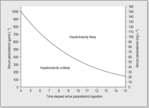
The paracetamol nomogram (Fig. 21.2.1), based on adult toxicity profiles, is extrapolated to children and predicts the potential for significant hepatotoxicity. The nomogram is applicable when a paracetamol level is taken between 4 and 16 hours post-acute ingestion. The nomogram cannot be utilised when the time of ingestion is unknown or in the case of staggered or chronic ingestions. Paracetamol treatment guidelines were reviewed by a representative panel of Clinical Toxicologists to the Australasian Poisons Information Centres and published in 2008. A major shift in management was the amalgamation of the high- and low-risk nomogram treatment lines into a single curve for both adults and children. This current line commences at 1000 μmol L–1 at 4 hours post-ingestion and has a half-life of 4 hours.

N-acetylcysteine (NAC), a glutathione precursor, is the antidote of choice in paracetamol poisoning. NAC is indicated in the setting of acute paracetamol ingestion, where the measured paracetamol level falls above the nomogram treatment line. The full course involves a loading dose of 150 mg kg–1 in dextrose over 15–60 minutes, followed by a second infusion of 50 mg kg–1 over 4 hours, and finally a 100 mg kg–1 infusion over 16 hours. NAC is preferably commenced within 8 hours of ingestion, but has been documented to be effective in adult patients when given up to 48 hours after a serious ingestion, and can even be considered later when hepatic failure is established. Anaphylactoid reactions, including rash, bronchospasm, pruritus, hypotension and tachycardia, occur in up to 15% of cases, and are most common during the second infusion. These reactions are managed similarly to other hypersensitivity reactions; the NAC infusion should be temporarily ceased and recommenced at half the rate.

As children usually ingest the elixir formulation rather than tablets, rapid absorption precludes the utility of oral activated charcoal. Activated charcoal can be administered for potentially toxic doses of the tablet formulation, if given within 1 hour. Children who present with established hepatic failure must receive prompt resuscitation and stabilisation. NAC is still indicated when hepatotoxicity is established. Coagulopathy and encephalopathy should be managed as from other causes of liver failure. These children are managed in the intensive care and should be assessed for potential liver transplantation.

Chronic over-dosing or repeated supratherapeutic ingestion is a significant problem. The treatment nomogram is not applicable to these situations and cases should be discussed with a toxicologist. Children with paracetamol levels below the nomogram treatment line after acute single ingestions can be cleared from a toxicological point of view. Parents or carers should be educated on correct paracetamol dosing and safe storage. Children with deliberate self-poisoning should be assessed by the mental health team.

Benzodiazepines

Benzodiazepine overdose is commonly seen in toddlers who ingest 1–2 tablets, or as part of a mixed overdose in adolescents. Benzodiazepines bind predominantly to the γ-aminobutyric acid (GABA) type-A receptor complex in the central nervous system (CNS) and enhance GABA activity to produce sedative, anxiolytic and anticonvulsant activity. The duration of sedation ranges from 4–36 hours, depending on the agent. Flumazenil is a competitive antagonist at benzodiazepine receptors. Drowsiness, slurred speech and ataxia are the most common manifestations. This may progress to coma and hypotension, hypothermia and respiratory depression in more significant ingestions. Death is rare unless other CNS depressants have been co-ingested.

Management is entirely supportive. Hypotension usually responds to fluid administration. GI decontamination is not indicated in pure benzodiazepine poisoning. A blood sugar level should be checked in all children with an altered level of consciousness. Other laboratory investigations are not routinely indicated. Chest radiography is only indicated if aspiration is suspected. A qualitative urine test for benzodiazepines may provide reassurance as to the aetiology of drowsiness in the setting of an unconscious patient without a clear history of ingestion. In some clinical scenarios, flumazenil may avert the need for intubation and mechanical support but its use should be discussed with a toxicologist. Flumazenil should be administered in boluses of 5–10 mcg kg-1 and titrated to clinical effect (respiratory rate and effort).

Most children can be discharged after 4–6 hours if vital signs are satisfactory and the child can walk unaided.

Opioids

Opioids are frequently available where family members suffer chronic pain, abuse drugs or are on drug-rehabilitation programmes. Iatrogenic intravenous overdosing of children is commonly a result of 10-fold errors in dose calculation.

Morphine and codeine are natural opium alkaloids. Other opioids are synthetic or semisynthetic analogues. The opioids act on various receptors on the brain, spinal cord and gastrointestinal tract as full or partial agonists or antagonists. Opioids are well absorbed by all routes except skin, are metabolised by the liver and are renally excreted. Some opioids (e.g. pethidine and diphenoxylate) have potent metabolites. Opioids vary in their duration of action and some are available as sustained release preparations (e.g. morphine). Toxicity is enhanced by co-ingestion of other sedative medications, which may be found in some cough remedies or analgesic preparations. Children are especially sensitive to the depressive effects of opioids.

Paediatric overdose of a parent’s methadone (long-acting opioid) syrup requires overnight admission for extended observation and monitoring. Antidiarrhoeal preparations contain diphenoxylate (with atropine) and produce delayed onset of symptoms, with numerous paediatric deaths reported. A major metabolite of diphenoxylate is more potent than the parent compound and undergoes enterohepatic circulation. Dextropropoxyphene has a membrane-stabilising effect on cardiac conducting tissue and may induce ventricular arrhythmias and heart block.

The classic features are of nausea and vomiting, drowsiness, pinpoint pupils, respiratory depression, and occasionally bradycardia and hypotension. Respiratory depression may lead to hypoxia and respiratory arrest. The histamine-releasing effects of some opioids may cause urticaria and hypotension.

Management of the opioid toxidrome is essentially supportive, with attention to airway and ventilation. All children with altered level of consciousness should have a blood sugar level checked. Activated charcoal may be considered for massive ingestions or long-acting preparations. Insertion of gastric tubes for charcoal administration is not recommended unless the child is intubated for other indications.

Naloxone, the antidote for opiate toxicity, is a competitive antagonist at opioid receptors. Naloxone (intravenously (IV) 0.01 mg kg−1, maximum 2 mg, as a bolus, repeated every few minutes till appropriate response) may be useful to reverse the neurological and respiratory depression, and should be titrated to respiratory rate and effort. Naloxone’s short therapeutic half-life of 30–60 minutes may necessitate a continuous infusion, in order to maintain the reversal and obviate the need for mechanical ventilation, particularly for ingestions of long-acting opiates.

All patients should be observed for a minimum of 6 hours. Methadone, dextropropoxyphene and sustained-release morphine sulfate may cause symptoms persisting for 24–48 hours so prolonged observation is necessary after ingestion of these agents.

Anticholinergics and antihistamines

Anticholinergic (antimuscarinic) poisoning can result from a diverse range of therapeutic substances, plants and natural remedies, many of which can be bought over the counter. Pharmaceuticals with prominent anticholinergic properties include first-generation antihistamines, antipsychotics and tricyclic antidepressants. Some plants, e.g. Jimsonweed, angel’s trumpet, and mushrooms contain alkaloids with potent anticholinergic effects.

The anticholinergic toxidrome is caused by competitive inhibition of the muscarinic receptor in the autonomic nervous system. The classic anticholinergic toxidrome is well described by the following rhyme:

Other anticholinergic effects include tachycardia, gastrointestinal ileus and urinary retention.

The management of anticholinergic poisoning includes attention to the ABCs with appropriate supportive therapy and monitoring of vital signs and mental state. Symptomatic patients should have IV access, a 12-lead electrocardiogram (ECG) and continuous cardiac monitoring. Benzodiazepines are useful in managing agitation or seizures. Physostigmine is a cholinesterase inhibitor which enters the CNS and is effective at reversing central anticholinergic delirium. Due to concerns regarding adverse cardiac side effects, the use of physostigmine should be discussed with a toxicologist. The initial dose in children is 0.02 mg kg−1 (maximum 0.5 mg) by slow IV push; doses may be repeated every 15 minutes.

Aggressive cooling measures may be required in severe hyperthermia. Urinary catheterisation is required for patients with urinary retention. Asymptomatic patients may be discharged after 6 hours. Patients with moderate or severe toxicity should be admitted to an intensive-care facility.

Antihistamine poisoning in children is of concern only for first-generation agents (e.g. promethazine), which have significant anticholinergic effects. Children should be observed for a minimum of 6 hours following ingestion. Management is supportive and the anticholinergic effects of antihistamines, while unpleasant, are generally not life threatening.

Hypotension should be treated with intravenous fluids. Convulsions and anticholinergic delirium are best managed with benzodiazepines. Continuous ECG monitoring is advised for symptomatic children and those with persistent tachycardia. Ventricular arrhythmias should be managed with sodium bicarbonate as the drug of choice for QRS prolongation with sodium-channel blocking antihistamines.

Corrosive ingestions

Most serious caustic ingestions involve strong acids and alkalis which account for a high number of presentations to the ED. The initial presentation and treatment are similar to other burns. Domestic bleaches and ammonia products generally cause minor injuries. Serious injuries result most often from the ingestion of drain and oven cleaners (NaOH, KOH). Dishwashing powder residue left in the dispenser of machines is a commonly accessed alkali that may cause serious injury.

The severity of burn depends on the nature, volume, pH and concentration of the agent and the duration of contact. Stomach contents may afford some protection from injury, but pylorospasm, oesophageal reflux and vomiting may exacerbate injury. Liquids may cause a circumferential injury and powders/granules or tablets may cause prolonged contact with a mucosal surface, with potential for linear burns, deep erosion and penetration. Acids cause superficial corrosion and a coagulative necrosis, and the extent of tissue penetration is limited by eschar formation. Alkalis start to burn immediately on contact and cause a liquefaction necrosis of fat and protein, penetrating deeply into tissues. Acids typically injure the stomach while alkalis damage the oropharynx and oesophagus.

Many children will be asymptomatic, especially if low-concentration household products are involved. Pain, drooling, dysphagia, vomiting and abdominal pain and haematemesis may occur. Airway compromise with laryngeal oedema, cough and bronchospasm may be seen after ingestion of high-concentration agents. Endoscopy provides the best guide to prognosis and management. The extent of injury is graded by the depth of ulceration and the presence of necrosis. Typically, after ingestion the mouth or oesophagus is red and ulceration follows within 24 hours. One-third of patients with oral burns have associated oesophageal lesions, whereas 10–15% of patients with oesophageal lesions have no oropharyngeal burns. Asymptomatic patients with no oral burns may have significant oesophageal injuries. Drooling and dysphagia persisting beyond 12–24 hours are reliable predictors for oesophageal scar formation and suggest the need for upper GI endoscopy.

Oesophageal perforation and mediastinitis may be suspected by chest pain, fever, pleural rub, dyspnoea. Abdominal pain, fever, peritonism and ileus may indicate gastric or abdominal oesophageal perforation. These signs may progress to septic shock, multiorgan failure and death. Large acid ingestions may be associated with hypotension, metabolic acidosis, haemolysis, nephrotoxicity and pulmonary oedema.

Late complications are infection, achlorhydria and stricture formation in 1–3%. All patients with full-thickness and 70% with deep ulceration will develop strictures. Eighty percent of all strictures occur within 2 months of ingestion and 99% within 1 year.

The management of caustic ingestions is aimed at limiting the extent of injury and preventing strictures and other complications. Immediate management consists of rinsing the skin with water or drinking water unless respiratory distress is notable or visceral perforation is suspected. Acids and alkalis do not bind to charcoal. Attempts to neutralise the substance are contraindicated, but dilution with water may possibly be helpful for acids and may reduce mucosal contact time in ingestion with particulate alkalis. Early treatment focuses on ensuring an adequate airway, intravenous fluid replacement, monitoring fluid balance, avoiding vomiting and adequate analgesia. Oesophagoscopy in the symptomatic patient guides further management. Patients with deep, especially circumferential burns of the oesophagus should be admitted to an intensive care unit and may require prolonged parenteral feeding and repeated endoscopic stricture dilatations. Early surgical intervention and prophylactic antibiotics are required if perforation or penetration is suspected clinically, on endoscopy or contrast radiography. Steroids have no proven benefit and may possibly increase the risk of perforation.

Asymptomatic patients should be advised to return if they develop respiratory difficulty, pain or dysphagia. All symptomatic children should be admitted for observation and potential endoscopy.

Ethanol

Ethanol is available in numerous household medicinals, mouthwashes and perfumery products as well as alcoholic drinks. All products marketed in Australia as ‘methylated spirits’ contain ethanol. Although frequently ingested by children, serious toxicity is uncommon.

Ethanol is well absorbed across gastrointestinal mucosa and respiratory tract, most within 30–60 minutes, and distributes to total body water. Children metabolise alcohol faster than adults. Only very small amounts are excreted unchanged in the urine and the breath. Hypoglycaemia is caused by depressed gluconeogenesis. The potentially fatal dose of alcohol for children is 4 mL kg−1 of absolute alcohol (e.g. 10 mL kg−1 for a 40% alcohol spirit), about half the dose required for adults. Quite low serum levels (>10 mmol L−1, >0.05% or >500 mg L−1) may produce clinically significant effects in children.

Ethanol acts on the reticular activating system to cause CNS depression. Low concentrations result in alterations of mood and thought processes, whereas higher concentrations affect cerebellar function, causing ataxia and slurred speech. Higher levels still depress all cortical function and brainstem activity, depressing respiratory drive and protective airway reflexes. Respiratory arrest or aspiration is a frequent cause of death. Facial flushing, excessive sweating and vomiting are common.

Management depends on the time elapsed since ingestion. Assess and secure the ABCs and correct electrolyte abnormalities and dehydration. Patients with severe CNS depression are at risk of aspiration and require airway protection. Blood glucose should be monitored. A blood alcohol level may be taken at least 1 hour post-ingestion if symptoms are present, although management is generally determined by the clinical state. Hypoglycaemia should be corrected with 5 mL kg−1 10% dextrose. Hypotension will usually respond to intravenous fluids and acidosis usually responds to correction of hypovolaemia and hypoglycaemia. Hypothermia should be corrected. Activated charcoal does not bind ethanol but may be considered if co-ingestants are suspected, provided the airway is protected. Gastric lavage is likely to be ineffective due to the rapid absorption from the stomach. Haemodialysis may be indicated in the extremely intoxicated child who is haemodynamically unstable, but this is uncommon. Admit all children who are clinically intoxicated until asymptomatic.

Rare and dangerous poisons

Although most substances are harmless to children in small amounts, a few pharmaceuticals and chemicals are extremely toxic in minute quantities. Table 21.2.1 lists drugs where ‘one pill can kill’ and chemicals where a sip or mouthful is potentially lethal.

Table 21.2.1 Potentially lethal ingestions in small amounts for a toddler

Pharmaceuticals Chemicals CCBs (diltiazem & verapamil) Organophosphate & carbamate pesticides Chloroquine & hydroxychloroquine Paraquat Clonidine Camphor Tricyclic antidepressants Naphthalene Sulfonylureas Toxic alcohols Amphetamines & ecstasy Essential oils

Salicylates

The incidence of acute salicylate poisoning has declined due to improvements in medication packaging, removal of aspirin from oral paediatric formulations and due to paracetamol now being the favoured over-the-counter analgesic. Methylsalicylate and salicylic acid are common in many topical preparations. Oil of Wintergreen containing methylsalicylate can be significantly toxic when ingested. Choline salicylate is a constituent of many teething gels.

Aspirin is rapidly absorbed from the upper gastrointestinal tract, with peak plasma concentrations at 1–3 hours after therapeutic doses. An ingestion of 150–300 mg kg–1 may result in mild to moderate toxicity. Ingestions over 500 mg kg–1 are potentially lethal. Oil of Wintergreen is 100% methylsalicylate and extremely small amounts may be lethal.

Aspirin is hydrolysed to form salicylic acid (salicylate). In large overdoses, the potential for pharmacobezoar formation in the gut may alter absorption kinetics. In therapeutic doses, salicylate is 85–95% plasma bound, but in overdose, free salicylate concentration rises as plasma protein binding is saturated. As metabolic pathways in the liver become saturated, the kidney becomes the main route of elimination. Clearance is markedly enhanced by an alkaline urinary pH.

Salicylate poisoning leads to the uncoupling of oxidative phosphorylation and anaerobic metabolism. The resulting lactic acidosis is more prominent in young children. Early signs of salicylism include a respiratory alkalosis from centrally driven hyperventilation and tinnitus. With increasing toxicity, confusion, hallucinations and seizures are reported. Metabolic derangements include temperature dysregulation, impaired glucose metabolism and transport as well as electrolyte abnormalities. Mixed picture acid–base derangement is a hallmark of severe salicylate poisoning, with serum pH below 7.3 being a late and ominous sign. Coagulopathy may result from competitive inhibition of synthesis of vitamin-K dependent factors. Non-cardiogenic pulmonary oedema has been reported, but the mechanism is unclear.

Investigations include baseline blood sugar level, electrolyte and renal function, acid–base status, coagulation panel and 6-hour salicylate level. Decontamination with activated charcoal is indicated in early presentations and may be considered in late presentations of enteric-coated preparations. Meticulous monitoring of fluid balance, temperature, glucose and electrolyte levels is recommended. In particular, potassium replacement is often required, along with maintenance of urine output.

Methods of enhancing elimination should be instituted following consultation with a toxicologist. Urinary alkalinisation with intravenous sodium bicarbonate infusion is known to enhance renal excretion of salicylate ions. The target urinary pH is at least 7.5 with maintenance bicarbonate doses ranging from 1–2 mmol kg−1 hr−1. Haemodialysis is indicated in severe toxicity with acidaemia, cardiorespiratory failure, renal impairment or CNS manifestations (coma, seizures). Complications of salicylate poisoning require aggressive supportive management. The development of non-cardiogenic pulmonary oedema often signals the need for invasive ventilation and haemodialysis. Seizures warrant benzodiazepine therapy and correction of any glucose or electrolyte derangement.

Observe all symptomatic children and those with ingestions of greater than 150 mg kg−1. Most patients will require admission for 6–12 hours for observation of clinical state and serum salicylate level.

Digoxin

Digoxin is a cardiac glycoside used for management of heart failure and supraventricular arrhythmias. Many plants contain digitalis glycosides (e.g. foxglove, oleander) and poisoning from these plants should be managed in a similar manner to digitoxicity. Acute digoxin poisoning in children is more often seen in the context of toddlers who obtain access to grandparents’ medication. Rarely, children with underlying cardiac disease on maintenance digoxin therapy can develop chronic toxicity and therapeutic drug monitoring is crucial. Chronic overdosing or renal impairment can lead to chronic digitoxicity.

Digoxin is well absorbed from the gastrointestinal tract and has a relatively large volume of distribution. Digoxin is predominantly excreted unchanged by the kidney, with an elimination half-life of about 36 hours. Digoxin inhibits the action of the cardiac Na/K ATPase pump and accumulation of sodium and calcium ions leads to intracellular depletion of potassium and hyperkalaemia. The slowing of conduction, as well as increased refractory period, through the AV node, enhanced automaticity of the Purkinje fibres and enhanced vagal tone leads to a multitude of arrhythmias including sinus bradycardia, sinoatrial arrest, conduction blocks, ventricular tachycardia and fibrillation.

Early signs of chronic toxicity include nausea, vomiting and diarrhoea. The child is often asymptomatic in acute poisoning, until haemodynamic instability from cardiac toxicity becomes clinically apparent. Patients can deteriorate suddenly and digitoxicity can produce a myriad of both brady- and tachyarrhythmias. Assess ABCs, secure IV access and continuously monitor blood pressure (BP) and ECG. Although digoxin is well bound by activated charcoal, repeated vomiting may reduce its effectiveness. Obtain a serum digoxin concentration and electrolytes. Serum potassium concentration should be monitored every 4 hours. Hyperkalaemia should be corrected to within upper limits of normal with sodium bicarbonate and insulin/dextrose. Calcium is relatively contraindicated due to potential for myocardial destabilisation.

Digoxin Fab antibodies are a specific and highly effective antidote in digoxin poisoning. Intravenous Fab fragments of digoxin-specific antibodies are first-line therapy for patients with cardiac arrhythmias with haemodynamic instability. The dosage is based on total body load, estimated from the serum digoxin concentration or from the ingested dose. Alternatively, a dose estimation can be made on the presumption that one vial of 40 mg will bind 0.6 mg of digoxin. A clinical response is seen in 20–30 minutes, with maximum effect at 2–4 hours. An empiric dose of 5 vials of digoxin Fab may be given IV over 20 minutes in severe life-threatening toxicity. It is important to note that subsequent digoxin levels post-treatment with antibodies are not interpretable and should not be performed.

Acute overdoses should be observed for a minimum of 12 hours or overnight. Symptomatic patients should be monitored in an intensive care unit or coronary care facility. Patients treated with Fab fragments should be monitored for subsequent hypokalaemia and for deterioration of pre-existing cardiac disease.

Calcium-channel blockers

Calcium-channel blockers (CCBs) are widely used in the treatment of hypertension, coronary artery disease and supraventricular tachyarrhythmias.

CCBs are rapidly absorbed from the gastrointestinal tract and have peak plasma concentrations ranging from 30 minutes (nifedipine) to 90 minutes (verapamil), but sustained-release preparations are associated with longer times to peak concentration and prolonged clinical effect. These agents inhibit the entry of calcium into the cells of cardiac and smooth muscle, decreasing the activity of the calcium-dependent actin-myosin ATPase. Dihydropyridines (represented by the prototype agent nifedipine) are more potent at peripheral vascular calcium channels and have little cardiac toxicity. Verapamil and diltiazem are highly toxic drugs where a single large dose tablet can cause profound cardiogenic shock in a toddler. Dihydropyridine CCBs are unlikely to cause hypotension or cardiac conduction abnormalities in small doses.

The features of cardiotoxicity include brady-arrhythmias, such as sinus bradycardia, varying degrees of AV block, and asystole. Myocardial depression may cause congestive failure or cardiogenic shock. Dose-dependent peripheral vasodilatation with hypotension may occur. Other manifestations include nausea and vomiting, lethargy, coma, seizures, hyperglycaemia and lactic acidosis.

Good supportive management is essential. Intravenous access should be secured, and blood glucose and electrolytes measured. ECG and BP should be continuously monitored. Activated charcoal is worth considering in patients that present early with a significant ingestion. Whole bowel irrigation with polyethylene glycol should be considered for large ingestions of sustained-release preparations.

Calcium is the initial antidote for hypotension and bradycardia (bolus: 10% calcium chloride 0.2 mL kg−1 or 10% calcium gluconate 0.6 mL kg−1). Atropine is likely to be ineffective. Catecholamine infusions (e.g. adrenaline (epinephrine) commencing at 1 mcg kg−1 min−1) may be required. Other inotropes that do not require calcium influx are potentially useful in managing intractable shock from CCBs. These include high-dose insulin (1–2 units kg−1 IV bolus; 1–2 units kg−1 hr−1 infusion) and glucagon (0.05–0.1 mg kg−1 IV bolus). Rarely, more extraordinary measures may be necessary such as transvenous pacing, cardiopulmonary bypass or aortic balloon pumps. Prolonged resuscitation and aggressive supportive care may allow the peak toxicity to pass and improve survival.

Symptomatic children should be monitored in an intensive-care setting. Observation for 24 hours is warranted for ingestion of sustained-release formulations.

β-Blockers

β-Blockers have wide clinical use in the treatment of cardiac conditions, hypertension, thyrotoxicosis and prophylaxis for migraine.

β-Blockers are class II antiarrhythmics, which act by competing with catecholamines at β-receptor sites. Different β-blockers have differing cardioselectivity, membrane-stabilising activity, partial agonist activity and lipid solubility. They are well absorbed from small intestine, with peak serum levels within 1–4 hours. The elimination half-life is less than 12 hours. They have a moderate to large volume of distribution. Highly lipid soluble drugs, such as propranolol, cross the blood–brain barrier and thus have more potent CNS effects. Propranolol is also known for its cardiac sodium channel blocking properties, which cause prolongation of the QRS complex.

The major clinical effects are the cardiovascular effects. Bradycardia may be sinus, junctional or ventricular and may progress to cardiac arrest. Hypotension results from bradycardia, myocardial depression and vasodilatation. Deterioration can be sudden and precipitous, particularly with propranolol, which can cause seizures, coma and wide complex arrhythmias. Hypoglycaemia may occur due to impaired gluconeogenesis and glycogenolysis. Bronchospasm is more likely in atopic subjects, and more prominent with non-selective agents.

Good supportive management is essential. Activated charcoal is the decontamination method of choice. Intravenous access should be secured, and blood glucose and electrolytes measured. ECG and BP should be continuously monitored. Cardiovascular effects should be treated with atropine, volume expansion and catecholamines. High-dose insulin enhances heart rate and myocardial contractility independently of β-receptor activation and is currently the preferred inotrope over glucagon. Doses are similar to those above in calcium channel blocker poisoning. Extreme measures such as transvenous pacing and cardiopulmonary bypass may be required in cases of intractable hypotension. Importantly, prolonged resuscitation and aggressive supportive care may allow the peak toxicity to pass and improve survival. Hypoglycaemia should be treated in the usual manner with 2.5–5 mL kg−1 10% dextrose. Seizures may respond to IV dextrose, even if blood glucose is normal. Benzodiazepines are the preferred anticonvulsant. Bronchoconstriction should be treated with inhaled β2-agonists. Symptomatic children should be monitored in an intensive-care setting.

Clonidine

Until recently clonidine was used primarily as an anti-hypertensive agent and accessibility to children was limited. The drug is now widely prescribed in the treatment of attention deficit hyperactivity disorder, conduct disorders, Tourette’s syndrome and for narcotic and alcohol withdrawal symptoms. Clonidine overdose is commonly seen in children with behavioural disorders, and their siblings who have access to clonidine.

Clonidine is a central α2-adrenoceptor agonist that acts on brainstem receptors, causing inhibition of sympathetic outflow. Its stimulation of peripheral α2-receptors on vascular smooth muscle may cause transient hypertension, but hypotension usually occurs subsequently. Clonidine also has opiate-like effects which may be mediated through mu receptors. It is rapidly absorbed and distributed, with peak plasma concentrations 60–80 minutes post-ingestion. The elimination half-life is 6–24 hours.

Clinical effects are seen 30–60 minutes after ingestion. Depression of the CNS with lethargy and impaired conscious state is the most frequent manifestation. Miosis and hypothermia may be observed. Symptoms are minimal with ingestions of under 10 mcg kg−1, but cardiovascular compromise with hypotension and bradycardia may occur after ingestion of 10–20 mcg kg−1. Respiratory depression and apnoea may be seen after ingestions of 20 mcg kg−1. There have been no reports of in-hospital paediatric deaths.

Treatment is largely supportive. Activated charcoal is only useful if given under 1 hour post-exposure. Hypotension should be treated with volume expansion and vasopressors. Hypertension is usually transient and, if treatment is required, a short-acting agent such as nitroprusside should be used. Atropine may be useful in the treatment of bradycardia. Naloxone therapy in clonidine poisoning is controversial and unreliable. There may be inconsistent reversal of the neurological, cardiovascular and respiratory effects after administration of naloxone.

Maximal toxicity is expected in the first 6 hours and children who show no symptoms at that stage can be discharged. Children with significant respiratory and cardiovascular compromise may require admission to an intensive-care unit for up to 24 hours.

Tricyclic antidepressants

Despite the declining prescription of tricyclic antidepressants (TCAs), the low therapeutic index and potential for lethal toxicity remain a concerning cause of paediatric morbidity and mortality.

TCAs are rapidly absorbed. TCAs have a high degree of protein binding and a large volume of distribution. Although different TCAs have different pharmacokinetic parameters, the effects in overdose are similar. Dothiepin is associated with the greatest lethality. Minor TCA toxicity is generally manifest by central and peripheral anticholinergic signs and the antiadrenergic effect of vasodilatation. More serious toxicity results from fast sodium-channel blockade in the myocardium, causing a wide variety of atrial and ventricular dysrhythmias, impaired contractility and impaired conduction with ECG changes of prolonged QT interval and widened QRS complexes.

Tricyclic ingestions of <5 mg kg−1 result in minimal toxicity and no treatment is required. Ingestions of 5–10 mg kg−1 may cause mild anticholinergic symptoms of drowsiness, ataxia, dilated pupils, ileus and urinary retention but life-threatening toxicity is unlikely. Ingestions over 10 mg kg−1 may cause life-threatening coma, seizures and cardiac dysrhythmias. There may be acidosis, hypokalaemia and inappropriate antidiuretic hormone secretion. Onset of symptoms is usually within 2 hours and persists for less than 12–24 hours.

The management of TCA poisoning includes attention to the ABCs, good supportive therapy and GI decontamination for potentially serious ingestions. Continuous cardiac monitoring, serial 12-lead ECGs and close observation of vital signs and mental state are required for all ingestions of >5 mg kg−1. Secure IV access. Airway protection should precede administration of charcoal if the patient is less than fully conscious.

Depressed conscious state is the best predictor of serious toxic complications (seizures, ventricular arrhythmias, hypotension and the need for mechanical ventilation) and the ECG limb lead QRS duration of 100 ms or greater is associated with an increased incidence of seizures and cardiotoxicity. Early intubation and hyperventilation to a serum pH of 7.45–7.55 may attenuate or prevent seizures and ventricular dysrhythmias. Intubate and mechanically ventilate all patients with rapidly decreasing conscious state, seizures and ventricular dysrhythmias. Hypotension should be treated with volume replacement and adrenaline (epinephrine) or noradrenaline (norepinephrine) infusion if required.

Sodium bicarbonate is regarded as a specific antidote in the treatment of the cardiovascular effects of TCA toxicity. It competitively overcomes sodium-channel blockade and its effect on serum pH appears to improve sodium-channel function. Ventricular dysrhythmias will usually respond to treatment with sodium bicarbonate (1–2 mEq kg−1 IV bolus, repeated till the QRS narrows or serum pH reaches 7.45–7.55). Refractory ventricular arrhythmias should be treated according to standard ACLS protocols avoiding type 1a and 1c antiarrhythmics. Lidocaine is safe. There is no documented evidence to support the use of phenytoin, which may aggravate hypotension and conduction problems due to its effect on fast sodium channels.

Seizures may be averted or attenuated by bicarbonate therapy. Benzodiazepines are the preferred agents to treat seizures but barbiturates may be required to treat refractory seizures. TCAs are not amenable to removal by extracorporeal methods due to their large volume of distribution. Quantitative analysis of TCA levels does not aid management but screening for other drugs should be considered in deliberate self-harm.

Patients who ingest >5 mg kg−1 TCA should be admitted for observation for at least 6 hours, but may be discharged at that time if the ECG remains normal and the child is well. TCA ingestions with significant CNS depression, seizures or significant cardiotoxicity should be admitted to an intensive-care facility.

Iron

Iron tablets are commonly available in the homes of toddlers but severe poisoning is uncommon.

The amount of elemental iron varies according to the formulation. Initial toxicity is due to the corrosive effects on the gastrointestinal tract. Iron is absorbed in the ferrous state and after oxidation to the ferric state becomes bound to ferritin. Toxicity occurs when ferritin and transferrin are saturated and serum iron exceeds the total iron-binding capacity (TIBC). High concentrations of intracellular iron cause mitochondrial dysfunction, interfering with mitochondrial processes, causing lactic acidosis and cell death.

Ingestions of less than 20 mg kg−1 elemental iron usually remain asymptomatic. Significant symptoms usually only occur in ingestions above 60 mg kg−1. Potentially lethal systemic toxicity may follow ingestions of greater than 100 mg kg−1 elemental iron. Serum iron peaks at 4–6 hours. Serum iron levels should be considered in conjunction with the clinical state.

Although four stages of iron poisoning are classically described, distinct phases may not be apparent with severe poisoning. In the initial 6 hours the gastric irritant effects predominate, with vomiting, diarrhoea and haematemesis or melaena. Circulating free iron may damage blood vessels and cause a transudate of fluid and hypotension. There may be a quiescent phase when the patient may appear to be improving, but about 12–24 hours after ingestion the physiological processes of cells are disrupted, leading to metabolic acidosis, gastrointestinal haemorrhage, altered mental state, pulmonary oedema, cardiovascular, hepatic and renal failure. The liver is particularly vulnerable and fulminant hepatic failure may cause hypoglycaemia, coagulopathy and death. At 4–6 weeks there may be stricture formation in the gastrointestinal tract due to scarring.

Symptomatic patients and those with ingestions greater than 60 mg kg−1 of elemental iron require laboratory investigations and an abdominal X-ray. Baseline electrolytes, renal function and a 4–6 hour iron level is recommended. Iron does not bind well to activated charcoal. Patients with ingestions potentially in excess of 60 mg kg−1 of elemental iron should have IV access and a blood glucose level. IV fluid resuscitation may be required and electrolytes and glucose should be monitored. Whole bowel irrigation with polyethylene glycol at 20 mL kg−1 hr−1 via a nasogastric tube is reserved for massive ingestions. A venous bicarbonate level should be performed and serum iron concentration is indicated at 4–6 hours after ingestion. The peripheral blood white cell count greater than 15 × 10 L−1and hyperglycaemia are suggestive of systemic toxicity.

Desferrioxamine (desferoxamine) binds unbound iron in the intravascular and extracellular space and the chelated complex is eliminated in the urine, imparting a pink-brown colour (vin-rose urine). The decision to use chelation therapy should be based on the combination of the patient’s clinical condition and serum iron concentration. IV desferrioxamine (15 mg kg−1 hr−1 to a maximum of 80 mg kg−1 per 24 hours) is indicated in patients with hypotension, shock, coma, convulsions or potentially if serum iron concentration is greater than 90 μmol L−1. Desferrioxamine infusion is usually required for 6–12 hours. The end-points of chelation therapy are clinical improvement and a reduction in free iron levels. Acid–base and electrolyte balance should be maintained and hepatic and renal function monitored. The chelated complex can be removed with haemodialysis should renal function be significantly impaired.

Asymptomatic children with ingestions of under 60 mg kg−1 may be observed at home. Symptomatic patients, or those with ingestions greater than 60 mg kg−1 of elemental iron, require further evaluation in hospital and an admission of 12–24 hours.

Toxic alcohols

Ethylene glycol is encountered in antifreeze compounds and radiator additives. Methanol is found in model aeroplane fuel and in home brewing concoctions. Toxic alcohols are rapidly absorbed from the GI tract and distribute to total body water.

Methanol is oxidised to formaldehyde by the rate-limiting enzyme alcohol dehydrogenase, and then aldehyde dehydrogenase converts the formaldehyde to formic acid (formate). Approximately 2% of methanol is excreted unchanged by the kidneys and a small amount is excreted via the lungs. The optic nerve is particularly susceptible to the toxic effects of formic acid. Lactate is produced from anaerobic glycolysis as a result of tissue hypoxia and a formate-induced inhibition of mitochondrial respiration. Ingestion of 1.5 mL of 100% methanol in a child weighing 10 kg would produce a potential peak plasma level of 6 mmol L–1 (0.02%, 20 mg dL−1), so a single mouthful is potentially lethal. Symptoms of methanol poisoning may be delayed with a 12–24 hour latent period because of the slow metabolism to formate. The most common presentation of intoxication consists of a triad of findings related to the GI tract, eyes and metabolic acidosis. Nausea and vomiting, epigastric abdominal pain, pancreatitis and GI bleeding may occur. Visual disturbance including blurred vision, central scotoma, yellow spots and complete blindness offer an important diagnostic clue.

Ethylene glycol depresses the CNS, but the hepatic metabolites glycoaldehyde, glycolic acid, glyoxylate and oxalate are responsible for toxicity. Formation of glycolic acid and some lactic acid is the primary cause of the delayed metabolic acidosis, which can occur 4–12 hours after ingestion. Oxalate is highly toxic, causing myocardial depression and acute renal tubular acidosis. Calcium oxalate crystals may be noted on examination of the urine. Ethanol has a 100-fold greater affinity for alcohol dehydrogenase than ethylene glycol, hence it is used to prevent metabolism of ethylene glycol into toxic metabolites. The initial symptoms of an acute ethylene glycol poisoning include those of alcohol intoxication with lethargy, slurred speech, nystagmus, ataxia and vomiting. Papilloedema occurs less frequently than with methanol poisoning. An elevated anion osmol gap acidosis may occur. Seizures, myoclonic jerks and tetanic contractions reflect hypocalcaemia. At 12–36 hours post-ingestion, progressive pulmonary oedema and congestive heart failure occur and may be followed by death due to cardiovascular collapse. If the child survives, renal insufficiency may ensue over the next 2–3 days.

Fomepizole and ethanol block alcohol dehydrogenase, which is involved in the metabolism of the parent compounds of methanol and ethylene glycol to their toxic metabolites. These antidotes do not prevent the toxic effects of the acid metabolites and are only useful if an osmolar gap exists. Fomepizole, which is difficult to source in Australasia, is expensive but easier to administer and monitor than IV ethanol, without the complications of profound hypoglycaemia, hepatotoxicity and inebriation that may occur with ethanol infusions.

The target serum ethanol concentration of 20 mmol L−1 (100 mg dL−1, 0.1%) will fully inhibit alcohol dehydrogenase. This can be difficult to achieve in children without advanced support of airway and ventilation. Ethanol is preferably administered orally or via gastric tube. The loading dose is 7.5 mL kg−1 of 10% ethanol in 5% glucose water over 30 minutes, followed by a maintenance dose of 0.8–1.5 mL kg−1 hr−1 of 10% ethanol. Serum ethanol and glucose levels should be monitored after the loading dose and frequently thereafter. Haemodialysis enhances elimination and is indicated for renal failure, visual impairment or severe metabolic acidosis. Asymptomatic children should have blood taken for determination of acid–base status, presence of osmolar gap and electrolytes. Admit all children who are clinically intoxicated until asymptomatic.

Essential oils

Essential oils are complex aromatic mixtures of alcohols, esters, aldehydes, ketones and turpenes widely used in perfumery, food flavourings, massage and alternative remedies. Eucalyptus oil is an essential oil commonly implicated in hospitalisations for childhood poisoning. Incidents usually involve vaporiser solutions, eucalyptus oil preparations and other medicinal preparations, which are freely available over the counter. Citronella oil is used as an insect repellent. Oil of turpentine has been largely replaced by white spirit and turpentine substitutes, which are less toxic.

Essential oils are complex mixtures of substances distilled from plant species, including oil of cloves, eucalyptus, citronella, lavender, peppermint, melaleuca (tea tree) and turpentine. The oils probably differ in the degree of toxicity but comparative data are lacking. The irritant effects are manifest by vomiting after ingestion and aspiration causing a chemical pneumonitis.

Essential oils are potentially very toxic. The breath, vomitus, urine and faeces smell strongly of the oil. Skin irritation may occur. Oil of turpentine has been reported to cause gastrointestinal irritation, central nervous system toxicity, hepatic and renal failure and metabolic acidosis. Eucalyptus oil toxicity has been reported to involve all major body systems and death has been reported in an adult after ingestion of 4 mL. CNS depression, seizures and gastrointestinal effects generally occur within 1 hour after ingestion and respiratory complications including respiratory depression, bronchospasm, aspiration pneumonitis and pulmonary oedema have been reported. In a retrospective hospital-based series of 41 paediatric cases of eucalyptus ingestion, 80% remained asymptomatic, eight children had transient symptoms prior to attendance at ED (vomiting in seven patients, respiratory distress in one patient) but only two children remained symptomatic on presentation to ED (one with drowsiness, hypertonia and hyper-reflexia and another with drowsiness and rash). Both children were discharged the following day.

As management is entirely supportive, assess and secure the ABCs. Benefit from oral activated charcoal or gastric lavage is dubious and not routinely recommended. A chest X-ray is indicated only if respiratory symptoms are apparent. Aspiration pneumonia is treated with respiratory support if required. Benzodiazepines are preferred for managing seizures.

With regards to eucalyptus oil, asymptomatic children can be discharged after 2 hours’ observation. Patients with impaired conscious state or respiratory distress on presentation should be admitted to an intensive-care unit.

Organophosphates and carbamates

Pesticide poisoning in children is a rare event in Australasia. Organophosphates and carbamates inactivate the enzyme acetylcholinesterase at cholinergic nerve terminals and neuromuscular junctions, resulting in the cholinergic toxidrome. Plasma (butyl) cholinesterase and red blood cell cholinesterase levels are surrogate markers for exposure and toxicity.

The cholinergic toxidrome involves excess secretions from muscarinic overstimulation, neuromuscular dysfunction and paralysis from nicotinic over-stimulation and central effects including delirium, seizures and eventually, coma. The onset, peak and duration of toxicity varies with each organophosphate.

In children, lethargy, coma and hypotonia are common early features of organophosphate toxicity. Excess secretions can be absent in children. Severe cases can progress rapidly to generalised weakness, coma, convulsions and respiratory failure. Organophosphates do not off-gas, unlike nerve agents, and do not cause secondary respiratory contamination of treating staff. They do, however, warrant the use of universal precautions including gown, gloves and goggles. Pesticides are often dissolved in hydrocarbon solvents and these chemicals give the characteristic odour, as well as causing symptoms in clinicians such as headaches and dyspnoea. Staff should be rotated regularly and the patient should be placed in a well ventilated resuscitation area.

Resuscitation and decontamination should be carried out concurrently. Vomitus and secretions should be washed off the skin with soap and water. Contaminated clothing should be removed and disposed into biohazard bins. The main treatment for organophosphate and carbamate poisoning is anticholinergic therapy with atropine. Charcoal decontamination or gastric lavage has not been proven to be effective. Atropine is indicated for the muscarinic symptoms of bradycardia and excess secretions. Bolus doses of atropine (0.05 mg kg−1, max 1–2 mg) should be repeated every 2–3 minutes until the end-points of normal heart rate, blood pressure and drying of secretions are reached. An atropine infusion may be necessary and should be discussed with a toxicologist. Tachycardia and dilated pupils following atropine therapy may indicate atropine toxicity. Oxime therapy in organophosphate poisoning is controversial and unproven. Pralidoxime, a cholinesterase reactivator at the neuromuscular junction, may be effective in re-establishing respiratory muscle and diaphragmatic function in some types of organophosphate poisoning. The loading dose is 25–50 mg kg−1 (maximum 2 g) infused IV over 30 minutes, followed by an infusion at 10–20 mg kg−1 hr−1 for up to 48 hours.

All children with possible or potential organophosphate or carbamate ingestion should be admitted for prolonged observation. Patients who remain asymptomatic in the ED after 12 hours, or overnight, may be discharged home for observation.

Oral hypoglycaemics

Sulfonylurea poisoning is uncommon in children, but even ingestions of a single tablet have led to significant morbidity and mortality, and the onset of symptoms may be delayed and prolonged. Children may access the tablets in the home of diabetic relatives. In Australasia, gliclazide, glipizide and glibenclamide are responsible for the majority of poisonings. Ingestion of the newer sustained release preparations of gliclazide warrant prolonged monitoring of blood glucose levels.

Sulfonylureas induce hypoglycaemia by stimulating endogenous insulin secretion. In contrast, biguanides do not cause hypoglycaemia, but can induce severe lactic acidosis. Children are more susceptible to hypoglycaemia than adults because of their increased metabolic rate and limited ability for gluconeogenesis.

Hypoglycaemic manifestations occur with palpitations, shaking, hunger, sweating, weakness and with increasing neuroglycopenia, confusion, coma and seizures occur. Long-term neurological disability and death may occur.

Following assessment and management of the ABCs, a blood glucose level should be performed immediately and checked hourly. An initial serum insulin level may be helpful in guiding subsequent management. Gastric decontamination with activated charcoal is not routinely warranted. Hypoglycaemia should be treated with dextrose 10% 5 mL kg−1 IV bolus. Glucagon is not recommended for sulfonylurea-induced hypoglycaemia.

Octreotide, a long-acting synthetic somatostatin analogue, inhibits secretion of insulin from the pancreas and may be the most appropriate method of stabilising blood glucose levels. Patients with persistent or recurrent hypoglycaemia requiring repeat bolus of dextrose should be given octreotide (1 mcg kg−1 IV bolus), followed by an octreotide infusion (250 mcg in 250 mL 5% dextrose at 1 mcg kg−1 hr−1, to maximum of 25 mcg hr−1). If octreotide is not available, a dextrose 10% infusion should be commenced at 1–2 mL kg−1 hr−1. Hourly blood glucose measurements are required until octreotide and/or dextrose infusions are ceased. Serum insulin levels may have a role in ongoing management.

Asymptomatic children should be observed for at least 8 hours, longer for sustained release preparations. Symptomatic children require intensive monitoring of blood sugar and clinical condition.

House fires

From a toxicological point of view, the main exposures relate to carbon monoxide (CO) and cyanide, derived from combustion products of nitrogen-containing polymers, both natural (wool and silk) and synthetic (polyurethane and polyacrylonitrile), which are used extensively in domestic furnishings. In children, carbon monoxide poisoning is often associated with other injuries, such as burns or smoke inhalation. The affinity of haemoglobin for carbon monoxide is 210-times its affinity for oxygen. Carbon monoxide dissolved in the plasma acts as a direct cellular poison reacting with other haem proteins, such as mitochondrial cytochromes, to disrupt cellular metabolism.

Although carboxyhaemoglobin (COHb) levels poorly correlate with symptoms or prognosis, patients with up to 20% of haemoglobin affected complain of headaches and nausea. At 20–40%, patients tire and become confused. COHb greater than 40% can result in ataxia, collapse, and coma. Death is preceded by cardiac arrhythmias, cerebral oedema, and severe metabolic acidosis. Standard oxygen saturation monitors are unreliable in the presence of COHb, with saturations of 100% occurring in the presence of significant hypoxia. Accurate oxyhaemoglobin saturation requires measurement with a co-oximeter. Conventional blood gas analysers can also be misleading.

Cyanide binds to ferric iron (Fe3+) in the cytochrome a-a3 complex, inhibiting its action and blocking the final step in oxidative phosphorylation. Aerobic metabolism is halted and carbohydrate metabolism is diverted to the production of lactic acid.

The diagnosis of cyanide poisoning requires a high index of suspicion as clinical signs are limited and made even more difficult with co-existing CO poisoning. Cardinal features are the presence of cyanosis with severe high anion gap metabolic acidosis and elevated lactate. Complications include coma, seizures and myocardial ischaemia. Treatment usually cannot wait until definitive diagnosis is made with cyanohaemoglobin levels.

Management of CO and cyanide poisoning involves high flow oxygen, supportive care and the potential use of cyanide antidotes. Unconscious patients require airway and ventilatory support and may warrant cerebral imaging in the event of trauma. Current evidence suggests that hydroxocobalamin is the most effective cyanide antidote with the fewest side effects.

Psychostimulants

Amphetamines and cocaine are psychomotor stimulants that promote central and peripheral sympathetic outflow. Ecstasy, 3,4-methylenedioxymethamfetamine (MDMA), is an amfetamine derivative and common drug of abuse. It produces typical amfetamine effects, such as locomotor stimulation, euphoria, excitement and stereotyped behaviour. Ecstasy has additional psychoactive effects that alter perception and mood.

Complications of amphetamine and ecstasy ingestion include coma, convulsions, arrhythmias, malignant hyperthermia, rhabdomyolysis, hypertension, and multiorgan failure. Cocaine also has sodium-channel blocking properties which can induce ventricular tachyarrhythmias. Hyponatraemia can be seen in ecstasy ingestion, leading to intractable seizures.

Patients who are asymptomatic should receive activated charcoal if ingestion has occurred within 1 hour. Blood pressure, temperature, and ECG monitoring should be instituted. Symptomatic patients and those with persistent tachycardia should be admitted to a monitored environment. Asymptomatic children may be discharged after 24 hours.

Patients with signs of cardiac or central nervous system toxicity require admission to the paediatric intensive care unit. Careful monitoring of haematological and biochemical parameters is essential. Hyperthermia may respond to fluid resuscitation and simple cooling measures; however, intractable cases should receive muscle paralysis and be ventilated in an intensive-care setting. Convulsions and agitation should be treated with benzodiazepines; phenytoin and neuroleptics should be avoided. Ventricular tachyarrhythmias are managed with sodium bicarbonate and benzodiazepines.