Sonographic Signs of Successful Injections

Published on 06/02/2015 by admin

Filed under Anesthesiology

Last modified 22/04/2025

Print this page

rate 1 star rate 2 star rate 3 star rate 4 star rate 5 star
Your rating: none, Average: 0 (0 votes)

This article have been viewed 927 times

13 Sonographic Signs of Successful Injections

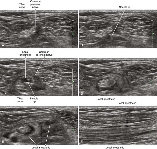
It seems simple enough to state that successful drug injections for regional blockade should surround the peripheral nerve. However, studies have reported that the doughnut sign, previously considered the gold standard for success, has a positive predictive value of only 90% for producing surgical anesthesia.1 It is therefore important to carefully consider multiple factors that constitute sonographic signs for success that can be evaluated after injection.

First, successful drug injections should clarify the nerve border. Most regional blocks are performed with nerves viewed in short axis to evaluate the circumferential distribution. If more than half of the nerve border is contacted by local anesthetic, it is unlikely there is an intervening fascial plane that will serve as a barrier to diffusion. Therefore, it is important that the injection round the corner of the nerve so that there is demonstrated curvature of the injection.

Second, successful drug injections will track along the nerve. Although the longitudinal distribution can be imaged with the nerve viewed in long axis, it is usually easier to slide the transducer along the nerve path with the nerve viewed in short axis (short-axis sliding assessment). If the local anesthetic truly tracks along the nerve, it will track along nerve divisions as well. This sign is especially useful for femoral and popliteal blocks because these block procedures are performed near points of nerve branching.

Third, peripheral nerves are often connected to adjacent structures, such as arteries or other peripheral nerves. Because they are covered in common connective tissue, successful injections should separate the connected structures. This is why practitioners often perform infraclavicular blocks or axillary blocks by placing the block needle tip between the axillary artery and the adjacent nerves. Understanding these connective tissue layers can provide a means of keeping the needle tip at a distance from the peripheral nerves.

Fourth, peripheral nerves are often more echogenic after injection of local anesthetic. This is because anechoic fluid has been injected into an attenuating sound field. This is not a perfect sign of success because anechoic fluid introduced anywhere between the nerve and the skin surface can cause this same effect.