Chapter 20
Soft tissue, peripheral nerveand brachial plexus injury
Beverley Wellington
New Victoria Hospital, Glasgow, UK
Introduction
The aim of this chapter is to provide evidence-based guidance for the assessment, investigation, surgical and conservative care, management and rehabilitation for patients who have sustained a soft tissue injury, peripheral nerve injury or injury to the brachial plexus. Each section will also provide a brief overview of the associated anatomy.
Soft tissue injuries
Soft tissue injuries have several mechanisms of injury but are often caused by either overuse of musculoskeletal soft tissue or a single specific injuring event. The impact on the patient of such injuries can be underestimated and it is important to make a full assessment and follow this with an effective plan of care. Evidence suggests that multiple risk factors, including a genetic predisposition, may be involved particularly in relation to the injuries to the Achilles tendon of the heel, rotator cuff tendons of the shoulder and cruciate ligaments of the knee. In the future, this may allow practitioners to consider genetic risk factors when assessing or treating individuals (Collins and Raleigh 2009).
Ligaments and tendons
Ligaments and tendons are fibrous tissues that have an important role in the musculoskeletal lever system. They are flexible bands of strong, dense connective tissue that connect the articular ends of bones or cartilages together, providing strength and mechanical stability to joints.
Tendons are inelastic fibrous connections between muscles and their points of insertion into bone, transmitting movement forces through joints. They are constructed of fibrous cords which are an extension of the fascia (the fibrous connective tissue found superficially beneath the skin and more deeply which forms a covering sheath for muscles and broad surfaces for muscle attachment). They have no elasticity and are of different lengths and thickness, making them very strong, but with few blood vessels and nerves. Tendons do not cope well with friction and are thus protected at areas of pressure or activity by bursae (sacs filled with a viscous fluid, cushioning movement of one bony part over another) (Hardy and Snaith 2011).
Sprains and strains
Soft tissue injuries are often treated in primary and secondary care settings. Strains and sprains are both soft tissue injures, but the tissue affected is different.
A sprain is an injury to a ligament supporting a joint. A twisting mechanism usually causes damage to fibres of the affected ligament and/or the joint capsule itself, resulting in decreased joint stability. Three grades of ligament injury/sprain are recognised (Altizer 2003):
- Grade I, Mild – stability is maintained, but can be decreased. The injury is often caused by a wrenching or twisting mechanism and commonly seen at the ankle. The site may be tender with bruising and mild oedema. The patient can usually walk, but with some discomfort.
- Grade II, Moderate – partial rupture or tear. Some fibres are torn, but some remain intact although with some loss of stability. More than one ligament may be torn, increasing the severity of the injury and affecting the treatment regime that follows. The joint is tender, painful and difficult to move usually with some swelling and bruising.
- Grade III, Severe – complete rupture with loss of continuity of one or several ligaments. This causes loss of stability and a weakened, painful joint with significantly decreased range of movement. The patient is unable to actively move the joint or to weight bear, and the injury has a significant impact on mobility and activities of living.
A strain is an injury caused by overstretching of the muscle or tendon and may be severe enough to cause a partial or complete tear. These may also be mild, moderate or severe:
- Mild – pain and stiffness lasting a few days
- Moderate – partial tears causing more pain and swelling, with bruising with symptoms lasting for 1–3 weeks
- Severe – complete tears resulting in significant swelling, bruising and pain and often resulting in the need for surgical repair.
Management and care
There are some general principles of management for all soft tissue injuries:
Pain management
The most obvious symptom of soft tissue injury is pain, caused by the traumatic damage to tissue, cell hypoxia and release of chemicals such as histamine, bradykinin and prostaglandin as well as pressure on local nerve endings from swelling in the tissue. Pain relief is usually provided with simple analgesia and non-steroidal anti-inflammatory drugs (Chapter 11). Cryotherapy (the application of cold) has also been shown to have a positive effect on pain relief (Airaksinen et al., 2003) as the cold reduces nerve conductivity.
Control of swelling
Swelling is due to the release of chemicals including prostaglandin, histamine and serotonin which increases the permeability of the cell membrane resulting in protein escaping into the interstitial space. This increases the osmotic pressure and causes increased oedema and associated pain and discomfort. The control of swelling is facilitated by rest, ice, compression and elevation.
Rest, ice, compression and elevation (mnemonic: RICE)
- Rest. It is important to maintain the normal correct anatomical alignment of the injured structures while immobilising the area until a full clinical examination and diagnosis is made. This will dictate the recovery time that can be expected and the subsequent plan of care. This may include a period of non-weight-bearing or supported weight-bearing with mobility aids, the application of a cast, brace, splint or supporting bandage and advice on when to return to physical activities.
- Ice. In an acute phase of injury, ice can help reduce pain and inflammation and muscle spasm. It decreases the inflammatory responses and cold-mediated vasoconstriction can help to decrease oedema. Protection of the injured area is important in avoiding ice burns. The ice is best applied frequently for moderate periods of time and for a minimum of three days (Airaksinen et al., 2003).
- Compression. This can be applied using elastic tubular bandage or equivalent with the aim of controlling the oedema at the injury site and to aid venous return. The evidence for this practice is, however, limited. Care is advised with the amount of compression applied to avoid other complications such as tourniquet effect and compartment syndrome. The aim is to assist with venous and lymphatic drainage and the dispersal of inflammatory fluid whilst preventing an accumulation of oedema.
- Elevation. This assists in lymphatic and venous drainage and reduces pain by reducing pressure on the capillaries and tissues. The injured part should be elevated above the level of the heart, but caution is advocated with any suspected compartment syndrome.
The goal of treatment, management and rehabilitation is to improve the condition of the injured area to allow a return of functional ability so that the patient can return to full daily activities. Depending on the level of mobility of the patient, significant nursing support may be needed along with assisted personal care.
Peripheral nerve injury
Peripheral nerve injuries are usually associated with another musculoskeletal injury such as a fracture and can have far-reaching effects for the patient. The practitioner needs an understanding of the peripheral nerve supply in order to understand the impact of nerve injury on the patient. Table 20.1 provides a summary of the origins of the main peripheral nerves of the arm.
Table 20.1 The origins of the main peripheral nerves of the arm. Reproduced with permission from Crown copyright
| Cord | Spinal nerve root | Nerve |
| Posterior | C5, C6 | Axillary |
| Posterior | C5, C6, C7, C8 | Radial |
| Lateral | C5, C6, C7 | Musculocutaneous |
| Lateral and medial | C6, C7, C8, T1 | Median |
| Medial | C8, T1 | Ulnar |
Radial nerve injury
The radial nerve is the most commonly injured nerve of the upper limb, often alongside fractures of the humerus. The nerve is derived from spinal nerve roots at level cervical 5 to 8 with contribution from thoracic 1 nerve root. The posterior cord of the brachial plexus is supplied from all 3 trunks of the plexus (upper, middle and lower) and terminates in both radial and axillary nerves. As the radial nerve travels into the arm its direction moves from medial to lateral and it enters near the spiral groove of the humerus running posterolaterally beneath the triceps. It then remains anterior to the humerus and passes anterior to the capitellum at the elbow before dividing at the level of the radial head. This position makes it prone to injury when the arm suffers trauma.
The radial nerve supplies the muscles on the posterior aspect of the arm and forearm. These include triceps brachii (the large muscle on the back of the arm principally responsible for extension of the elbow joint), brachioradialis (muscle of the forearm that acts to flex the forearm at the elbow and facilitates both pronation and supination) and the main muscles that control movements at the wrist as well as sensation to the posterior aspect of the arm and forearm, the radial side of the posterior hand and the dorsum of the fingers.
The causes of radial nerve injury are closely associated with its anatomical position:
- Radial nerve (and axillary nerve) injury may be caused by incorrectly administered intramuscular injection into the deltoid muscle.
- Radial nerve palsy can be caused by pressure damage from a spiral fracture of the distal shaft of humerus or from a cast applied too tightly around the mid humerus.
- Radial nerve neurapraxia can occur following dislocation of the radial head creating a traction-type injury.
- Radial nerve compression occurs when the arm is left hanging over the side or back of a chair, bed or trolley (often associated with alcohol intoxication and poor sleep posturing). It may also be compressed by the use of axillary/shoulder crutches.
- Radial nerve palsy can occur post-operatively due to prolonged use of or poor technique in the use of a tourniquet or blood pressure cuff.
- Other causes include compression by a limb cast, direct trauma to the nerve from an open wound, tumour and idiopathic neuritis.
Patients usually present with weakness in their hand grip and pinch and may also have sensory changes or loss over the dorsum of the thumb and first web space, although this may be minimal due to overlapping of the sensory innervations by other adjacent nerves. Any severe injury causing paralysis of the wrist extensor muscles results in wrist drop, inability to extend the wrist and fingers with a flexion of the hand at the wrist with flaccidity. Conservative management of the wrist drop is provided with a correctly fitting wrist drop support/splint and specific physiotherapy exercises.
Ulnar nerve injury
The ulnar nerve is derived from spinal nerve roots at level cervical 8 to thoracic 1. The medial cord of the brachial plexus is supplied from the lower trunk of the plexus and supplies the ulnar nerve and part of the median nerves. As the ulnar nerve travels into the arm it descends along the posteromedial aspect of the humerus. In the forearm, it enters the anterior (flexor) compartment through the two heads of flexor carpi ulnaris and runs alongside the ulna bone. The nerve also supplies the anteromedial muscles of the forearm and most of the muscles of the hand including the medial side of the hand, little finger and medial half of ring finger.
The causes of ulnar nerve injury are closely associated with its anatomical position and it can be:
- trapped or pinched as it passes through the cubital tunnel at the elbow
- damaged in association with fractures to the medial epicondyle of the humerus
- stretched or compressed when the forearm is extended and pronated.
Patients usually experience paraesthesia in the fourth and fifth digits. They may also be unable to spread the fingers apart, flex the metacarpophalangeal joints or extend the interphalangeal joints. Severe entrapment or complete severing of the ulnar nerve can present with a ‘claw hand’ and hypothenar wasting with an inability to flex the thumb. If cubital tunnel syndrome is diagnosed then surgical management with nerve decompression may be undertaken. Conservative management of ulnar clawing of the fingers is provided with splinting to maintain functional position and specific exercises to prevent further joint stiffness.
Median nerve injury
The median nerve is derived from spinal nerve roots at level cervical 5 to thoracic 1. The medial cord of the brachial plexus is supplied from the lower trunk of the plexus (C8 and T1) and the lateral cord is supplied from the upper and middle trunks of the plexus (C5 to C7). The nerve enters the arm from the axilla and then passes vertically down and alongside the brachial artery along the medial side of the arm between the biceps brachii and brachialis muscles. It moves from being lateral to the artery to lie anterior to the elbow joint and then crosses anteriorly to run medial to the artery in the distal arm and into the cubital fossa, travelling between the flexor muscles before entering the hand through the carpal tunnel. This nerve supplies most of the flexor muscles in the forearm which activate pronation and wrist and finger flexion. In the hand it supplies motor innervation to the first and second lumbrical muscles and also supplies the muscles of the thenar eminence which activate thumb opposition along with sensation to the skin of the lateral side of the palm side of the thumb, the index and middle finger and the lateral half of the ring finger.
The causes of median nerve injury are closely associated with its anatomical position and include:
- Above the elbow, an injury may result in loss of pronation and a reduction in flexion of the hand at the wrist.
- At the wrist, an injury by compression at the carpal tunnel causes carpal tunnel syndrome.
- Severing the median nerve causes median ‘claw hand.’
- In the hand, the thenar muscles are paralysed and will atrophy over time causing a loss to thumb opposition and flexion.
Patients with carpal tunnel syndrome usually experience paraesthesia (pins and needles and tingling) in the thumb, the index finger, the middle finger and half of the ring finger. They may also present with dull aching and discomfort in the hand, forearm or upper arm. There may also be dry skin, swelling or changes in the skin colour of the hand and eventually weakness in the thumb when trying to bend it at a right angle away from the palm (abduction) with weakness and wasting of the muscles in the thumb.
If carpal tunnel syndrome is diagnosed then surgical management with a nerve decompression may be undertaken. Conservative management is provided with splinting to maintain functional position and specific physiotherapist-led exercises to prevent further joint stiffness. Corticosteroid injection into the joint may provide some temporary relief.
Sciatic nerve injury
The sciatic nerve arises from the sacral plexus from the roots of spinal nerves L4 and L5 and S1 to S3. It is actually two nerves (the tibial and common fibular) that are bound together with a sheath of connective tissue before dividing around the knee region. Injury can result from improperly performed injections into the buttock resulting in sensory loss. It is more commonly associated with pelvic injuries, specifically fractures of the acetabulum with posterior dislocation of the hip joint with displacement through the greater sciatic notch. It can also be damaged during hip arthroplasty surgery when surgical exposure can put the nerve at risk of being damaged or when the hip is dislocated and after the prosthesis is inserted the nerve may be stretched. More rarely the nerve may be damaged by the growth of a Schwannoma (a tumour – usually benign – of the tissue that covers nerves) on the nerve sheath. These tumours develop from a type of cell called a Schwann cell.
The symptoms of sciatic nerve damage include paraesthesia over the nerves’ dermatomal distribution. They will have a loss of sensation below the knee (except in the medial border of the foot supplied by the saphenous nerve). The patient may also have severe weakness or paralysis of the hamstring muscles causing an inability to flex the knee with a weakness of ankle dorsiflexion resulting in plantar flexion (foot drop). Management of sciatic nerve injuries is usually conservative with the use of orthotics, splints, walking aids and lifestyle adaptation advice.
All peripheral nerve injuries require a complete patient history and thorough clinical examination (see also Chapter 7) to establish the level of the injury. The most commonly used tool to grade muscle power and sensory function is the Medical Research Council (MRC) scale (Table 20.2). Best practice in the management of nerve injuries is discussed in Box 20.1
Table 20.2 MRC scale for assessment of muscle power (Medical Research Council 1981)
| Motor function | |
| M 0 | No muscle contraction is visible |
| M 1 | Return of perceptible contraction in proximal muscles |
| M 2 | Return of perceptible contraction in proximal and distal muscles |
| M 3 | Return of proximal and distal muscle power to act against resistance |
| M 4 | Return of function as stage 3 + independent movement possible |
| M 5 | Complete recovery |
| Sensory function within the autonomous area | |
| S 0 | Absence of sensibility |
| S 1 | Return of deep cutaneous pain |
| S 2 | Return of some degree of superficial cutaneous pain and touch |
| S 3 | Return of some degree of superficial cutaneous pain and touch with disappearance of previous overreaction |
| S 3/4 | As stage 3 with some recovery of 2-point discrimination |
| S 4/5 | Complete recovery |
Box 20.1 Evidence digest. Reproduced with permission from British Orthopaedic Association
Brachial plexus injuries
The anatomy of the brachial plexus
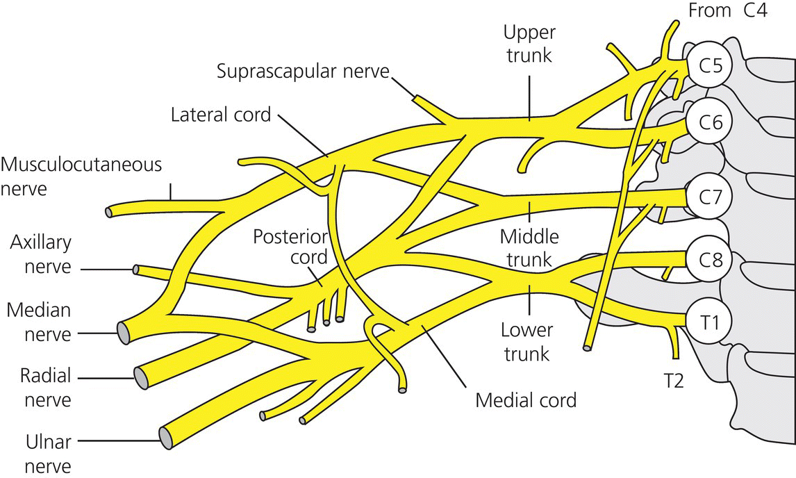
A plexus is a network of nerves or blood vessels and brachial is an adjective relating to or affecting the arm. The brachial plexus is a network of nerves that is formed from the union of five spinal nerves: the anterior (ventral) rami of spinal nerves from the four lowest cervical roots (C5, C6, C7 and C8) and the first thoracic root (T1) (Figure 20.1). The spinal nerves result from a fusion of many smaller spinal rootlets that have all passed through cells in the spinal ganglion. The spinal nerves exit from the vertebral foramen and then undergo a complex joining and division of nerve fibres to form the brachial plexus. This network of nerves extends inferiorly and laterally on either side of the last four cervical and first thoracic vertebrae before passing above the first rib posterior to the clavicle and then entering the axilla. The brachial plexus descends into the posterior triangle of the neck between the scalenus anterior and medius muscles.

Figure 20.1 The brachial Plexus(With permission: Scottish National Brachial Plexus Injury Service, Information for patients, Dec 2012. Medical Illustration Services, NHS Greater Glasgow and Clyde)
The nerves form three trunks:
- upper (from C5 and 6)
- middle (C7)
- lower (C8 and T1).
Each trunk divides into two branches: anterior and posterior. The anterior branch forms the two anterior cords: lateral and medial. The musculocutaneous nerve and the lateral root of the median nerve stem from the lateral cord. The medial cord gives rise to the medial root of the median nerve, the ulnar nerve, the medial cutaneous nerve and the medial antebrachial cutaneous nerve. The lateral and medial roots of the median nerve unite to form the median nerve. The three posterior branches (divisions) unite to form the posterior cord which gives rise to the axillary and radial nerve (Tortora and Grabowski 2003).
The brachial plexus provides the entire nerve supply of the shoulders and upper limbs. Five important nerves arise from the brachial plexus (each nerve has sensory and motor components):
- axillary nerve – supplies the deltoid and teres minor muscles
- musculocutaneous nerve – supplies the flexor muscles of the arm
- radial nerve – supplies the muscles of the posterior aspect of the arm and forearm
- median nerve – supplies most of the muscles of the anterior forearm and some of the muscle of the hand
- ulnar nerve – supplies the anteromedial muscles of the forearm and most of the muscles of the hand.
Mechanisms of injury
Brachial plexus injuries are caused by damage to some or all of the nerves. Injuries can be classified in different ways and are often referred to as being open or closed injuries or high impact or low impact injuries:
- Open injuries occur as a result of a penetrating wound that can lacerate the nerves e.g. injury from an assault by a broken bottle, knife or falling through a glass door.
- Closed injuries occur as a result of either high impact injury or low impact injury.
- High impact injuries are often associated with motor vehicle collisions and cause a traction injury from a violent stretching or pulling force between the clavicle and the shoulder girdle. The plexus can also be compressed from injured and damaged tissues in the vicinity or from adjacent bony injury such as comminuted fractures of the clavicle, causing local haematoma formation (Krishnan et al., 2008). There are also conditions in babies which are obstetric in origin causing brachial plexus palsies known as Erb’s palsy or Klumpke’s paralysis.
- Low impact injuries are usually the result of blunt trauma to the neck and upper limb causing crushing of the nerves. Rarely there may be radiation-induced damage to the plexus causing a brachial radiopathy from locally treated malignancies (e.g. breast, sarcoma) or a malignant infiltration into the area.
If the injury was sustained due to a high velocity accident e.g. a motorcycle collision, the likelihood of a more serious pathology is much greater than when the injury is sustained from a comparably low velocity fall. Patients involved in high velocity accidents are also more likely to sustain other injuries such as head injuries, spinal and upper limb fractures and vascular damage (see Chapters 16 and 19 for further information). These other injuries have to be considered when prioritising care for the newly injured patient.
Grades of injury
Patients who have sustained an injury or damage to the brachial plexus will present with motor and sensory loss in all or part of the upper limb depending on the extent of the injury. The damage to the brachial plexus nerves can be classified into four different grades (SNBPI Information for Patients 2012):
- Pre-ganglionic tear − Nerve root avulsion. The nerves are torn away from their roots in the spinal cord. The nerve root cannot be rejoined to the cord. Some function of the arm will be permanently lost. Any possibility of surgery may mean that nerves may be transferred from other areas to improve function.
- Post-ganglionic tear – Neurotmesis. The nerve has been stretched to breaking point and has been snapped or torn (similar to an overstretched elastic band). Ruptures will not heal without surgery.
- Severe lesion in-continuity – Axonotmesis. The nerve is stretched but remains intact, damaged but not torn apart. The nerves may recover to a variable degree on their own, but this may take some months. This type of injury may not require surgical treatment.
- Mild lesion in-continuity – Neurapraxia. With this injury the nerve is minimally stretched or compressed with no structural damage. The sensitive nerve fibres temporarily stop working but will usually recover without surgery.
The number and combination of nerves injured are very variable. It should be noted that some patients can present with a combination of root avulsions, post-ganglionic tears and lesions in-continuity. Injuries can also be classified depending on the anatomical region of the plexus that is affected e.g. supraclavicular or infraclavicular.
Supraclavicular injuries
Supraclavicular injuries can be caused by a traction injury to the brachial plexus e.g. in a motorcycle accident where the head is flexed sideways and the shoulder girdle is depressed or through direct trauma e.g. a knife injury or gunshot wound. Common patterns of supraclavicular injury can be subdivided into three groups:
- Upper plexus – from C5, C6 (+/– C7 and +/– C8). If C7 and C8 are involved the roots are sometimes avulsed. There is less likelihood that the roots of C5 and C6 will be avulsed.
- Total plexus – there is damage to all nerve roots. C5, C6 may have post-ganglionic ruptures (neurotmesis) with the roots of C8 and T1 avulsed.
- Lower plexus – the roots of C8 and TI are avulsed but C5 and C6 are working normally.
A high velocity accident is more likely to cause avulsion of the nerve roots from the spinal cord. Patients presenting with avulsion injuries usually complain of an instantaneous onset of pain. This is commonly described as a deep burning pain with frequent shocks of shooting pains throughout the day. The pain is caused by deafferentation of the dorsal horn of the spinal cord, which means that without input from the periphery, pain information passes from the dorsal horn to the brain unmodulated. Interestingly, these patients usually do not have problems with sleep disturbance due to pain.
There are a number of clinical factors that indicate a more serious lesion:
- high impact injury
- burning or shooting pains present since the time of injury
- possible Horner’s sign.
Horner’s sign (syndrome) – symptoms include a constricted pupil (miosis), drooping of the upper eyelid (ptosis) and absence of sweating of the face on the affected side (anhydrosis). These symptoms are due to a disorder of the sympathetic nerves.
Infraclavicular injuries
This type of injury can affect any one or all of the peripheral nerves. The most common presentations are:
- a complete lesion (neurotmesis)
- damage to the axillary nerve
- damage to the musculocutaneous nerve.
Injuries are usually caused by excessive traction of the brachial plexus e.g. following shoulder dislocation or in conjunction with a fracture of the humerus.
It is especially important to check with patients presenting following shoulder dislocation that the disruption of shoulder movement is not caused by a tear in the rotator cuff. Where there has been a severe infraclavicular injury affecting several peripheral nerves, the surgeon may choose to reconstruct only some of the peripheral nerves. This could be because the gap between the damaged nerve ends is too wide to successfully bridge. If a nerve is irreparable it is sometimes used to reconstruct another peripheral nerve.
There are some clinical factors that indicate a relatively mild lesion:
- low impact injury
- no pain
- Tinel’s sign
- absent Horner’s sign.
Tinel’s sign is a method of checking the regeneration or activity of a nerve by direct tapping over the pathway of the nerve sheath to elicit a distal tingling sensation (axonal regeneration advances by approximately 1 mm per day) (Hems 2000).
Diagnosis and investigations
A thorough clinical examination is essential, noting all nerve actions and muscle power using the Medical Research Council (MRC) Scale for Muscle Strength (Table 20.2). This should be documented in detail according to muscle group. X-rays of the cervical spine, shoulder and clavicle may be taken to exclude any skeletal/bony injury. A MRI scan will often help to confirm the diagnosis. The location of any root avulsion can sometimes be seen on the scan image if there is a meningocele (sack filled with cerebrospinal fluid leaking from the spinal cord). Neurophysiology or electrical nerve conduction tests record the passage of electrical signals along nerves in the limbs using small electrical pulses on the skin. This may include a recording of the electrical activity of muscles using fine needles. These tests can be used to diagnose a variety of nerve or muscle problems.
Management of a patient with a brachial plexus injury
Surgical management
If there is a diagnosed nerve injury that is suitable for surgical intervention, there are a few options available. It is possible to repair damaged nerves. In order to have a chance of success this surgery must be performed within a few months of the injury. Exploratory surgery is advised to ascertain the anatomical state of the damaged nerve and then the most appropriate surgical procedure is performed. There are a number of factors that are collectively considered prior to surgery, including the level or grade of injury, the age and fitness of the patient and any other co-morbidities.
Surgical options include:
- Direct nerve suturing – if there is minor damage to the nerve and the ends are in approximation and can be directly micro-sutured/glued.
- Nerve graft – usually when the nerves are torn, the damaged segment of nerve either side of the injury must be removed and repaired using grafts from elsewhere. Common nerve donor sites are the cutaneous nerves in the forearm or the sural nerve in the lower leg. These are sensory nerves and will leave a feeling of paraesthesia or numbness over the nerve distribution area, but have little effect on function. The nerve graft acts as a guide through which new nerve fibres can grow and cross the gap caused by the injury. Growth is very slow, recovery time is lengthy and complete recovery may be impossible due to the way that each individual microscopic nerve fibre grows.
- Nerve Transfer – undamaged nerves in the area that are performing less valuable functions can be transferred to other parts of the brachial plexus to try and regain some function within the limb. As the nerves used in this transfer start to recover, the patient needs to work very hard at retraining these nerves to move the arm and initially they may have to engage different movements to make the arm function.
These primary surgical options are undertaken as soon as is reasonably possible after full investigations and preparation of the patient. Recovery following primary surgery varies according to the individual, the level of injury and the type of surgery performed. There may be a period of complete immobilisation of the arm in a sling or a plan for immediate physiotherapy. It is important to follow the postoperative care plan as advised by the surgeon and ensure the patient is fully informed at every stage. The need for later or secondary surgical procedures is entirely dependent on the individual’s recovery from injury or primary surgery. The main aim of any surgery is to improve functional ability and secondary surgery is often not considered until two years after injury or primary surgery. This is a recognised time scale for ultimate recovery potential to be reached, although it is known that some nerve regeneration can continue for a longer period of time.
Secondary surgical procedures fall into three categories:
- bony/joint fusion (arthrodesis) – commonly wrist or shoulder
- tendon transfer
- muscle transfer.
Arthrodesis
The joints most commonly fused are the wrist and the shoulder. Bony fusions will only be considered when there is no chance of further useful recovery. This is one of the most common secondary operations undertaken. It is performed at the shoulder because the patient has poor shoulder control but has gained other functional return in the hand and elbow. The main aim of this surgery is to stabilise the shoulder to optimise elbow and hand function but allow some active movement of the upper limb through the scapulothoracic joint (Sousa et al., 2011). The patient must have good thoracoscapular muscle power (e.g. upper trapezius, serratus anterior) to be suitable for this type of surgery. Fusion of the joint may also be helpful in relieving pain.
Following shoulder arthrodesis the joint is immobilised in an abduction brace for at least six weeks. Patients are given advice about the position, function and appearance of the brace. Once the brace is removed they can start passive and active movements. The increase in range of movement usually progresses quite quickly, with the expectation that they will be able to achieve between 60 and 90 degrees of elevation and abduction. The patient is warned that they will have loss of medial rotation and putting their ‘hand behind back’ and that the arm will hang in a slightly abducted position. It is vital to manage the patient’s expectations carefully and ensure they are thoroughly informed about expected improvements and expected limitations.
Arthrodesis is performed at the wrist to correct the effects of muscle dysfunction. The fusion of the wrist joint can assist in maximising finger function by stabilising the wrist in a neutral position. It can provide the possibility of synchronising movements and enable easier tasks such as lifting and holding objects using the hand (Terzis and Barmpitsioti 2009).
Tendon transfer
Tendon transfer surgery is most commonly performed in the hand and is necessary when the function of a specific muscle is lost because of nerve injury. If a nerve is injured and cannot be repaired, the nerve no longer sends signals to particular muscles. Those muscles are paralysed and their function is lost. Tendon transfer surgery can be used to attempt to replace that function by leaving the origin of the muscle in place and moving the tendon insertion (attachment) to a different position. It can be sutured into a different bone or a different tendon. When the muscle fires after the insertion point has been moved it will produce a different action to previously, depending on where it has been inserted. Patients who present only with avulsion of the lower trunk (C8–T1) nerve roots may eventually be considered for tendon transfer. In order for this to be successful it is important to teach the patient how to maintain range of joint movement and to maximise the strength in the muscle groups that are still functional.
Tendons used for transfer must be of good quality (Grade 4 or better). It is important that tendon transfers in the hand are planned so that pinch and grip will be improved. Rehabilitation involves the re-education of function, occasionally with trick movements or with the co-ordination of other movements e.g. wrist extension with finger flexion.
Muscle transfer
A variety of muscles may be used as free transfers or transplantations and this surgery is often performed by a plastic surgeon. The aim is often to restore elbow flexion or wrist extension. The muscles used include latissimus dorsi and gracilis.
Non-surgical management
Some patients will have suffered temporary damage to the conduction of the nerve e.g. a neurapraxia or an axonotmesis or those who have had a virus causing a brachial neuritis. These injuries/pathologies can take from several months to over a year to recover and it is essential that the patient understands this and the importance of maintaining range of joint movement while waiting for recovery. It may be necessary to provide some form of splinting to aid function and/or to maintain hand position. Early signs of recovery are sometimes difficult to detect and this highlights the importance of accurate record keeping. Once a flicker of muscle contraction can be detected the patient should then commence prescribed exercises to maximise this improvement. This can include muscle stimulation and gravity-assisted exercises. Muscle stimulation is an option that can be helpful at this stage. When the muscles that have not been working start to show signs of working, it will often be a very small flicker. In the early recovery phase this may not be enough to move the joints involved so it can be difficult for the individual to exercise. Muscle stimulation works by using a device that electrically stimulates the muscles with pads placed on the skin over the muscle that is to work. It stimulates the muscles to contract and can help to give the feeling of moving them again, but it does not replace an exercise programme.
Rehabilitation and ongoing care and support
Many patients will experience pain of a neuropathic nature which can be difficult to treat and requires specific neuro-analgesics. Some of these drugs have another mode of use as antidepressants or antiepileptics. The use of other techniques in addition to drug therapy can be useful to help the patient ‘live with’ the pain. These may include psychological exercises such as relaxation, visualisation of pain and guided imagery, cognitive behavioural therapy and specific directed counselling. General pain management issues are considered in more detail in Chapter 11.
Acceptance of the effects of the injury and changes to body image should be addressed with counselling support. The purpose of therapy is to focus on the effects of pain on behaviour, mood, function and activity. Intervention involves setting goals such as being more active, returning to previous activities and how to use the pain relief achieved to reach these goals. The aim is to improve the individual’s ability to function and enhance their ability to cope with the pain. The use of a TENS device may also be useful. This is a small portable electrical device which is designed to help relieve pain. It works by sending a harmless electrical current through pads that are placed on the skin. This is felt as pins and needles and these sensations can help to block pain messages. It can be used on various parts of the body but only on skin that has normal feeling.
Current developments in relieving deafferentation pain following brachial plexus injury are starting to filter into clinical practice. The dorsal root entry zone (DREZ) procedure is performed as a last resort for resolution of intractable pain associated with some neuropathic pain syndromes. This invasive procedure is performed via a myelotomy or laminectomy. Thermo-coagulation (the use of heat generated by an electric current to destroy tissue) is selectively applied and destroys the posterolateral aspect of the spinal cord corresponding to the area through which dorsal (sensory) root fibres enter the cord itself. When used in appropriately selected patients, particularly in those with post-traumatic avulsion of the brachial plexus, the DREZ procedure may produce lasting pain relief (in a majority of patients), but carries some significant potential side effects including loss of feeling and temperature sensitivity (the pathway is blocked at the spinal cord and messages no longer cross over), weakness and a loss of proprioception (Blaauw et al., 2008).
Physical activity of any type is advantageous for a number of reasons:
- It is known to release endorphins into the bloodstream which can act as natural analgesics and help to improve the patient’s mood.
- It is good for the body to stay in good physical condition to help with the healing process.
- If an individual enjoyed sports or other physical activities before their injury it is very helpful to return to them as soon as possible to fulfil their enjoyment.
There are many ways of adapting activities to allow a return to them even with an injured or non-functioning upper limb. Participation in sport has well known benefits to health, wellbeing and self-esteem. The majority of activities can be adapted to allow participation. Patients have been reported as being able to return to a variety of sport, leisure and hobby activities using adaptations including fishing, playing guitar, running, cycling, playing snooker and gardening. Quality of life issues for patients with brachial plexus injury are considered in Box 20.2.
Box 20.2 Evidence digest. Reproduced with permission from Elsevier
There may be a need for an orthosis to be fitted to aid in supporting a flail or paralysed arm that is not functional. Specialised orthoses that work using biomechanical principles may be provided on an individual basis according to the level of injury and functional ability.
The aims of orthoses are to:
- Reduce shoulder pain (as subluxation from poorly controlled shoulder musculature can be problematic).
- Allow improved positioning of the hand for functional activities (the ability to ‘crank’ the orthosis from a position of elbow extension to flexion is possible).
- Improve cosmetic appearance of the upper limb (which may ‘sit’ in a more neutral position).
The patient may also require adaptations or modifications to a vehicle so that they may safely drive it. These may include steering wheel ‘rotators’, change to automatic transmission, positioning of indicator switches etc.
Even though brachial plexus injury is uncommon, practitioners working in the trauma setting are likely to care for patients in the early stages of their recovery. The complexity of brachial plexus injuries can provide a challenge to the healthcare professional. A holistic approach to the assessment, treatment, care and support of the patient and their family is best provided by an interdisciplinary team with specialist knowledge and expertise. Team members will include consultant medical staff, nurse specialists and therapy practitioners as well as other professionals such as psychologists, pain specialists and orthotists. In the UK, for example, such care is currently provided in only three specialist brachial plexus injuries units who receive referrals from around the country.
Suggested further reading
- Anscomb, S.J. (2007) Managing sprains and strains. Practice Nurse, 33(5), 44, 46, 48–49.
- Bailey, R., Kaskutas, V., Fox, I., Baum, C.M. and Mackinnon, S.E. (2009) Effect of upper extremity nerve damage on activity participation, pain, depression and quality of life. Journal of Hand Surgery, 34(a), 1682–1688.
- Elton, S.G. and Rizzo, M. (2008) Management of radial nerve injury associated with humeral shaft fractures: an evidence-based approach. Journal of Reconstructive Microsurgery, 24(8), 569–573.
- Norris, B.L. and Kellam, J.F. (1997) Soft tissue injuries associated with high-energy extremity trauma: principles of management. Journal of the American Academy of Orthopaedic Surgeons, 5(1), 37–46.
- Wardrope, J., Barron, D., Draycott, S. and Sloan, J. (2008) Soft tissue injuries: principles of biomechanics, physiotherapy and imaging. Emergency Medicine Journal, 25, 158–162.
References
- Airaksinen, O.V., Kyrklund, N., Latvala, K. et al. (2003) Efficacy of cold gel for soft tissue injuries. American Journal of Sports Medicine, 31(5), 680–684.
- Altizer, L. (2003) Strains and sprains. Orthopaedic Nursing, 22 (6), 404–409.
- Blaauw, G., Muhlig, R.S., and Vredeveld, J.W. (2008) Management of brachial plexus injuries. Advances and Technical Standards in Neurosurgery, 33, 201–231.
- British Orthopaedic Association (BOA) (2011) the Management of Nerve Injuries: A Guide to Good Practice. BOA, London. Available at: http://www.boa.ac.uk/Publications/Documents/The%20Management%20of%20Nerve%20Injuries.pdf (accessed 7 April 2014).
- Collins, M. and Raleigh, S.M. (2009) Genetic risk factors for musculoskeletal soft tissue injuries. Medicine and Sport Science 54: 136–149.
- Hardy, M. and Snaith, B. (2011) Musculoskeletal Trauma. Churchill Livingstone, Edinburgh.
- Hems, T.E.J. (2000) Nerves, in Bailey and Love’s Short Practice of Surgery, 23rd edn (eds R.C.G. Russell, N.S. Williams and C.J.K. Bulstrode), Arnold, London, pp. 530–568.
- Krishnan, K.G., Mucha, D., Gupta, R., and Schackert, G. (2008) Brachial plexus compression caused by recurrent clavicular non-union and space-occupying pseudoarthrosis: definitive reconstruction using free vascularised bone flap – a series of eight cases. Neurosurgery, 62(5 Suppl 2), 461–469; discussion 469–470.
- Medical Research Council (1981) Aids to the Examination of the Peripheral Nervous System. Memorandum 45. Her Majesty’s Stationery Office, London.
- Scottish National Brachial Plexus Injury Service (SNBPI) (2012) Information for Patients. Available at: www.brachialplexus.scot.nhs.uk (accessed 7 April 2014).
- Sousa, R., Pereira, A., Massada, M. et al. (2011) Shoulder arthrodesis in adult brachial plexus injury: what is the optimal position? Journal of Hand Surgery (European Volume), 36(7), 541–547.
- Terzis, J.K., and Barmpitsioti, A. (2009). Wrist fusion in posttraumatic brachial plexus palsy. Plastic and Reconstructive Surgery, 124(6), 2027.
- Tortora, G.J. and Grabowski, S.R. (2003) Principles of Anatomy and Physiology. John Wiley and Sons Inc., New York
- Wellington, B. (2009) Quality of life issues for patients following traumatic brachial plexus injury – Part 1. Journal of Orthopaedic Nursing, 13(4), 194–200.
- Wellington, B. (2010) Quality of life issues for patients following traumatic brachial plexus injury – Part 2. International Journal of Orthopaedic and Trauma Nursing, 14(1), 5–11.


