[level-membership-for-dermatology-category]
Skin cancer – Malignant melanoma
Malignant melanoma is a malignant tumour of melanocytes, usually arising in the epidermis. It is the most lethal of the main skin tumours and has increased in incidence over the last three decades. The important pathogenic role of excessive ultraviolet (UV) radiation exposure has been the subject of public education campaigns. Genetics may be important, and up to 5% of patients have a family history of malignant melanoma.
Clinical presentation
Four main clinicopathological variants are recognized. These are described below.
Superficial spreading malignant melanoma
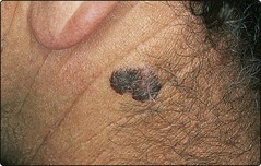
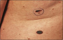
This type accounts for 50% of all British cases, shows a female preponderance and is commonest on the lower leg. The tumour is macular and shows variable pigmentation, often with regression (Fig. 1).
Lentigo malignant melanoma
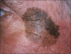
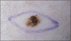
Malignant melanoma developing in a longstanding lentigo maligna (Fig. 2) constitutes 15% of UK cases. A lentigo maligna arises in sun-damaged skin, often on the face of an elderly person who has spent many years in an outdoor occupation.
Acral lentiginous malignant melanoma
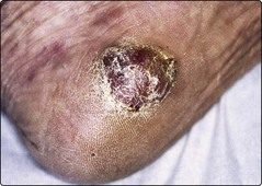
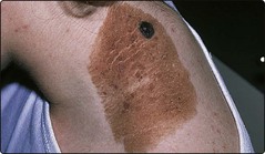
The acral lentiginous type makes up 1 in 10 of British cases, but is the commonest form in dark-skinned races. The tumour affects the palms, soles (Fig. 3) and nail beds, is often diagnosed late and has poor survival figures.


Staging
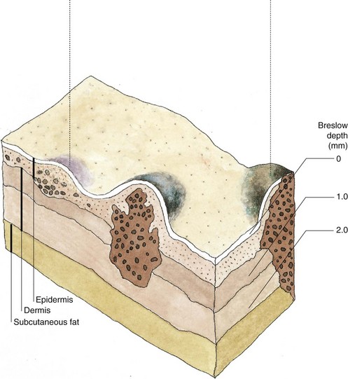
Malignant melanomas usually progress through two phases: horizontal growth in the epidermis then vertical invasion of the dermis (Fig. 5).
Aetiopathogenesis
The main risk factor that increases risk of melanoma is exposure to UV radiation. Some people are more at risk of melanoma than others (Fig. 6). Histological evidence of a pre-existing melanocytic naevus is found in 30% of malignant melanomas but, with the exception of dysplastic or congenital naevi (Fig. 7), the risk of change in a common melanocytic naevus is small.

Diagnosis
Any of the following changes in a naevus or pigmented lesion may suggest malignant melanoma:


Prognosis
The prognosis relates to the tumour depth. The approximate 5-year survival rates are:


Examples of thin and thick tumours are given in Figures 8 and 9.
Management
2. lymphatic – either in the regional lymph nodes or in transit in the lymphatics draining from the tumour to the nodes
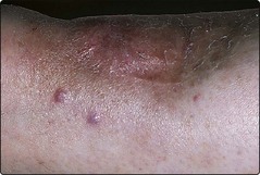
Fig. 10 Hypomelanotic recurrent malignant melanoma.
Pink papules of recurrent tumour are evident at the edge of a previously excised and grafted site.
Routine sentinel node biopsy (p. 115) or elective lymph node dissection is not recommended as a standard procedure at present. Radiotherapy is of limited use. Interferon-alpha may increase survival in patients with tumours more than 1.5 mm thick. For metastatic disease, chemotherapy with dacarbazine is the current standard but has limited effectiveness and significant toxicity. New therapies including ipilimumab, a monoclonal antibody targeting the negative T cell regulator molecule, CTLA-4, has been shown to improve survival in advanced melanoma, and BRAF kinase inhibitors in BRAF mutated melanomas have shown promise.
Prevention and public education


[/level-membership-for-dermatology-category][not-level-membership-for-dermatology-category]
Skin cancer – Malignant melanoma
Malignant melanoma is a malignant tumour of melanocytes, usually arising in the epidermis. It is the most lethal of the main skin tumours and has increased in incidence over the last three decades. The important pathogenic role of excessive ultraviolet (UV) radiation exposure has been the subject of public education campaigns. Genetics may be important, and up to 5% of patients have a family history of malignant melanoma.
Clinical presentation
Four main clinicopathological variants are recognized. These are described below.
Superficial spreading malignant melanoma
This type accounts for 50% of all British cases, shows a female preponderance and is commonest on the lower leg. The tumour is macular and shows variable pigmentation, often with regression (Fig. 1).
Lentigo malignant melanoma
Malignant melanoma developing in a longstanding lentigo maligna (Fig. 2) constitutes 15% of UK cases. A lentigo maligna arises in sun-damaged skin, often on the face of an elderly person who has spent many years in an outdoor occupation.
Acral lentiginous malignant melanoma
The acral lentiginous type makes up 1 in 10 of British cases, but is the commonest form in dark-skinned races. The tumour affects the palms, soles (Fig. 3) and nail beds, is often diagnosed late and has poor survival figures.
