Skin and Subcutaneous Tissue


Anatomy and Physiology
Skin
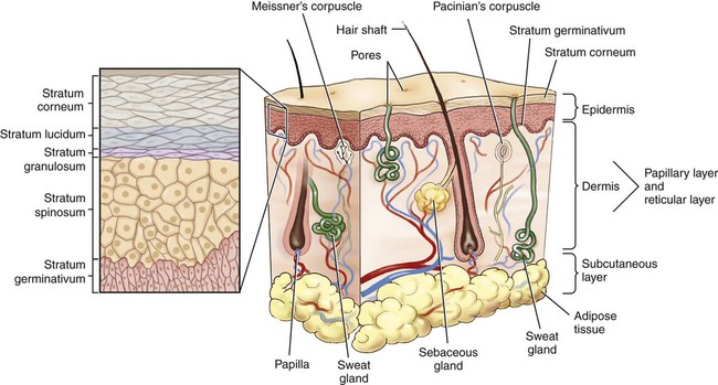
The skin is composed of two layers: the epidermis, which forms the outermost layer, and the dermis or corium, the inner layer (Fig. 4-1). The dermis is attached to a layer of connective tissue called the hypodermis or the subcutaneous layer, which is mainly composed of fat (adipose tissue).
 Note
Note
This chapter includes all the anatomy necessary to assign ICD-10 skin and subcutaneous tissue codes, including detail on sebaceous glands, sweat glands, and the dermis. See Appendix F for a complete list of body parts and how they should be coded.
 Be Careful!
Accessory Structures
Nails
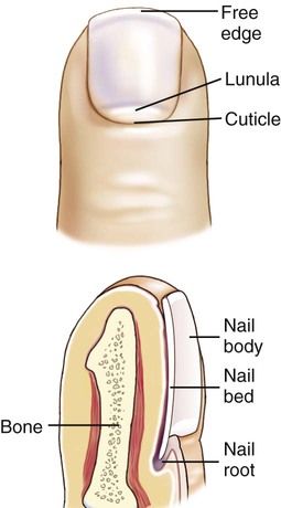
Nails cover and thus protect the dorsal surfaces of the distal bones of the fingers and toes. The part that is visible is the nail body (also called the nail plate), whereas the nail root (matrix) is in a groove under a small fold of skin at the base of the nail. The nail bed is the highly vascular tissue under the nail that appears pink when the blood is oxygenated or blue/purple when it is oxygen deficient (Fig. 4-2). The moonlike white area in the base of the nail is called the lunula (meaning little moon), behind which new growth occurs. The small fold of skin above the lower part of the nail is called the cuticle, or eponychium. The paronychium is the fold of skin that is near the sides of the nail.
 Be Careful!
 Exercise 1:
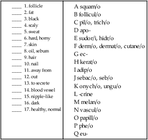
 To practice labeling the structure of the skin, click on Label It.
 Exercise 2:
Operative Report
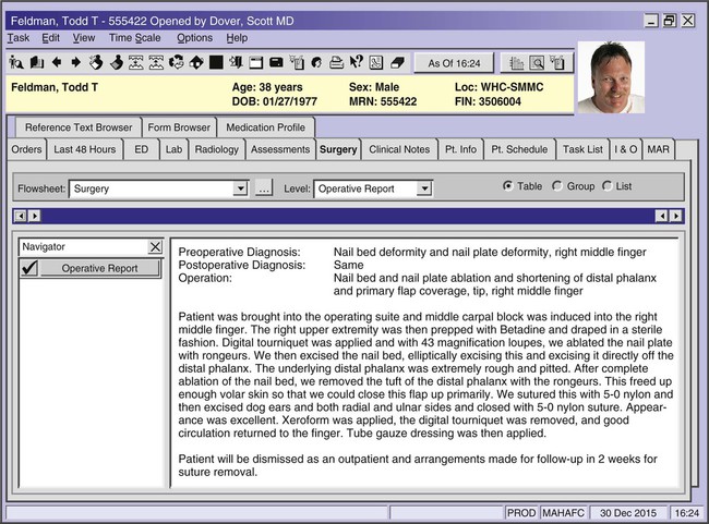
Using the above operative report above, answer the following questions:
1. What are the nail bed and nail plate?
___________________________________________________________________________________________________
2. Where is the “volar skin” located? (Refer to Chapter 2 if you’ve forgotten.) __________________________
3. What is a “digital tourniquet,” and why do you think it was used?
4. From your knowledge of the anatomy of a nail, what, if any, parts of the nail do you think remain?
___________________________________________________________________________________________________
Combining Forms for the Anatomy of the Integumentary System
| Meaning | Combining Form |
| base, bottom | bas/o |
| black, dark | melan/o, phe/o |
| fat | adip/o, lip/o |
| follicle | follicul/o |
| hair | trich/o, pil/o |
| hard, horny | kerat/o, corne/o |
| nail | onych/o, ungu/o |
| papilla, nipple | papill/o |
| scaly | squam/o |
| sebum, oil | seb/o, sebac/o |
| skin | derm/o, dermat/o, cut/o, cutane/o |
| sweat | hidr/o, sudor/i |
| vessel | vascul/o |
Prefixes for the Anatomy of the Integumentary System
| Prefix | Meaning |
| a- | no, not, without |
| apo- | separate, away from |
| ec- | out, outward |
| epi- | above |
| eu- | healthy, normal |
| hypo-, sub- | under, below |
| par- | near |
Suffixes for the Anatomy of the Integumentary System
| Suffix | Meaning |
| -al, -ar, -ous, -ic | pertaining to |
| -crine | to secrete |
| -cyte | cell |
| -ferous | pertaining to carrying |
| -ium | structure |
 You can review the anatomy of the integumentary system by clicking on Body Spectrum, then Integumentary.
Pathology
Skin Lesions
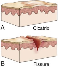
A skin lesion is any visible, localized abnormality of skin tissue. It can be described as either primary or secondary. Primary lesions (Fig. 4-3) are early skin changes that have not yet undergone natural evolution or change caused by manipulation. Secondary lesions (Fig. 4-4) are the result of natural evolution or manipulation of a primary lesion.
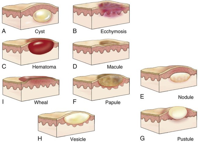
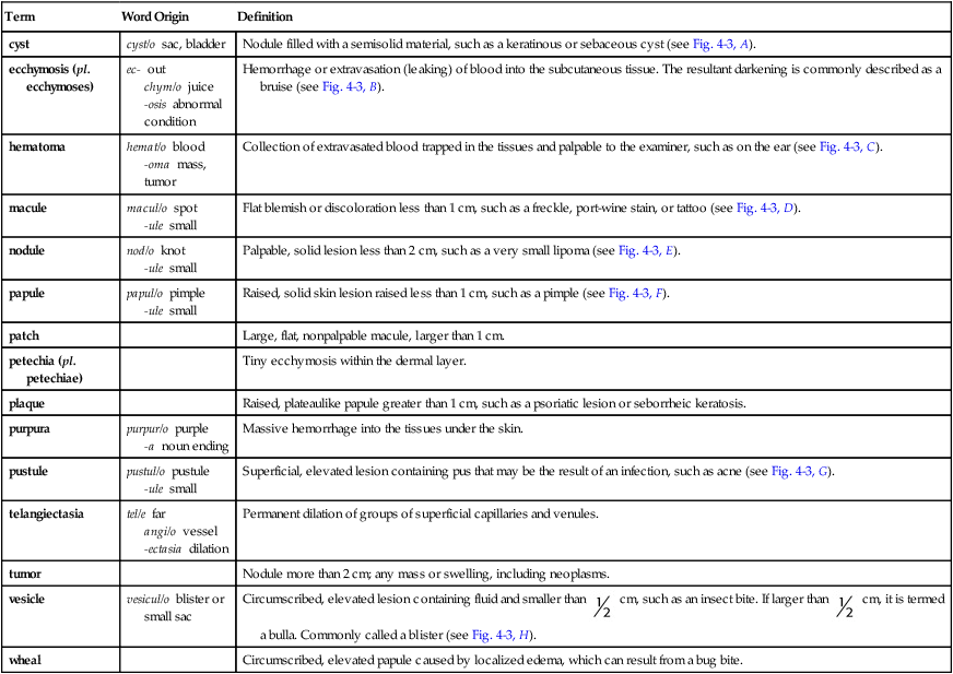
Terms Related to Primary Skin Lesions
| Term | Word Origin | Definition |
| cyst | cyst/o sac, bladder | Nodule filled with a semisolid material, such as a keratinous or sebaceous cyst (see Fig. 4-3, A). |
| ecchymosis (pl. ecchymoses) | ec- out chym/o juice -osis abnormal condition |
Hemorrhage or extravasation (leaking) of blood into the subcutaneous tissue. The resultant darkening is commonly described as a bruise (see Fig. 4-3, B). |
| hematoma | hemat/o blood -oma mass, tumor |
Collection of extravasated blood trapped in the tissues and palpable to the examiner, such as on the ear (see Fig. 4-3, C). |
| macule | macul/o spot -ule small |
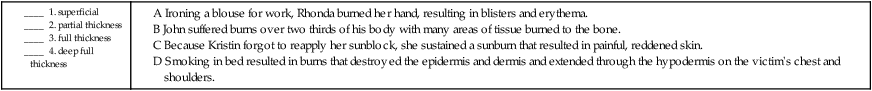
Flat blemish or discoloration less than 1 cm, such as a freckle, port-wine stain, or tattoo (see Fig. 4-3, D). |
| nodule | nod/o knot -ule small |
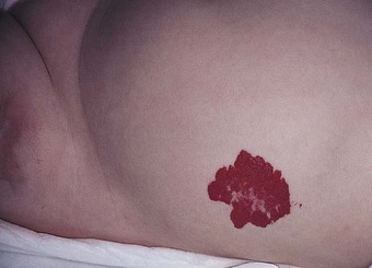
Palpable, solid lesion less than 2 cm, such as a very small lipoma (see Fig. 4-3, E). |
| papule | papul/o pimple -ule small |
Raised, solid skin lesion raised less than 1 cm, such as a pimple (see Fig. 4-3, F). |
| patch | Large, flat, nonpalpable macule, larger than 1 cm. | |
| petechia (pl. petechiae) | Tiny ecchymosis within the dermal layer. | |
| plaque | Raised, plateaulike papule greater than 1 cm, such as a psoriatic lesion or seborrheic keratosis. | |
| purpura | purpur/o purple -a noun ending |
Massive hemorrhage into the tissues under the skin. |
| pustule | pustul/o pustule -ule small |
Superficial, elevated lesion containing pus that may be the result of an infection, such as acne (see Fig. 4-3, G). |
| telangiectasia | tel/e far angi/o vessel -ectasia dilation |
Permanent dilation of groups of superficial capillaries and venules. |
| tumor | Nodule more than 2 cm; any mass or swelling, including neoplasms. | |
| vesicle | vesicul/o blister or small sac | Circumscribed, elevated lesion containing fluid and smaller than  cm, such as an insect bite. If larger than cm, such as an insect bite. If larger than  cm, it is termed a bulla. Commonly called a blister (see Fig. 4-3, H). cm, it is termed a bulla. Commonly called a blister (see Fig. 4-3, H). |
| wheal | Circumscribed, elevated papule caused by localized edema, which can result from a bug bite. |
Terms Related to Secondary Skin Lesions
| Term | Word Origin | Definition |
| atrophy | a- no, not, without -trophy process of nourishment |
Paper-thin, wasted skin often occurring in the aged or as stretch marks (striae) from rapid weight gain. |
| cicatrix (pl. cicatrices) | A scar—an area of fibrous tissue that replaces normal skin after destruction of some of the dermis (see Fig. 4-4, A). | |
| eschar | eschar/o scab | Dried serum, blood, and/or pus. May occur in inflammatory and infectious diseases, such as impetigo, or as the result of a burn. Also called a scab. |
| fissure | Cracklike lesion of the skin, such as an anal fissure (see Fig. 4-4, B). | |
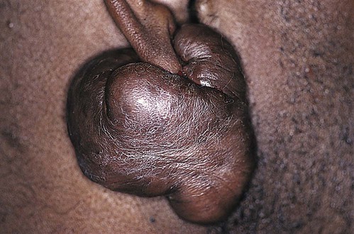
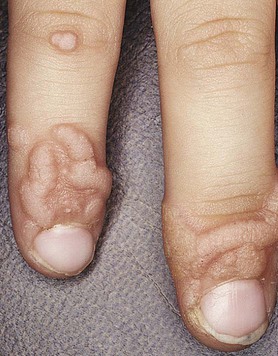
| keloid | Type of scar that is an overgrowth of tissue at the site of injury in excess of the amount of tissue necessary to repair the wound. The extra tissue is partially due to an accumulation of collagen at the site (Fig. 4-5). | |
| ulcer | Circumscribed, craterlike lesion of the skin or mucous membrane resulting from necrosis, or tissue death, that can accompany an inflammatory, infectious, or malignant process. An example is a pressure ulcer seen sometimes in bedridden patients (see Fig. 4-14). |
 Exercise 3:
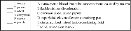
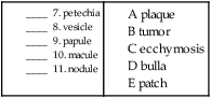
Match the smaller version of a primary skin lesion with the larger version.
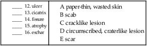
Match the secondary lesions with their definitions.
Terms Related to Infections of the Skin and Subcutaneous Tissue (LØØ-LØ8) and Bullous Disorders (L1Ø-L14)
| Term | Word Origin | Definition |
| abscess | A collection of pus in any part of the body, formed by a localized infection. | |
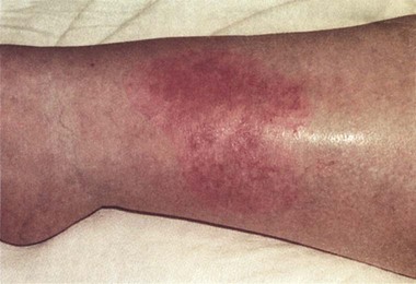
| cellulitis | cellul/o cell -itis inflammation |
Diffuse, spreading, acute inflammation within solid tissues. The most common cause is a Streptococcus pyogenes infection (Fig. 4-6). |
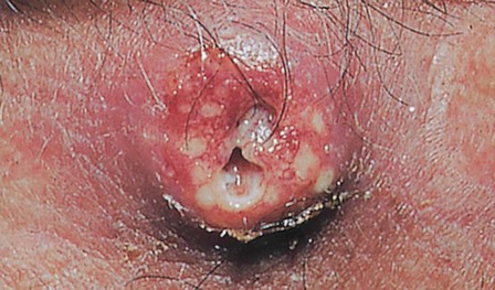
| furuncle | Localized, suppurative staphylococcal skin infection originating in a gland or hair follicle and characterized by pain, redness, and swelling (Fig. 4-7). If two or more furuncles are connected by subcutaneous pockets, it is termed a carbuncle. | |
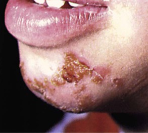
| impetigo | Superficial vesiculopustular skin infection, normally seen in children, but possible in adults (Fig. 4-8). | |
| omphalitis | omphal/o navel -itis inflammation |
Inflammation of the navel. |
| onychia | onych/o nail -ia condition |
Inflammation of the fingernail. Also called onychitis. |
| paronychia | par- beside, near onych/o nail -ia condition |
Infection of the skin beside the nail. |
| pemphigus | Autoimmune disorder characterized by large blisters of the skin and mucous membranes. | |
| pilonidal cyst | pil/o hair nid/o nest -al pertaining to |
Growth of hair in a cyst in the sacral region. |
| pyoderma | py/o pus -derma skin condition |
A purulent (containing pus) bacterial skin disease. |
Terms Related to Dermatitis and Eczema (L2Ø-L3Ø)
| Term | Word Origin | Definition |
| atopic dermatitis | a- no, not, without top/o place, location -ic pertaining to dermat/o skin -itis inflammation |
Chronic, pruritic, superficial inflammation of the skin usually associated with a family history of allergic disorders. |
| contact dermatitis | dermat/o skin -itis inflammation |
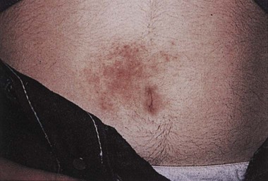
Irritated or allergic response of the skin that can lead to an acute or chronic inflammation (Fig. 4-9). |
| eczema | Superficial inflammation of the skin, characterized by vesicles, weeping, and pruritus. Also called dermatitis. | |
| pruritus | prurit/o itching -us noun ending |
Itching. May be described as occurring in a specific body area (e.g., ani, scroti, vulvae). |
| seborrheic dermatitis | seb/o sebum -rrheic pertaining to discharge dermat/o skin -itis inflammation |
Inflammatory scaling disease of the scalp and face. In newborns, this is known as cradle cap or seborrheic capitis. |
Terms Related to Papulosquamous (L4Ø-L45) Urticaria and Erythema (L49-L54) and Radiation-Related Disorders of the Skin and Subcutaneous Tissue (L55-L59)
| Term | Word Origin | Definition |
| actinic keratosis | act/i rays -in substance -ic pertaining to kerat/o hard, horny -osis abnormal condition |
Skin lesions caused by prolonged sun exposure. |
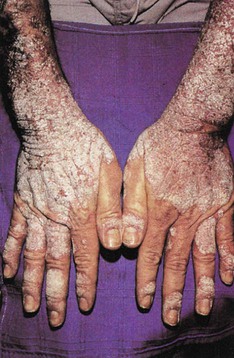
| psoriasis | Common chronic skin disorder characterized by circumscribed, salmon-red patches covered by thick, dry, silvery scales that are the result of excessive development of epithelial cells (Fig. 4-10). | |
| urticaria | Skin condition characterized by wheals. May be caused by allergies, insect bites, drugs, or stress. Also called hives. |
 Exercise 4:
Various Skin Diseases and Disorders
Fill in the blanks with the correct terms from the list below.
1. Autoimmune skin disorder characterized by large blisters __________________________________________
2. An irritated or allergic response of the skin that can lead to an acute or chronic inflammation
__________________________________________________________________________________________________
3. Diffuse spreading inflammation within solid tissues _______________________________________________
4. Inflammatory scaling disease of the scalp and face _________________________________________________
5. Term that means itching _________________________________________________________________________
6. Chronic skin disorder characterized by circumscribed salmon-red patches ___________________________
7. Growth of hair in a cyst in the sacral region of the skin ____________________________________________
8. A superficial vesiculopustular skin infection normally seen in children ______________________________
9. Skin lesion caused by prolonged sun exposure _____________________________________________________
10. Skin condition characterized by wheals ___________________________________________________________
11. Localized, suppurative staphylococcal skin infection in a gland or hair follicle _______________________
12. Chronic, pruritic superficial inflammation of the skin, usually associated with a family history of allergic disorders ________________________________________________________________________________
13. inflammation of the navel ________________________________________________________________________
14. pertaining to a hair nest _________________________________________________________________________
15. condition of nail ________________________________________________________________________________
16. condition beside the nail _________________________________________________________________________
Terms Related to Disorders of Skin Appendages (L6Ø-L75)
| Term | Word Origin | Definition |
| acne vulgaris | vulgar/o common -is noun ending |
Inflammatory disease of the sebaceous glands characterized by papules, pustules, inflamed nodules, and comedones (sing. comedo), which are plugs of sebum that partially or completely block a pore. Blackheads are open comedones, and whiteheads are closed comedones. |
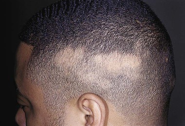
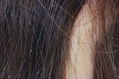
| alopecia | Hair loss, resulting from genetic factors, aging, or disease (Fig. 4-11). | |
| anhidrosis | an- no, not, without hidr/o sweat -osis abnormal condition |
A condition in which a person produces little or no sweat. |
| bromhidrosis | brom/o odor, stench hidr/o sweat -osis abnormal condition |
Disorder of the apocrine sweat glands; a condition of abnormal body odor. Note that the combining vowel o is dropped in this term. |
| folliculitis | follicul/o follicle -itis inflammation |
Inflammation of the hair follicles, which may be superficial or deep, acute or chronic. |
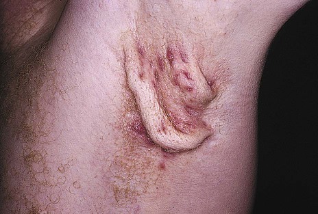
| hidradenitis suppurativa | hidraden/o sweat gland -itis inflammation |
Inflammation of the sweat glands (Fig. 4-12). |
| hyperhidrosis | hyper- excessive hidr/o sweat -osis abnormal condition |
Excessive perspiration caused by heat, strong emotion, menopause, hyperthyroidism, or infection. |
| hypertrichosis | hyper- excessive trich/o hair -osis abnormal condition |
Abnormal condition of excessive hair; also known as hirsutism. |
| keratinous cyst | kerat/o hard, horny -in substance -ous pertaining to |
Benign cavity lined by keratinizing epithelium and filled with sebum and epithelial debris. Also called a sebaceous cyst. |
| milia | Tiny, superficial keratinous cysts caused by clogged oil ducts. | |
| miliaria | Minute vesicles and papules, often with surrounding erythema (redness), caused by occlusion of sweat ducts during times of exposure to heat and high humidity. | |
| onychocryptosis | onych/o nail crypt- hidden -osis abnormal condition |
Abnormal condition of hidden (ingrown) nail. |
| onychodystrophy | onych/o nail dys- abnormal -trophy nourishment, development |
Abnormally developed finger or toe nails. |
| onychogryphosis | onych/o nail gryph/o curved -osis abnormal condition |
Abnormally thickened and curved finger or toe nails. |
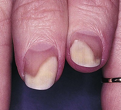
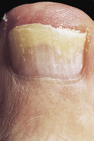
| onycholysis | onych/o nail -lysis loosening |
Separation of the nail plate from the nail bed (Fig. 4-13). |
| onychomalacia | onych/o nail -malacia softening |
Softening of the nails. |
| poliosis | poli/o gray -osis abnormal condition |
Localized patch of gray or white hair due to lack of melanin caused by disease. |
 Be Careful!
Paronychium is the structure that surrounds the nail. Paronychia is an infection of the nail.
 Be Careful!
 Be Careful!
Hidr/o with an i means sweat; hydr/o with a y means water. Both are pronounced HYE droh.
 Exercise 5:
Disorders of Skin Appendages
1. abnormal condition of curved nail ________________________________________________________________
2. softening of the nail ______________________________________________________________________________
3. abnormal condition of hidden nail ________________________________________________________________
4. abnormal condition of excessive hair ______________________________________________________________
5. inflammation of a hair follicle ____________________________________________________________________
6. abnormal condition of gray _______________________________________________________________________
7. abnormal condition of no sweating ________________________________________________________________
8. abnormal condition of sweat odor _________________________________________________________________
9. inflammation of a sweat gland ____________________________________________________________________
10. abnormal nail development _______________________________________________________________________
Terms Related to Other Disorders of the Skin and Subcutaneous Tissue (L8Ø-L99)
| Term | Word Origin | Definition |
| callus | Common, painless thickening of the stratum corneum at locations of external pressure or friction. | |
| chloasma | Increased pigmentation on face, often during pregnancy. Also called melasma and “mask of pregnancy.” | |
| corn | Horny mass of condensed epithelial cells overlying a bony prominence as the result of pressure or friction; also referred to as a clavus. | |
| discoid lupus erythematosus (DLE) | disc/o disc -oid like lupus erythemat/o red -osus pertaining to |
Chronic, inflammatory autoimmune condition that is limited to the skin. Characterized by red, round-shaped lesions. Also referred to as discoid erythematosus. |
| dyschromia | dys- abnormal chrom/o color -ia condition |
Abnormality of skin pigmentation. Hyperchromia is abnormally increased pigmentation. Hypochromia is abnormally decreased pigmentation. |
| ichthyosis | ichthy/o fish -osis abnormal condition |
Category of dry skin that has the scaly appearance of a fish. It ranges from mild to severe. The mild form is known as xeroderma. |
| lentigo | Darkened macules usually caused by sun exposure. Also called age spots. | |
| leukoderma | leuk/o white -derma skin condition |
Loss of skin pigmentation. |
| pressure ulcer | Inflammation, ulcer, or sore in the skin over a bony prominence. Most often seen in aged, debilitated, cachectic (wasted), or immobilized patients; pressure sores or ulcers are graded by stages of severity. The highest stage, stage 4, involves muscles, fat, and bone. Also known as a bedsore, decubitus ulcer, or pressure sore (Fig. 4-14). | |
| seborrheic keratosis | seb/o sebum -rrheic pertaining to discharge kerat/o hard, horny -osis abnormal condition |
Benign, circumscribed, pigmented, superficial warty skin lesion. |
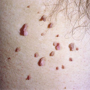
| skin tags | Small, soft, pedunculated (with a stalk) lesions that are harmless outgrowths of epidermal and dermal tissue, usually occurring on the neck, eyelids, armpits, and groin; usually occur in multiples. Also known as acrochordons (Fig. 4-15). | |
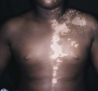
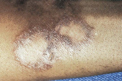
| vitiligo | Benign acquired disease of unknown origin, consisting of irregular patches of various sizes lacking in pigment (Fig. 4-16). | |
| xerosis cutis | xer/o dry -osis abnormal condition cut/o skin -is noun ending |
Dry skin. |
 CM Guideline Alert
 Exercise 6:
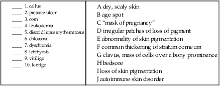
Terms Related to Viral Infections Characterized by Skin and Mucous Membrane Lesions (BØØ-BØ9)
| Term | Word Origin | Definition |
| exanthematous diseases | exanthemat/o rash -ous pertaining to |
Generally, viral diseases characterized by a specific type of rash (exanthem). The main ones are measles, rubella, fifth disease, roseola, and chickenpox. |
| herpes simplex virus (HSV) | Viral infection characterized by clusters of small vesicles filled with clear fluid on raised inflammatory bases on the skin or mucosa. HSV-1 causes fever blisters (herpetic stomatitis) and keratitis, an inflammation of the cornea. HSV-2 is more commonly known as genital herpes. | |
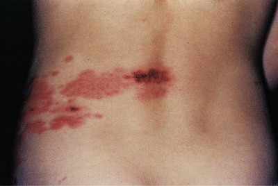
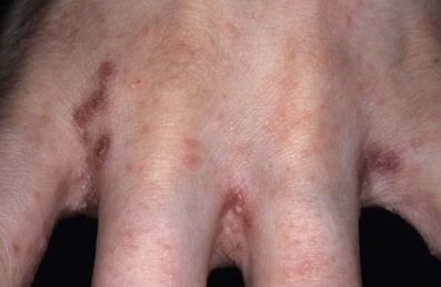
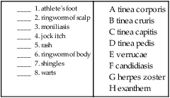
| herpes zoster | Acute, painful rash caused by reactivation of the latent varicella-zoster virus. Also known as shingles (Fig. 4-17). | |
| verruca (pl. verrucae) | Common, contagious epithelial growths usually appearing on the skin of the hands, feet, legs, and face; can be caused by any of 60 types of the human papillomavirus (HPV) (Fig. 4-18). Also called warts. |
Terms Related to Mycoses (B35-B49)
| Term | Word Origin | Definition |
| candidiasis | Yeast infection in moist, occluded areas of the skin (armpits, inner thighs, underneath pendulous breasts) and mucous membranes. Also called moniliasis. | |
| dermatomycosis | dermat/o skin myc/o fungus -osis abnormal condition |
Fungal infection of the skin. Also called dermatophytosis. |
| onychomycosis | onych/o nail myc/o fungus -osis abnormal condition |
Abnormal condition of nail fungus. Also called tinea unguium (Fig. 4-19). |
| tinea capitis | capit/o head -is structure |
Fungal infection of the scalp; also known as scalp ringworm. |
| tinea corporis | corpor/o body -is structure |
Ringworm of the body, manifested by pink to red papulosquamous annular (ringlike) plaques with raised borders; also known as ringworm (Fig. 4-20). |
| tinea cruris | crur/o leg -is structure |
A fungal infection that occurs mainly on external genitalia and upper legs in males, particularly in warm weather; also known as jock itch. |
| tinea pedis | ped/o foot -is structure |
Fungal infection of the foot; also known as athlete’s foot. |
Terms Related to Pediculosis, Acariasis, and Other Infestations (B85-B89)
| Term | Word Origin | Definition |
| pediculosis | pedicul/i lice -osis abnormal condition |
Parasitic infestation with lice, involving the head, body, or genital area (Fig. 4-21). |
| scabies | Parasitic infestation caused by mites; characterized by pruritic papular rash (Fig. 4-22). |
 Exercise 7:
Viral Infections, Mycoses, and Infestations
Match these fungal or yeast infections with their definitions or synonyms.
 CM Guideline Alert
 CM Guideline Alert
Burns
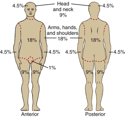
Burns are injuries to tissues that result from exposure to thermal, chemical, electrical, or radioactive agents (covered in the Injury, Poisoning and Certain Other Consequences of External Causes). They may be classified into four different degrees of severity, depending on the layers of the skin that are damaged. Coders must categorize burns higher than second degree according to the “rule of nines” (Fig. 4-23) that divides the body into percentages that are, for the most part, multiples of nine: the head and neck equal 9%, each upper limb 9%, each lower limb 18%, the front and back of the torso 36%, and the genital area 1%. Fig. 4-24 is an illustration of the different degrees of burns.
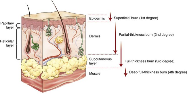
• Superficial burn: Burn in which only the first layer of the skin, the epidermis, is damaged; also known as a first-degree burn. Characterized by redness (erythema), tenderness, and hyperesthesia, with no scar development.
• Partial-thickness burn: Burn in which only the first and second layers of the skin (epidermis and part of the dermis) are affected; sometimes called a second-degree burn. If the burn extends to the papillary level, it is classified as a superficial partial-thickness burn. If it extends farther, to the reticular layer, it is classified as a deep partial-thickness burn. Characterized by redness, blisters, and pain, with possible scar development.
• Full-thickness burn: Burn that damages the epidermis, dermis, and subcutaneous tissue; also known as a third-degree burn. Pain is not present because the nerve endings in the skin have been destroyed. Skin appearance may be deep red, pale gray, brown, or black. Scar formation is likely.
• Deep full-thickness burn: Although not a universally accepted category, some burn specialists use this category to describe a rare burn that extends beyond the subcutaneous tissue into the muscle and bone. Also called a fourth-degree burn.
 Exercise 8:
Burns
Match the characteristics of the burns listed with their degree.
A Ironing a blouse for work, Rhonda burned her hand, resulting in blisters and erythema.
B John suffered burns over two thirds of his body with many areas of tissue burned to the bone.
C Because Kristin forgot to reapply her sunblock, she sustained a sunburn that resulted in painful, reddened skin.
D Smoking in bed resulted in burns that destroyed the epidermis and dermis and extended through the hypodermis on the victim’s chest and shoulders.
As in our other body system chapters, the following neoplasm terms are included here to complete the body system.
Terms Related to Benign Skin Growths (D1Ø-D36)
| Term | Word Origin | Definition |
| angioma | angi/o vessel -oma tumor, mass |
Localized vascular lesion that includes hemangiomas (Fig. 4-25), vascular nevi, and lymphangiomas. |
| dermatofibroma | dermat/o skin fibr/o fiber -oma tumor, mass |
Fibrous tumor of the skin that is painless, round, firm, and usually found on the extremities. |
| dysplastic nevus (pl. nevi) | dys- abnormal plast/o formation -ic pertaining to nev/o birthmark -us structure, thing |
A nevus is a pigmented lesion often present at birth. It is also called a mole. Various abnormal changes of a pigmented congenital skin blemish give rise to concern for progression to malignancy. Changes of concern are categorized as ABCDE. Asymmetry Borders, irregular Colors, changes or uneven pigmentation Diameter, increasing size or >6 mm Elevation |
| lipoma | lip/o fat -oma tumor, mass |
Fatty tumor that is a soft, movable, subcutaneous nodule (Fig. 4-26). |
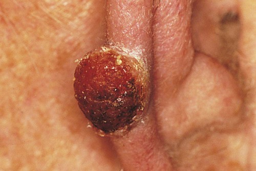
Terms Related to Malignant Neoplasms (CØØ-C96)
| Term | Word Origin | Definition |
| basal cell carcinoma (BCC) | bas/o base -al pertaining to carcinoma cancer of epithelial origin |
The most common form of skin cancer, it originates in the stratum germinativum of the epidermis. It usually occurs on the face as a result of sun exposure and rarely metastasizes (spreads to distant sites). |
| Kaposi’s sarcoma (KS) | sarcoma connective tissue cancer | A rare form of skin cancer that takes the form of red/blue/brown/purple nodules, usually on the extremities. One form appears most often in patients with deficient immune systems. |
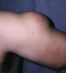
| malignant melanoma | melan/o black, dark -oma tumor, mass |
This cancerous tumor arises from mutated melanocytes. This particular cancer is the leading cause of death from all skin diseases (Fig. 4-27). |
| squamous cell carcinoma (SCC) | squam/o scaly -ous pertaining to carcinoma cancer of epithelial origin |
The second most common type of skin cancer, also caused by sun exposure, but developing from squamous cells (Fig. 4-28). |
 Exercise 9:
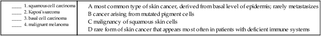
5. dermatofibroma __________________________________________________________________________________
6. angioma __________________________________________________________________________________________
7. lipoma ___________________________________________________________________________________________
Procedures
 PCS Guideline Alert
 PCS Guideline Alert
 PCS Guideline Alert
| Term | Word Origin | Definition |
| excisional biopsy | Biopsy (bx) in which the entire tumor may be removed with borders as a means of diagnosis and treatment. | |
| exfoliation | Scraping or shaving off samples of friable (easily crushed) lesions for a laboratory examination called exfoliative cytology. | |
| incisional biopsy | Biopsy in which larger tissue samples may be obtained by excising a wedge of tissue and suturing the incision. | |
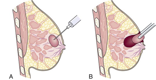
| needle aspiration | Aspiration of fluid from lesions to obtain samples for culture and examination (Fig. 4-29). | |
| punch biopsy | Biopsy in which a tubular punch is inserted through to the subcutaneous tissue, and the tissue is cut off at the base (Fig. 4-30). |
Terms Related to Laboratory Tests
| Term | Word Origin | Definition |
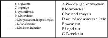
| bacterial analyses | Culture and serology of lesions to help diagnose such disorders as impetigo. | |
| fungal tests | Cultures of scrapings of lesions used to identify fungal infections, such as tinea pedis, tinea capitis, and tinea cruris. | |
| sweat tests | Laboratory test for abnormally high levels of sodium and chloride present in the perspiration of persons with cystic fibrosis. | |
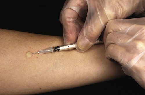
| tuberculosis (TB) skin tests | Intradermal test (e.g., Mantoux test) using purified protein derivative (PPD) to test for either dormant or active tuberculosis; much more accurate test than the multiple puncture tine test, which had been used for screening purposes (Fig. 4-31). | |
| Tzanck test tzahnk |
Microscopic examination of lesions for the purpose of diagnosing herpes zoster and herpes simplex. | |
| viral culture | Sampling of vesicular fluid for the purpose of identifying viruses. | |
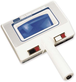
| Wood’s light examination | Method used to identify a variety of skin infections through the use of a Wood’s light, which produces ultraviolet light; tinea capitis and Pseudomonas infections in burns are two of the disorders it can reveal (Fig. 4-32). | |
| wound and abscess cultures | Lab samplings that can identify pathogens in wounds, such as diabetic or decubitus ulcers, postoperative wounds, or abscesses. |
 Exercise 10:
Biopsies and Laboratory Tests
Fill in the blanks with the correct terms from the list below.
1. An entire tumor is removed in a/an _________________ biopsy.
2. Fluid from a lesion is aspirated to obtain samples for culture in a/an _________________ biopsy.
3. A wedge of tissue is removed and the incision is sutured in a/an _________________ biopsy.
4. Samples of friable lesions are scraped or shaved off in __________________________________.
5. A cylindrical punch is inserted into the subcutaneous tissue layer, and the tissue is cut off at the base in a/an __________________________________ biopsy.
Match the following disorders with the tests that may be used to diagnose them.
 Exercise 11:
Progress Note
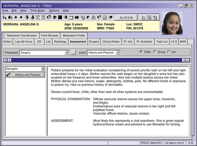
Review the progress note above and answer the following questions.
1. How do we know that her rash is itchy?
___________________________________________________________________________________________________
2. What phrase tells us that she has had no skin inflammations diagnosed in the past?
___________________________________________________________________________________________________
3. What characteristic would “vesicular” lesions have?
___________________________________________________________________________________________________
4. “Exanthem” is a medical term for what?
___________________________________________________________________________________________________
Terms Related to Grafting Techniques and Other Therapies
| Term | Word Origin | Definition |
| allograft | allo- other | Harvest of skin from another human donor for temporary transplant until an autograft is available. Also called a homograft. |
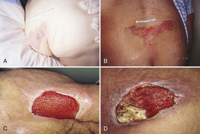
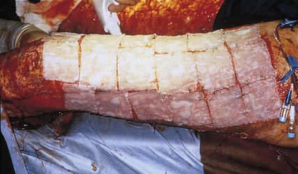
| autograft | auto- self | Harvest of the patient’s own skin for transplant (Fig. 4-33). |
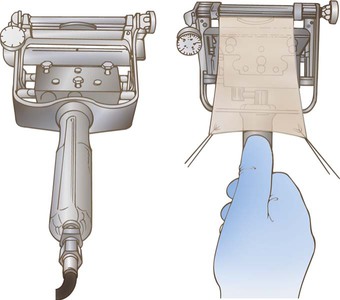
| dermatome | dermat/o skin -tome instrument to cut |
Instrument used to remove split-skin grafts (Fig. 4-34). |
| flap | Section of skin transferred from one location to an immediately adjacent one. Also called a skin graft. | |
| laser therapy | Procedure to repair or destroy tissue, particularly in the removal of tattoos, warts, port-wine stains, and psoriatic lesions. | |
| occlusive therapy | occlus/o to close -ive pertaining to |
Use of a nonporous, occlusive dressing to cover a treated area to enhance the absorption and effectiveness of a medication; used to treat psoriasis, lupus erythematosus, and chronic hand dermatitis. |
| psoralen plus ultraviolet A (PUVA) therapy | Directing a type of ultraviolet light (UV) onto psoriatic lesions. | |
| skin grafting (SG) | Skin transplant performed when normal skin cover has been lost as a result of burns, ulcers, or operations to remove cancerous tissue. | |
| full-thickness graft | Free skin graft in which full portions of both the epidermis and the dermis are used. | |
| split-thickness skin graft (STSG) | Skin graft in which the epidermis and parts of the dermis are used. | |
| xenograft | xen/o foreign | Temporary skin graft from another species, often a pig, used until an autograft is available. Also called a heterograft. |
 Note
NoteTerms Related to Tissue Removal and Other Procedures
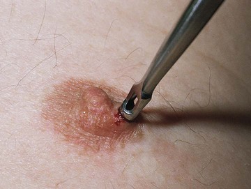
| Term | Word Origin | Definition |
| cauterization | cauter/i burn -zation process of |
Destruction of tissue by burning with heat. |
| cryosurgery | cry/o extreme cold | Destruction of tissue through the use of extreme cold, usually liquid nitrogen. |
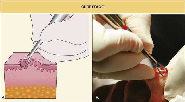
| curettage | Scraping of material from the wall of a cavity or other surface to obtain tissue for microscopic examination; this is done with an instrument called a curette (Fig. 4-35). | |
| débridement | First step in wound treatment, involving removal of dirt, foreign bodies (FB), damaged tissue, and cellular debris from the wound or burn to prevent infection and to promote healing. | |
| escharotomy | eschar/o scab -tomy cutting |
Surgical incision into necrotic tissue resulting from a severe burn. This may be necessary to prevent edema leading to ischemia (loss of blood flow) in underlying tissue. |
| incision and drainage (I&D) | Cutting open and removing the contents of a wound, cyst, or other lesion. | |
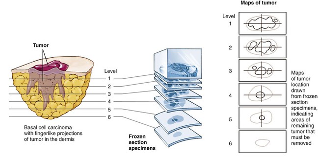
| Mohs surgery | Repeated removal and microscopic examination of layers of a tumor until no cancerous cells are present (Fig. 4-36). | |
| onychectomy | onych/o nail -ectomy cutting out |
Removal of nail usually to treat trauma. The procedure allows the nail to regrow normally. |
| onychoplasty | onych/o nail -plasty surgically forming |
Surgical treatment usually including removal of the nail matrix to treat ingrown toenails. |
| onychotomy | onych/o nail -tomy cutting |
Cutting the nail to drain the subungual hematoma. |
| shaving (paring) | Slicing of thin sheets of tissue to remove lesions. |
Terms Related to Cosmetic Procedures
| Term | Word Origin | Definition |
| blepharoplasty | blephar/o eyelid –plasty surgically forming |
Surgical repair of the eyelid. |
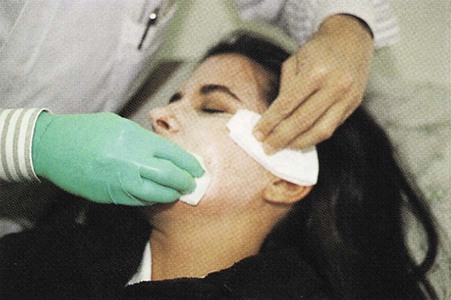
| chemical peel | Use of a mild acid to produce a superficial burn; normally done to remove wrinkles (Fig. 4-37). | |
| dermabrasion | derm/o skin -abrasion scraping |
Surgical procedure to resurface the skin; used to remove acne scars, nevi, wrinkles, and tattoos. |
| dermatoplasty | dermat/o skin -plasty surgically forming |
Transplant of living skin to correct effects of injury, operation, or disease. |
| lipectomy | lip/o fat -ectomy cutting out |
Removal of fatty tissue. |
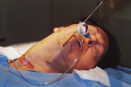
| liposuction | lip/o fat | Technique for removing adipose tissue with a suction pump device (Fig. 4-38). |
| rhytidectomy | rhytid/o wrinkle -ectomy cutting out |
Surgical operation to remove wrinkles. Commonly known as a “face-lift.” |
 Exercise 12:
Grafting, Tissue Removal, Cosmetic, and Other Procedures
1. Explain the differences among the following:
A autograft ______________________________________________________________________________________
B allograft ______________________________________________________________________________________
C xenograft _____________________________________________________________________________________
2. Which type of graft includes the epidermis and the dermis? _________________________________________
3. What instrument is used to cut skin for grafting? ___________________________________________________
Fill in the blanks with the correct terms from the list below.
4. _________________ is used to destroy tattoos.
5. Removing dirt, foreign bodies, damaged tissue, and cellular debris from a wound is called _________________.
6. The destruction of tissue by burning with heat is called _______________.
7. The destruction of tissue through the use of extreme cold is called _________________.
8. Scraping of material from the wall of a cavity is called _________________.
10. Another term for paring is _________________.
11. Covering an area to enhance absorption of medicine is called _________________.
12. Removal of a tumor by layers is called _________________.
13. cutting out wrinkles _____________________________________________________________________________
14. cutting out fat __________________________________________________________________________________
15. surgically forming the eyelid _____________________________________________________________________
16. scraping of skin _________________________________________________________________________________
17. surgically forming the skin _______________________________________________________________________
18. surgically forming a nail _________________________________________________
Pharmacology
anesthetic agents: Reduce pain and discomfort; some can be given topically on an affected area. Examples include lidocaine (Xylocaine) and benzocaine (Orajel).
antibacterials: Prevent and treat bacterial growth. Topical agents such as erythromycin (Ery 2% Pads) and clindamycin (Benzaclin) are used to treat acne. Triple antibiotic ointment containing bacitracin, polymixin B, and neomycin (Neosporin), silver sulfadiazine (Silvadene), and mupirocin (Bactroban) are used to prevent and treat skin or wound infections. Oral agents for the treatment of acne include erythromycin (Ery-Tab), tetracycline (Sumycin), and minocycline (Minocin).
antifungals: Treat fungal infections. Topical agents include nystatin (Nystop), butenafine (Lotrimin Ultra), ciclopirox (Loprox), and econazole (Ecoza).
antihistamines: Lessen itching by reducing an allergic response. Diphenhydramine (Benadryl) is available in oral and topical formulations. Other oral agents include chlorpheniramine (Chlor-Trimeton), cetirizine (Zyrtec), and loratadine (Claritin).
antiinflammatories: Reduce inflammation and pain. Oral agents include prednisone and aspirin; topical agents include hydrocortisone (Cortizone), fluocinonide (Vanos), and triamcinolone (Kenalog).
antipsoriatics: Treat psoriasis. Examples include anthralin (Drithocreme) and calcipotriene (Dovonex).
antiseptics: Topical agents used to prevent infection by destroying microbials. Examples include iodine and chlorhexidine (Peridex).
antivirals: Reduce the effect of viruses. Examples include valacyclovir (Valtrex) and acyclovir (Zovirax) for the treatment of herpes simplex virus (cold sores or genital herpes) and herpes zoster (shingles).
emollients: Topical substances that soften and moisturize the skin. Emollients come in the form of lotions, creams, ointments, and bath additives. A couple of well-known OTC products containing emollients are Aveeno and Eucerin.
immunomodulators or immunosuppressants: Agents that suppress the body’s immune system. Topical agents such as pimecrolimus (Elidel) and tacrolimus (Protopic) are used to treat atopic dermatitis and eczema.
keratolytics: Topical substances used to break down hardened skin and shed the top layer of dead skin to treat warts, calluses, corns, acne, rosacea, and psoriasis. Examples include salicylic acid, cantharidin, and podofilox (Condylox).
pediculicides: Destroy lice. Examples include malathion (Ovide), lindane (Kwell), and permethrin (Nix).
protectives: Topicals with sun protection factors (SPFs) that protect the skin against ultraviolet A and B in sunlight. A wide variety of these are available OTC.
retinoids: Derived from vitamin A, retinoids alter the growth of the top layer of skin and may be used to treat acne, reduce wrinkles, and treat psoriasis. Examples include tretinoin (Retin-A), isotretinoin (Claravis), and tazarotene (Tazorac).
scabicides: Destroy mites and scabies. Examples include permethrin (Elimite) and crotamiton (Eurax).
 Exercise 13:
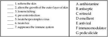
Recognizing Suffixes for PCS
Suffixes and Root Operations for the Skin and Subcutaneous Tissue
| Suffix | Root Operation |
| -abrasion | Extraction |
| -ectomy | Excision, resection, alteration |
| -graft | Repair |
| -plasty | Repair, replacement, reposition, supplement |
| -tomy | Drainage |
| Abbreviation | Meaning |
| BCC | basal cell carcinoma |
| bx | biopsy |
| DLE | discoid lupus erythematosus |
| FB | foreign body |
| HPV | human papillomavirus |
| HSV-1 | herpes simplex virus 1 |
| HSV-2 | herpes simplex virus 2 |
| I&D | incision and drainage |
| KS | Kaposi’s sarcoma |
| PPD | purified protein derivative |
| PUVA | psoralen plus ultraviolet A |
| SCC | squamous cell carcinoma |
| SG | skin graft |
| STSG | split-thickness skin graft |
| TB | tuberculosis |
 Exercise 14:
ED Record

Using the ED record above, answer the questions below.
1. Describe a superficial partial-thickness burn. _______________________________________________________
__________________________________________________________________________________________________
2. What are the characteristics that make this a second-degree burn?
__________________________________________________________________________________________________
__________________________________________________________________________________________________
 Go to the Evolve website to interactively:


