SIEA Flap Breast Reconstruction

Published on 22/05/2015 by admin

Filed under Plastic Reconstructive Surgery

Last modified 22/04/2025

Print this page

rate 1 star rate 2 star rate 3 star rate 4 star rate 5 star
Your rating: none, Average: 0 (0 votes)

This article have been viewed 1433 times

CHAPTER 10 SIEA Flap Breast Reconstruction

image

Introduction

Breast reconstruction utilizing autologous abdominal tissue has long been considered the gold standard to which all other techniques have been compared. In more recent years, free tissue transfer of the abdominal adipocutaneous compartment has emerged as an essential tool for breast reconstruction in the armamentarium of the reconstructive surgeon. The use of abdominal tissue for breast reconstruction has the advantage of replacing the breast tissue which is composed mainly of soft fat and glandular tissue with tissue similar in composition and texture. The free transverse rectus abdominis muscle (free TRAM), deep inferior epigastric perforator (DIEP) and the superficial inferior epigastric artery (SIEA) flaps have come into view as the main free flaps utilizing abdominal tissue for this exact purpose.

The desire to retain maximal strength and integrity of the abdominal wall musculature and fascial layers, while still making the most of its unique tissue characteristics, led reconstructive surgeons to explore the SIEA flap as a premier modality for reconstructive breast surgery. During surgical dissection of the SIEA flap the rectus fascia is neither excised nor incised thus maintaining its preoperative strength.

The abdominal donor site benefits the patient in two additional aspects: first a vastly improved abdominal contour, and second an easily hidden low-lying abdominal bikini-line scar, both of these features being the main reasons patients seek to undergo a cosmetic abdominoplasty procedure.

Historically, the first microvascular transfer of abdominal tissue based on the SIEA pedicle is credited to Antia and Buch in 1971,1 using it to reconstruct a soft tissue deformity of the face. Since then the SIEA flap has often been described for various reconstructive purposes. Holmström2 first reported the use of a free SIEA flap for breast reconstruction, but it was not until 1991 that Grotting3 popularized the SIEA flap for immediate breast reconstruction. Since then the microvascular transfer of the SIEA flap has been reported by several groups.49

Indications

In our institute for breast reconstruction the lower abdominal tissue is the favored donor site. The SIEA flap is our flap of choice when the appropriate criteria for its use are met; if these criteria are not met we will use the DIEP flap. Therefore, we have devised an algorithm for choosing the SIEA flap,7 a detailed overview of the different parameters involved in choosing the appropriate flap is presented later on in this chapter. When patients are not candidates for abdominal tissue reconstruction we will reconstruct the breast with either a superior gluteal artery perforator flap (SGAP), a latissimus dorsi flap combined with an implant, or infrequently with a breast implant only (Boxes 10.1 and 10.2).

Patient Selection and Preoperative Consultation

Women who are candidates for breast reconstruction are seen in consultation at least twice prior to surgery. The first consult evolves thorough medical and surgical history review and a complete physical examination. Specific points addressed are breast cancer staging, breast imaging studies, previous and/or planned breast surgical interventions, prior chemotherapy, and radiation treatments (emphasizing any local condition or radiation that might affect the breast skin and/or recipient vessels). A family history of breast cancer, bleeding tendency or hypercoagulable state is also noted.

Upon physical exam the chest, axilla and abdomen are thoroughly examined. The chest wall, breast skin and axilla are inspected for scars, masses, and any radiation damage. Lymphedema and range of motion are also examined in the relevant upper extremity. The abdominal wall is examined for the amount of excess tissue available for reconstruction, presence of diastasis recti, hernias, masses and scars.

It is important to note Pfannenstiel scars when considering using the SIEA flap. Laterally-extending low transverse abdominal scars (e.g., Pfannenstiel) will transect the SIEA and SIEV (superficial inferior epigastric vein), while more centered transverse scars might sever only the SIEV; in both scenarios we will usually prefer to use the DIEP flap. Hence, only in cases where a short Pfannenstiel scar is present, transecting neither the SIEA nor SIEV, will we consider the SIEA flap for reconstruction.

We refrain from operating on actively smoking patients and encourage all active smokers to quit smoking for at least 2 months prior to surgery and 6 weeks after. If question exists regarding compliance we will defer the microsurgical intervention.

An up-to-date breast imaging evaluation, preferably MRI, is required for all surgical candidates. High risk and elderly patients should receive surgical clearance by their primary physician and/or cardiologist as indicated.

Obviously, the patient’s oncologic status and breast cancer stage are taken into consideration. The ideal patient is one with early stage cancer who is not a candidate for radiation therapy. However, we will not decline a patient who presents with more advanced stage tumor, and we plan to perform reconstruction in a delayed fashion after completion of the necessary adjuvant therapy (radiation and chemotherapy). We traditionally delay breast reconstruction for 6 months from completion of radiation therapy to allow for fibrosis and involutional changes to take place.

Operative Technique (Box 10.3)

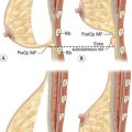
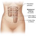
Surgical anatomy (Fig. 10.1)

From a vascular point of view the SIEA flap is anatomically considerably different from the free TRAM and DIEP flaps, which are based on the deep inferior epigastric perforator system that consists of large caliber musculocutaneous perforators. The SIEA flap is perfused by a subdermal vascular plexus system. This superficial system includes significantly smaller vessels nourished by the superficial inferior epigastric axial vessels. Contrary to the free TRAM and DIEP flaps that are based on perforating vessels, the SIEA flap is actually a direct axial adipocutaneous flap. Since the superficial inferior epigastric vessels do not perforate through a muscle or a septum it is not a true perforator flap, a common misnomer.

The SIEA flap territory is mainly the lower abdomen as perfused by the SIEA, and it overlaps laterally with the superficial circumflex iliac artery (SCIA). The SIEA originates approximately 2–5 cm below the inguinal ligament from the common femoral artery. The SIEA originates as either an independent trunk or more commonly shares a common trunk with the circumflex iliac artery, with various other variants of origin from the deep femoral artery and pudendal artery.

Inferior to the inguinal ligament the vessels lie deep to Scarpa’s fascia, as they course superiorly they penetrate Scarpa’s fascia and lie in the superficial subcutaneous tissue. The SIEA and its venae comitantes typically run laterally to the lateral deep inferior epigastric perforator row. The venous drainage of the SIEA flap abdominal territory is mainly via the superficial inferior epigastric vein (SIEV) which empties into the saphenous bulb, and via two venae comitantes running with the SIEA emptying into the femoral vein or occasionally the saphenous bulb. The average length of the main pedicle (artery and two venae comitantes) varies from 5–8 cm, with an arterial diameter range of 1.1–1.9 mm, depending on the site of measurement. The sensory innervation of the SIEA flap is derived from the 10th to 12th intercostal nerves.1013

In Taylor and Daniel’s original anatomical study of the SIEA in 100 cadavers, they found the SIEA to be totally absent in 35% of their dissections; they also noted that the origin of the SIEA flap in the remaining cadavers was variable with 48% originating as a common trunk with the superficial circumflex from the common femoral artery, while 17% originated directly from the common femoral artery.10 Our published series of 278 clinical dissections during breast reconstruction procedures correlates with this data, showing absence of the SIEA in 42% (n = 118).7 Further assessment of the remaining 58% (n = 160) that did have an obvious artery, revealed that 54% (n = 87) of the arteries had an external diameter greater than or equal to 1.5 mm at the level of the lower abdominal incision. Using our criteria for choosing a SIEA flap, requiring the artery to have an external diameter of 1.5 mm or more at the level of the lower abdominal incision line, the SIEA was adequate for use in approximately 31% of all our cases.

Debate exists in the literature concerning the reliability of tissue perfusion and venous drainage when attempting to harvest unipedicled SIEA flaps across the midline. While a few anatomical and clinical studies have commented on the unpredictability of the arterial perfusion and venous drainage of SIEA flaps across the midline,5,6,14,15 others have demonstrated the possibility of reliably transferring this extended adipocutaneous tissue.8,9,11 In our experience we have observed that the amount of tissue reliably harvested across the midline in an SIEA flap varies with each patient, and we make that determination by intraoperatively evaluating flap perfusion. Therefore, in some patients the zone of demarcation is present at a point roughly corresponding to the medial border of the contralateral rectus muscle, while in others the demarcation is seen at the lateral border of the contralateral rectus muscle.
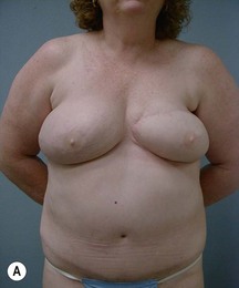
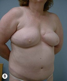
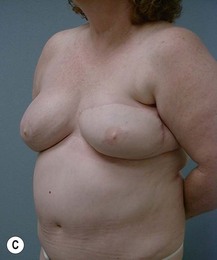
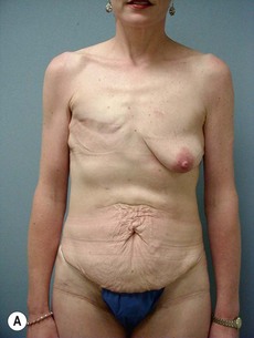
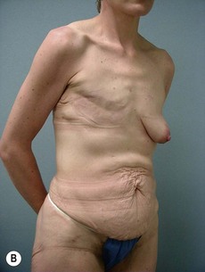
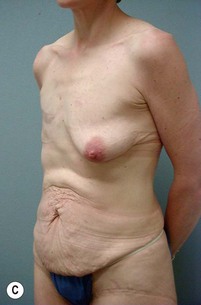
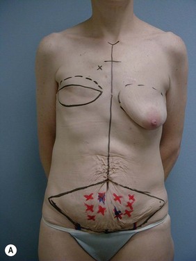
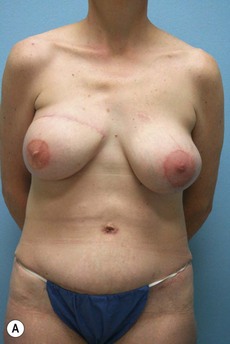
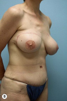
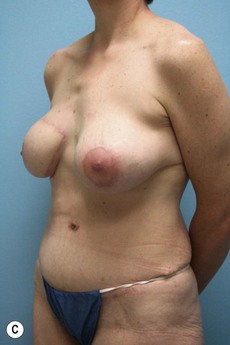
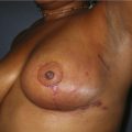
Case Presentations I and II (Figs 10.210.7)

Two case studies are now presented, which illustrate the following procedures.
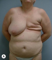
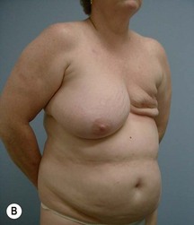
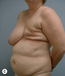
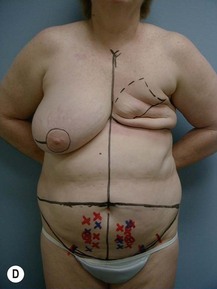
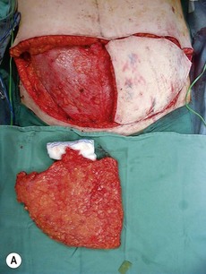
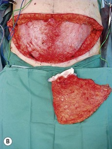
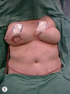
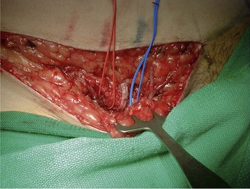
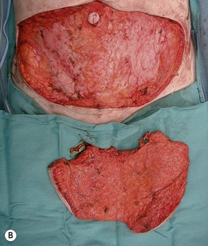
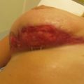
Preoperative markings (Figs 10.2d and 10.6a)

Preoperative vascular imaging may be performed to both accurately map the vessels, and to help establish whether or not the diameter of the SIEA is adequate. This can be done using either Duplex ultrasonography or contrast enhanced CAT scan. We do not routinely use preoperative imaging at our institution, we reserve this for patients with extensive abdominal scarring.

The patient is marked in the preoperative area at bedside on the morning of surgery. Marking is begun with the patient in the supine position. At this point, marking of the abdominal site sets the stage for possible harvest of both the SIEA flap and the DIEP flap. The selection between an SIEA flap versus a DIEP flap depends mainly on the vessel anatomy encountered during surgery, as an algorithm for choosing between the two and will be discussed later. Both hemiabdomens are marked in unilateral and bilateral cases allowing for both ipsilateral and/or contralateral flap harvest as indicated.

Using a hand-held pencil Doppler; we initially mark the SIEA with its accompanying venae comitantes, which usually course around midway between the symphysis pubis and the anterior superior iliac spine (ASIS) on the lower abdomen just above the inguinal crease. Also, the more medially located SIEV is mapped and marked. Next, periumbilical arterial and venous perforators are mapped and marked on both hemi-abdomens. Doppler signals from both the lateral and medial row deep inferior epigastric perforators are marked.

A crucial marking is that of the lower incision line, this line should lie low enough so as to possibly capture the SIEA at a suitable caliber. As pointed out earlier, one of our criteria for choosing an SIEA flap is arterial caliber of 1.5 mm or greater at the lower abdominal incision line. However, one should take into account the likely possibility of using a DIEP flap, thus necessitating the capturing of periumbilical perforators in the flap design while still allowing for acceptable tension during the donor site closure.

For most of our cases the lower abdominal line is centrally marked just superior to the pubic hair line, and extends laterally in a linear curving line superior to the inguinal crease toward, and ending at the ASIS. The upper abdominal incision line is a gradually descending curved line marked at around one centimeter above the umbilicus joining the lateral extension of the lower markings. The midline is also marked at this point relative to the midline of mons pubis. The average size of the SIEA flap is around 14 cm vertically and 17 cm on each side from the midline.

Marking is then continued with the patient standing. In immediate reconstructive cases the inframammary fold (IMF) and superior breast pole are marked bilaterally. If a skin sparing mastectomy is to be performed the proposed peri-areolar line is marked at this stage. In unilateral delayed reconstruction the IMF and superior breast pole are marked to match the contralateral breast, while in bilateral cases proposed IMF lines are marked. Previous mastectomy scars are also outlined.

The midline extending from the suprasternal notch to the umbilicus is marked as are the second and third intercostal spaces, which are palpated and marked with an ‘X’ at this stage for locating the recipient vessels.

Operative procedure

Flap harvest (Figs 10.3a–b and 10.6b)

The SIEA flap harvest is initiated by carefully incising the inferior aspect of the abdominal flap, at this stage just deeper to the dermis, starting on the ipsilateral side of the reconstructed breast. The subcutaneous dissection is carefully performed, as not to injure the SIEV which is frequently quite superficial. The SIEA and SIEV are identified and separated from the surrounding tissue. The SIEA is then preserved with vessel loops and explored at the lower line of skin incision.

We have previously published our algorithm for choosing the SIEA flap in breast reconstruction.7 In this review of our clinical experience with the SIEA flap, we have noted that whenever the diameter of the SIEA was smaller than 1.5 mm as the pedicle entered the flap (lower line of abdominal incision), there exists an increased chance of vascular compromise. Thus, when opting to use the SIEA flap we precede in the following manner; using a vessel sizer provided with the venous coupler, we measure the SIEA’s external diameter; if its external diameter is larger than 1.5 mm and there is a visible, palpable and audible Doppler pulse, the artery and its venae comitantes are dissected in a retrograde fashion. We do not necessarily dissect the vessels to their origin; rather we continue the dissection as long as we can perceive an increase in the vessel diameter. Once a point is reached at which the diameter is constant there is no need for further dissection since pedicle length is rarely an issue with SIEA flaps (contrary to DIEP flaps). Another key point to keep in mind during pedicle dissection is not to skeletonize the vessels. Preferably a narrow cuff of tissue (e.g., fat) is left just deep to the vessels to protect the vascular pedicle. One should refrain from adding too much surrounding tissue that could unnecessarily lead to prolonged seroma formation due to removal of lymphatic nodes and ducts. Only the distalmost part of the vessels is fully skeletonized, at the area closest to the anastomotic site.

Additionally, in cases in which the SIEA is borderline in caliber, we also look at the SIEV caliber before making the final decision between the SIEA and DIEP flaps. When a sizable SIEV is present we assume dominance of the superficial draining system and strongly consider using the SIEA flap, and not the DIEP flap, despite a non-remarkable SIEA caliber.

Since we published our original description of the algorithm for choosing the SIEA flap7 we have changed our preferred hemi-abdomen for unilateral reconstructions, and are now preferentially using the ipsilateral side. The rationale for preferring an ipsilateral flap is threefold. First, the SIEA pedicle enters the abdominal flap on its lateral aspect (usually lateral to the lateral row of the DIEA perforators), thus, since the flaps are rotated 180° for their final inset, vessel proximity to the recipient internal mammary vessels is preferable in ipsilateral flaps. Second, the final positioning of the least reliable zone of blood supply (across the midline) will be at the lateral aspect of the reconstructed breast and not medially (should there be any flap loss preferably it should be laterally). Lastly, suturing of the flap at the previous umbilical excision site will cone and shape the breast at the desired position laterally. In unilateral cases both SIEA’s are explored before making the final decision as to the preferred pedicle side which is mainly based on arterial caliber.

Naturally, in unilateral cases where the ipsilateral external diameter is smaller than 1.5 mm or the pulse is weak, we explore the contralateral SIEA. If the contralateral SIEA is adequate, we preserve it and use the contralateral SIEA flap. If, however, both superficial systems are too small or display a weak pulse at the level of the lower abdominal incision we assume a dominance of the deep perforator system and revert to the use of DIEP flaps. As in the SIEA flaps, also in DIEP flaps, preference is given to the ipsilateral side. The lateral perforator row is explored followed by the medial row on the ipsilateral side, if inadequate; the contralateral side perforators are then analyzed.

In bilateral cases the selection criteria are similar and dictate the use of an ipsilateral SIEA flap or an ipsilateral DIEP flap. One should keep in mind that a combination of SIEA flap for one side and a DIEP flap for the other is a rational alternative, when taking into account the reduced donor site morbidity compared to bilateral DIEP or free TRAM flaps.

Once the SIEA flap has been chosen, and the vessels are preserved, we progress quite rapidly with the dissection. In the majority of cases the upper incision line starts at the ASIS and extends to just above the umbilicus on each hemi-abdomen. The incision is deepened down through the adipose tissue to the loose areolar tissue plane above the abdominal muscular fascia. The flap is elevated from lateral to medial, using Bovie cautery to ensure meticulous hemostasis is obtained, thus not obscuring accurate surgical planes, making it easier not to incise any fascia.

Next, the umbilicus is delivered from the surrounding flap tissue; with the aid of two skin hooks to elevate the umbilicus, a circumumbilical incision is performed and carried down to the rectus sheath plane. One should make sure enough blood supply is maintained with the umbilical stalk by avoiding the skeletonization of the umbilical stalk. The abdominal flap is then dissected apart from the umbilicus. It is essential at this stage to be alert to the possible existence of periumbilical hernias, which should be noted and repaired at the abdominal closure stage.

As previously mentioned special consideration should be given to unilateral reconstructions regarding the amount of abdominal tissue needed to match the contralateral side. In cases where more tissue is needed than can be obtained from a hemi-flap, we will still consider using the SIEA flap because in the majority of cases the SIEA flap perfusion is reliable across the midline up to the lateral row of the contralateral deep perforators. In our experience with more than one hundred SIEA flaps we have not encountered increased incidences of fat necrosis (partial flap loss) using this guideline. More so, upon reviewing the patient data base we have recognized that the average percentage of flap used was 66.5% of the total abdominal tissue available (zones I to IV). It is our observation that as long as zone IV and any poorly perfused zone III tissue are excised while the flap is perfused by its vascular pedicle at the donor site, we do not have to define the limit of the flap at the midline or in terms of its weight.

If concern still exists regarding reliable perfusion of the SIEA flap we will test the ability of the superficial system to support the flap while still having the option to revert to a DIEP flap. To accomplish this, we maintain two to three perforators from the ipsilateral deep system, and then we simply clamp any deep perforator encountered and the contralateral SIEA and SIEV (if still not ligated) with vascular clamps, thus maintaining flap perfusion solely on the ipsilateral superficial system. We then visually assess the flap for signs of poor perfusion.

Using this simple test, in cases where resection of the underperfused tissue will not leave enough flap tissue for reconstruction, we have not ‘burnt the bridge’ of using the deep perforator system, which is more likely to support more tissue across the midline.

When reconstructing bilateral cases the abdominal tissue is split along the midline into two hemi-flaps of similar size. After completing the anastomosis of the first side attention is then turned to the superficial inferior epigastric vein and artery of the second side. Once more, as in the first side the vessels are examined for their adequacy according to the previously described criteria, and the algorithm for choosing the SIEA is implemented once more. We have performed many bilateral reconstructive cases in which one flap was based on the superficial system while its counterpart was based on the deep perforating system. This type of reconstruction has the advantage of not compromising the abdominal fascia on both sides compared to a bilateral DIEP or bilateral free TRAM.

After complete elevation of the flap from the abdominal wall (still attached to the vascular pedicle) we will mark the superficial aspect of the vascular pedicle for future reference. The flap edges are then stapled back into place and attention is turned to the mastectomy defects.

Managing the mastectomy skin

We can usually obtain the best cosmetic results in patients who undergo skin-sparing mastectomy with immediate reconstruction, since the flap merely serves as a neo-filler for the recently emptied skin envelope. We also fabricate a small skin paddle, which will serve as the platform for nipple reconstruction and the area to be tattooed as the areola after the flap has healed.

As previously mentioned, in cases in which irradiation is part of the treatment regimen, we will defer reconstruction to 6 months after completion of treatment and will opt for delayed reconstruction. When confronted with delayed reconstruction, depending on the type of mastectomy performed and whether or not the mastectomy skin had been irradiated, one could potentially encounter a few reconstructive scenarios. In cases when a skin-sparing mastectomy was performed and no irradiation was given or the skin changes after irradiation are judged to be minimal, we will attempt to raise the mastectomy skin flaps and use them as the skin envelope for the flap. If, however, not enough skin is left to recreate the skin envelope (as in a simple mastectomy) and/or there are noticeable skin irradiation changes, we usually prefer to remove the skin below the mastectomy scar, leaving any subcutaneous tissue as an additional layer under the flap and over the chest wall. We will then use a bigger skin paddle to reconstruct most of the missing skin envelope.

In both immediate and delayed reconstructions the height and integrity of the IMF is revised, and the lateral border of the breast pocket is re-created by interrupted sutures as needed. Since the upper abdominal tissue will inevitably be pulled downward to close the abdominal donor site, it will tend to pull the chest skin along. It is important to consider this and to compensate accordingly by adequately positioning the IMF, in certain cases even raising the IMF by 1–2 cm.

Vessel anastomosis

The mastectomy skin is gently retracted to provide adequate exposure of the internal mammary vessels for anastomosis. Now, under microscope magnification the internal mammary vessels are definitively primed for anastomosis.

Once the internal mammary vessels are prepared, the SIEA, venae comitantes, and SIEV are ligated and the flap is brought up to the ipsilateral mastectomy field (depending on the adequacy of the ipsilateral side). The flap is turned 180° and appropriately positioned for anastomosis. The flap is then stapled to the surrounding skin, having the SIEA vessels align with the internal mammary artery and vein, and covered with a moist laparotomy sponge. Next the distal part of the IMA and internal mammary vein (IMV) are ligated as distally as possible leaving enough length for comfortable positioning of vascular clamps.

Most often we will begin with the venous anastomosis of the IMV and the SIEA venae comitantes by using an appropriately measured coupling device. Depending on the caliber and length of the veins one can choose between the medial or lateral IMV. We have found that the medial IMV is more commonly used since it is a better size match (wider caliber) to the venae comitantes. However, when the medial and lateral veins are similar in caliber we will prefer to use the lateral IMV since it tends to be longer. Preferably the venae comitantes were harvested distal to their point of merging; hence the anastomosis is completed to the merged vessel.

Although initially we used to anastomose the IMV to the SIEV, we have abandoned this technique and routinely anastomose to the venae comitantes. We have found that both the vessel caliber and the proximity of the venae comitantes, which naturally accompany the artery (as apposed to the SIEV that exits the flap in a less favorable orientation) are anatomically more convenient and carry a lower risk of vessel kinking compared to when we utilized the SIEV. Hence to date we reserve the SIEV primarily for cases in which the venae comitantes are inadequate or a second backup venous anastomosis is needed (rarely a problem with SIEA flaps that typically have excellent venous drainage).

Subsequently the IMA and SIEA ends are brought into proximity with a double opposing clamp for the arterial anastomosis which is completed in standard fashion using a running or interrupted 9-0 nylon sutures. Upon completion of both vascular anastomoses an internal Doppler cuff is positioned around the venous anastomosis.

Flap inset

Once the anastomosis is complete, attention is turned to the flap inset. Attention should be focused on the way the SIEA pedicle is oriented on the chest wall, and it is at this stage that the previous marking of the superficial side of the pedicle becomes helpful. First, the pedicle should not twist on itself throughout its course into the flap. Second, the SIEA pedicle travels from within the intercostal space and up into the superficial layer of the flap, this causes a steep curvature (step off) over the chest wall and pectoralis major muscle. Ensure the pedicle is not kinked or compressed under the flap at this point. Moreover, when the overlying skin has been irradiated, there is a higher likelihood of increased skin tension on the flap and the more superficial location of the SIEA pedicle. Every effort should be made to properly orient the pedicle at this critical stage, the appropriate time should be taken to re-explore any change in the internal Doppler signal prior to making the final flap inset. Interrupted 2-0 Monocryl sutures are positioned between the flap and chest wall usually on the superior–lateral and inferior aspects to anchor the flap in position.

During the final stages of flap inset the patient’s bed is flexed into the semi-sitting position. This is done to assess the final positioning of the reconstructed breast on the chest wall, check for breast symmetry and aid in the abdominal donor site closure. It is also of paramount importance to check for any changes in Doppler signals while the patient is seated.

Pitfalls – Avoidance and Correction

The SIEA pedicle and flap inset

One of the most critical stages of the operation is the final inset of the SIEA flap. As previously noted, the anatomy of the SIEA pedicle is unique, and differs from the vascular pedicle of the DIEP/free TRAM (and most other free flaps), in the manner in which the vessels enter the flap. Contrary to the above mentioned flaps in which the pedicle enters the flap from beneath, the SIEA pedicle enters the flap from its inferior edge lying almost horizontally at the superficial subcutaneous tissue.

This anatomical variation is important to understand for proper pedicle orientation while the flap is being inset. The internal mammary vessels are the primary recipient vessels, lying deep within the intercostal space. Anatomically these recipient vessels are positioned more favorably for the DIEP and free TRAM pedicles as their pedicles travel on the undersurface of the flap and there is less tendency for pedicle kinking as the pedicle travels from the flap to the site of anastomosis. The SIEA pedicle however has to transition from a more superficial plane to a deeper one, making it more susceptible to kinking, malrotation and compression under the flap and overlying skin.

Aside from cautious positioning of the flap on the pedicle one should also create a large enough opening in the pectoralis muscle to prevent inadvertent compression of the pedicle by the muscle. In addition, the overlying surrounding skin above the area of anastomosis should be widely lifted from the underlying pectoralis muscle to relieve any constricting forces transmitted from the skin to the flap and onto the superficial lying pedicle. This is especially true in delayed reconstruction where no glandular resection is performed at the stage of reconstruction, and in cases in which the skin is even more constricting due to previous irradiation.

References

1 Antia NH, Buch VI. Transfer of an abdominal dermo-fat graft by direct anastomosis of blood vessels. Br J Plast Surg. 1971;24:15-19.

2 Holmström H. The free abdominoplasty flap and its use in breast reconstruction. An experimental study and clinical case report. Scand J Plast Reconstr Surg. 1979;13:423-427.

3 Grotting JC. The free abdominoplasty flap for immediate breast reconstruction. Ann Plast Surg. 1991;27:351-354.

4 Arnez ZM, Khan U, Pogorelec D, Planinsek F. Breast reconstruction using the free superficial inferior epigastric artery (SIEA) flap. Br J Plast Surg. 1999;52:276-279.

5 Chevray PM. Breast reconstruction with superficial inferior epigastric artery flaps: a prospective comparison with TRAM and DIEP flaps. Plast Reconstr Surg. 2004;114:1077-1083.

6 Granzow JW, Levine JL, Chiu ES, Allen RJ. Breast reconstruction using perforator flaps. J Surg Oncol. 2006;94:441-454.

7 Spiegel AJ, Khan FN. An intraoperative algorithm for use of the SIEA flap for breast reconstruction. Plast Reconstr Surg. 2007;120:1450-1459.

8 Volpe AG, Rothkopf DM, Walton RL. The versatile superficial inferior epigastric flap for breast reconstruction. Ann Plast Surg. 1994;32:113-117.

9 Ulusal BG, Cheng MH, Wei FC, Ho-Asjoe M, Song D. Breast reconstruction using the entire transverse abdominal adipocutaneous flap based on unilateral superficial or deep inferior epigastric vessels. Plast Reconstr Surg. 2006;117:1395-1403.

10 Taylor GI, Daniel RK. The anatomy of several free flap donor sites. Plast Reconstr Surg. 1975;56:243-253.

11 Hester TRJr, Nahai F, Beegle PE, Bostwick J. Blood supply of the abdomen revisited, with emphasis on the superficial inferior epigastric artery. Plast Reconstr Surg. 1984;4:657-670.

12 Reardon CM, O’Ceallaigh S, O’Sullivan ST. An anatomical study of the superficial inferior epigastric vessels in humans. Br J Plast Surg. 2004;57:515-519.

13 Stern HS, Nahai F. The versatile superficial inferior epigastric artery free flap. Br J Plast Surg. 1992;45:270-274.

14 Holm C, Mayr M, Höfter E, Ninkovic M. The versatility of the SIEA flap: a clinical assessment of the vascular territory of the superficial epigastric inferior artery. J Plast Reconstr Aesthet Surg. 2007;60:946-951.

15 Schaverien M, Saint-Cyr M, Arbique G, Brown SA. Arterial and venous anatomies of the deep inferior epigastric perforator and superficial inferior epigastric artery flaps. Plast Reconstr Surg. 2008;121:1909-1919.