5 Shoulder girdle
Clavicle (Figs 5.1 and 5.2)
Type
Long bone, but it has no medullary cavity and is ossified from a membrane.
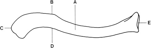
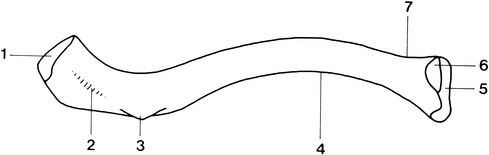
Position
Runs horizontally from the base of the neck to the shoulder and is subcutaneous throughout.
Main parts
Scapula (Figs 5.5 and 5.6)
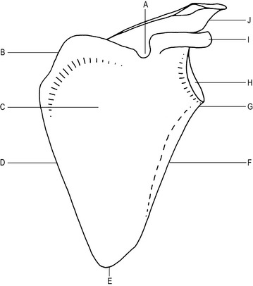

Articulations
Glenoid cavity of the scapula with the head of the humerus to form the shoulder joint.
Main parts
Lateral aspect
Ossification
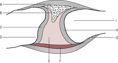
Shoulder joint (Figs 5.9 and 5.10)
Type
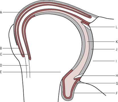
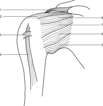
Strengthening ligaments
Tendons
These reinforce the fibrous capsule, forming the ‘rotator cuff’:
Supraspinatus tendon attached to the superior aspect of the greater tuberosity of the humerus.
Infraspinatus tendon attached to the middle of the greater tuberosity of the humerus.
Teres minor attached to the lower, posterior aspect of the greater tuberosity of the humerus.
Subscapularis tendon attached to the anterior aspect of the lesser tuberosity of the humerus.
Intracapsular structures
Tendon of biceps, long head
Movements
Flexion by the pectoralis major and the anterior fibres of the deltoid.
Extension by the posterior fibres of the deltoid and teres major, assisted by the latissimus dorsi.
Adduction by the pectoralis major and latissimus dorsi, assisted by the teres major.
Lateral rotation by the posterior fibres of the deltoid, infraspinatus and teres minor.
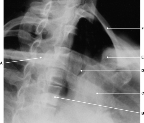
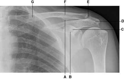
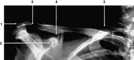
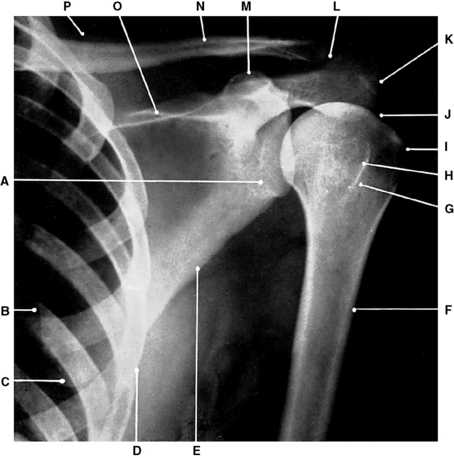
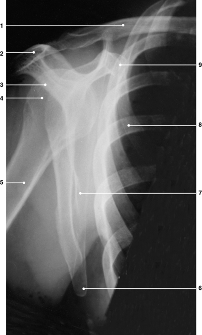
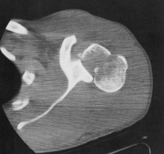
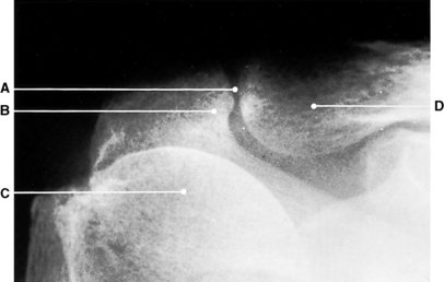
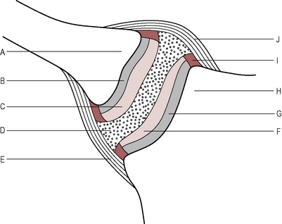
Radiographic appearances of the shoulder joint (Figs 5.11 to 5.15)
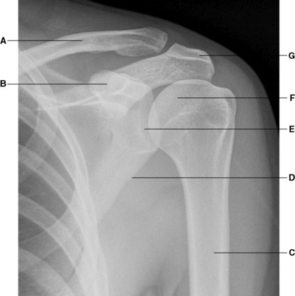
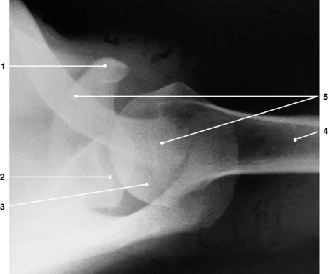
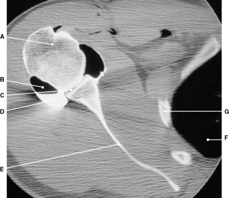
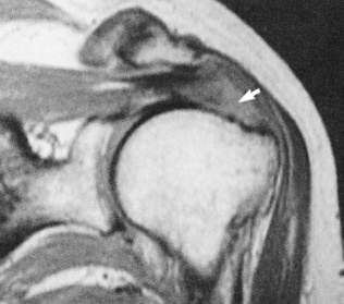
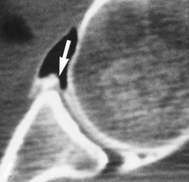
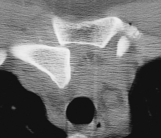
Fig. 5.15 Shoulder, abnormal glenoid labrum (arrowed). CT arthrotomogram.
(From Resnick Kransdorf, 2005.)
Acromioclavicular joint (Fig. 5.17)
Sternoclavicular joint (Fig. 5.19)
Movements
Anterior movement in a horizontal direction.
Blood supply
Internal mammary arteries and suprascapular arteries, which are branches of the subclavian artery.