CHAPTER 2 Shoulder Disorder: From Dysfunction to the Lesion
“From dysfunction to lesion” is a very complex concept that involves knowledge and interpretation of different parameters that could alter a function and bring about a short-term and more often a long-term anatomic lesion. Initially, an ultrastructural lesion may evolve with distinctive peculiarities, and can be clinical or subclinical. It can show up with subjective and objective symptoms only under certain circumstances and according to intrinsic and extrinsic factors combined.1
Over the past few decades, this syndrome has been increasingly diagnosed. It was first described in the early 20th century. In 1931, Meyer2 proposed that rotator cuff tears occurred secondary to friction against the undersurface of the acromion and described corresponding lesions on the undersurface of the acromion and greater tuberosity. However, he did not implicate the acromion directly. Codman,3 in 1934, defined the critical zone where most degenerative changes occur as the portion of the rotator cuff located 1 cm medial to the insertion of the supraspinatus on the greater tuberosity. Armstrong4 introduced the term supraspinatus syndrome.
Neer and Welsh5 have described subacromial impingement syndrome as a distinct clinical entity and hypothesized that the rotator cuff is impinged on by the anterior third of the acromion, coracoacromial ligament, and acromioclavicular joint, rather than merely by the lateral aspect of the acromion. The clinical diagnosis of impingement syndrome is commonly based on findings defined by the impingement sign and test. The patient’s history typically includes pain at night and positional discomfort referred as a painful arc. The clinical presentation may be confusing, and it is important to differentiate subacromial impingement syndrome from other conditions that may cause symptoms in the shoulder. Especially in young patients and in overhead athletes, the diagnosis of impingement should be made carefully.
Partial or complete resection of the acromion has been reported to be a promising method in the treatment of rotator cuff syndrome. On the other hand, the disappointing results of complete and lateral acromionectomy prompted Neer6 to focus on the undersurface of the acromion as the offending area. He developed the technique of anterior acromioplasty, which includes acromioclavicular resection arthroplasty, when indicated, to correct impingement by decompressing the subacromial space. This procedure has been the gold standard for the treatment of impingement and still represents the main procedure for many surgeons.
CAUSATIVE FACTORS OF IMPINGEMENT SYNDROME
Many causes have been proposed for subacromial impingement syndrome. These factors can be broadly classified as intrinsic or intratendinous factors, which are related to the intrinsic theory on the origin of impingement, and extrinsic or extratendinous factors, which are related to the mechanical theory. They can be further characterized as primary or secondary. A primary cause, either intrinsic or extrinsic, results in the impingement process by decreasing the subacromial space or by causing a degenerative process of the rotator cuff tendons. A secondary cause is the result of another process, such as instability, neurologic injury, tight posterior capsule of the glenohumeral joint, or muscle dysfunction.
Intrinsic Theory
Degenerative Tendinopathy
Ozaki and colleagues7 have studied the pathologic changes on the undersurface of the acromion as associated with tears of the rotator cuff in 200 cadaveric shoulder. They suggested that rotator cuff tears or injuries are the result of intrinsic rather than extrinsic causes associated with impingement, as advocated by Neer.8 They found that although a lesion in the anterior third of the undersurface of the acromion was always associated with a rotator cuff tear, the reverse was not true, and they concluded that the pathogenesis of most cuff tears was probably an intrinsic process.
They concluded that the pathogenesis of most tears is probably a degenerative process. Ogata and Uhthoff9 have suggested that tendon degeneration is the primary cause of partial tears of the rotator cuff, and that they might allow proximal migration of the humeral head, which could result in impingement and lead to complete tears of the rotator cuff.
Muscle Dysfunction
It has been observed that an intrinsic contractile tension overload on the muscle rather than primary impingement is the major factor in the cause of rotator cuff tendinitis. When the arm is in the overhead position, eccentric contraction of the supraspinatus decelerates internal rotation and adduction of the arm, causing an overload. This phenomenon is most dramatic in those involved in overhead sports, and it may also occur in manual workers who use overhead motions in their work. The proximal migration of the humeral head has also been associated with muscle fatigue, injury, and degenerative changes in the rotator cuff tendons. Bigliani and Levine10 have noted that resection of the coracoacromial ligament should be avoided in this situation because it may not relieve the impingement, but could allow for additional proximal migration of the humeral head. Decrease in proprioceptive sense with muscle fatigue might play a role in decreasing athletic performance and in fatigue-related shoulder dysfunction.
Imbalance of the rotator cuff muscles in athletes, who have developed this as a result of training or sport activities, has generally been found to be a predisposing factor or a consequence of the impingement syndrome. Brox and coworkers11 have reported that surgery and supervised exercise improve rotator cuff disease equally and significantly compared with placebo, suggesting the importance of considering this factor.
Extrinsic (Mechanical) Theory
Shape of the Acromion
Acromial morphology and differences in the shape and slope of the acromion as a potential source of symptoms in the shoulder have often been observed. Neer6 focused on the cause and effect relationship between acromial morphology and subacromial impingement. He proposed that variations in the shape and slope of the anterior aspect of the acromion were responsible for subacromial impingement and associated tears of the rotator cuff. A spur protruding into the subacromial space10 apparently was thought to be caused by tensile forces on the coracoacromial ligament. In another study, Morrison and Bigliani12 evaluated supraspinatus outlet radiographs and found that 80% of 82 patients who had a tear of the rotator cuff visible on an arthrogram had a type III acromion (hook acromion).
The classification system described by Bigliani and colleagues13 has been cited widely in the literature, but investigators have questioned its reliability. Zuckerman and associates14 reported low interobserver reliability during the evaluation of 110 anatomic specimens to determine acromial shape according to this classification.13 Jacobson and coworkers15 also reported low interobserver reliability when the system was used to evaluate acromial morphology as seen on a supraspinatus outlet view. They also questioned the correlation between acromial morphology and tears of the rotator cuff. The classification of acromial morphology on the basis of a subacromial outlet projection has reportedly been difficult because of individual differences in the supraspinatus outlet angle. Some investigators have stated that fluoroscopic control is necessary for a proper supraspinatus outlet view.
Wuh and Snyder16 have modified their classification system13 by addressing the thickness as well as the shape of the acromion. Three types of acromion were identified: type A (<8 mm), type B (8 to 12 mm) and type C (>12 mm).
Toivonen and colleagues17 measured the acromial angle, which is in accordance with the hypothesis proposed by Morrison and Bigliani12 of an association between acromion type III and rotator cuff tears. Aoki and associates18 studied 130 cadaveric shoulders and found that acromions with spur formation had a flatter slope and were associated with increased pitting on the surface of the greater tuberosity. They also showed that the prevalence of spurs in the subacromial space increased with age and noted a decreased alpha angle (also called acromial tilt) in the patients with impingement.
Degeneration of the Acromioclavicular Joint
Neer6,8 proposed that degeneration of the acromioclavicular joint may contribute to subacromial impingement. This hypothesis is supported by several other authors. Osteophytes protruding inferiorly from the undersurface of a degenerative acromioclavicular joint can contribute to impingement narrowing the supraspinatus outlet. Kessel and Watson19 found that one third of the patients in their study had lesions of the supraspinatus tendon, usually associated with degeneration of the acromioclavicular joint. Penny and Welsh20 subsequently found that osteoarthritis of the acromioclavicular joint may lead to failure of operative treatment of subacromial impingement. However, resection of the acromioclavicular joint should be performed only if the patient has symptoms in the joint region and if osteophytes contribute to the impingement.10
Coracoid Impingement
Coracoid impingement along the more medial aspect of the coracoacromial arch is less common, but has been reported.21 In patients with coracoid impingement, the pain is usually located on the anteromedial aspect of the shoulder and is felt in the arm and forearm. Forward elevation and internal rotation may elicit pain.10 In a recent study, cine magnetic resonance imaging was used to measure the interval between the coracoid process and the lesser tuberosity. In the asymptomatic control group, the average interval between the coracoid process and the lesser tuberosity was 11 mm, whereas in symptomatic patients the interval was found to be 6 mm. Gerber and coworkers22 have reported that coracoid impingement can be idiopathic, iatrogenic, or traumatic. As a choice for operative treatment, Dines and colleagues23 have recommended coracohumeral decompression.
Impingement by the Coracoacromial Ligament
A number of investigators6,8,24 have also implied the coracoacromial ligament as a source of impingement. McLaughlin25 observed the condition termed snapping shoulder and concluded that the coracoacromial ligament is an offending structure in painful shoulders.
Soslowsky and colleagues26 found statistically significant changes in the geometric dimensions of the lateral band of the coracoacromial ligament, which is the region most likely to impinge on the rotator cuff. In another study, they found significant changes in the material properties of the ligament. Sarkar and associates27 and Uhthoff and coworkers24 reported that histologic studies of specimens of the coracoacromial ligament from patients who had impingement syndrome revealed only degenerative changes without thickening.
Os Acromiale
Os acromiale is an unfused distal acromial epiphysis first described in 1863 by Gruber.28 Folliasson29 classified the lesion into four distinct types according to the anatomic location, with mesoacromion being the most common type. The prevalence of os acromiale, as reported in both radiographic and anatomic studies, has varied a great deal, with a range from 1% to 15%. It is difficult to detect an os acromiale on routine anteroposterior plain x-rays and an axillary view may thus be needed.10 An association between os acromiale, impingement syndrome, and rotator cuff tears has been reported. Impingement may occur because the unfused epiphysis on the anterior aspect of the acromion may be hypermobile and may tilt anteriorly as a result of its attachment to the coracoacromial ligament. Hertel and colleagues30 have recommended stable fusion of a sizeable and hypermobile os acromiale.
Tight Posterior Capsule
Harryman and associates31 have stated that oblique glenohumeral translations are not the result of ligament insufficiency or laxity. Instead, translation results when the capsule is asymmetrically tight. Asymmetrical tightness is thought to cause anterior and superior migration of the humeral head during forward elevation of the shoulder, possibly contributing to impingement.
Glenohumeral joint inflexibility can also create abnormal biomechanics of the scapula. Posterior shoulder inflexibility caused by capsular or muscular tightness affects the smooth motion of the glenohumeral joint31 and creates a wind-up effect so that the glenoid and scapula actually get pulled in a forward and inferior direction by the moving and rotating arm. This can create an excessive amount of protraction of the scapula on the thorax as the arm continues into the horizontally adducted position in follow-through. Because of the geometry of the upper aspect of the thorax, the more the scapula is protracted in follow-through, the farther it and its acromion move anteriorly and inferiorly around the thorax.
Reeves32 has proposed that the stiff shoulder be classified as a frozen shoulder or post-traumatic stiff shoulder. The term frozen shoulder, introduced by Codman33 in 1934, refers to no traumatic stiffness of the glenohumeral joint capsule.
Although many different causes have been proposed for frozen shoulder, the common thread in most causal theories is the presence of inflammation. On the other hand, a post-traumatic stiff shoulder may originate from a traumatic extracapsular process; however, subsequent capsular contracture may soon develop. Each of these diagnoses may exhibit a different underlying pathology, history, and treatment.
Scapulothoracic Dyskinesia
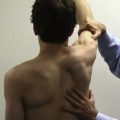
The dynamic scapulothoracic stability and importance of the core stability strongly indicate that those mechanisms, when altered, can lead to shoulder dysfunction. The shoulder is a complex mechanical structure containing several joints connecting the humerus, scapula, clavicle, and sternum. The scapula slides over the dorsal part of the thorax; it can glide over the so-called scapulothoracic gliding plane. It is a closed-chain mechanism. The relationship between the rotations of the humerus and scapula is commonly referred to as the scapulohumeral rhythm. The scapular motion strongly affects the mechanical energy delivered by muscles and the metabolic cost required to obtain the desired force. At the same time, the scapula has different roles; it is a functional part of the glenohumeral joint, retracting and protracting along the thoracic wall and elevating the acromion. It is a site for muscle attachment and a link in the proximal to distal sequencing of velocity, energy, and force that allows the most appropriate shoulder function.34
Function, the end result of the kinetic chain, can be defined as optimal anatomy acted on by physiologic muscle activations to produce optimal biomechanical forces and motions. Core stability is essential for the maximum efficiency of shoulder function. A functional definition of core stability is the ability to control the trunk over the pelvis to allow the coordinated sequenced activation of body part to produce, transfer, and control force and motion to the terminal segments in integrated body activities to obtain the desired work or athletic task.35 This definition implies patterned sequences for force generation and transfer, proximal stability for distal mobility, and control in three dimensions.
Muscle activation in kinetic chain function is based on preprogrammed patterns that are task-oriented, specific for different activities, and improved by repetition. Core muscle activation is used to generate rotational torques around the spine and provides stiffness to the entire central mass, making a rigid cylinder that confers a long lever arm around which rotation can occur and against which muscles can be stabilized as they contract.36
One of the most important abnormalities in scapular biomechanics is actually the loss of the link function in the kinetic chain. If the scapula becomes deficient in motion or position, transmission of the large generated forces from the lower to the upper extremity is impaired. This creates a deficiency in resultant maximum force, which can be delivered to the hand or creates a situation of catch-up, in which the more distal links have to work more actively to compensate for the loss of the proximally generated force. This can impair the function of the distal links because they do not have the size, muscle cross-sectional area, or time to develop these larger forces efficiently. Kibler’s34 calculations have shown that a 20% decrease in kinetic energy delivered from the hip and trunk to the arm necessitates an 80% increase in mass or a 34% increase in rotational velocity at the shoulder to deliver the same amount of resultant force to the hand. This required adaptation can cause overload problems with repeated use.
Adaptation Mechanisms: Allostasis.
The adaptation mechanism in this case is first neuromuscular, aimed at temporarily maintaining a work- or sports-related function. In addition to overloading some muscle groups, they stress joints, ligaments, and tendons to work outside the save zones, placing these structures at risk. All this can lead to the concept of allostasis introduced by Sterling and Eyer.37
To adapt to or ultimately survive a stressful situation, animals and humans must be able to change their behavior and physiology. Allostasis is the coordinated process that promotes such adaptation and increases the chance of survival. For example, in response to an intensive emotional stressor, the sympathetic branch of the autonomic nervous system is activated and the adrenal medulla secretes epinephrine (adrenaline). This prepares the individual for intense and vigorous physical activity by mobilizing energy resources. The blood flow is redirected to the skeletal muscles, heart, and brain. The heart and respiratory rates, as well as arousal and vigilance, increase. The behavior is changed toward, for example, an appropriate level of aggressiveness—the fight-or-flight response described by Cannon.38 When the adaptation to a stressor is successful, the allostatic responses protect the body and help the individual to cope effectively.
This example initially can seem excessive for the shoulder, but a careful evaluation can help in understanding how every body system survives, thanks to function-dependent replacement mechanisms that have to meet different versatility characteristics. However, so as not to impose unnecessary wear and tear on the organism, the physiologic responses should return to baseline (resting values) quickly after the challenge or threat has terminated, and a period of rest and recovery should follow before a new challenge is encountered. The evolutionary basis for this defense reaction is very old, but in modern society mental and social stressors probably elicit it more often than physical threats requiring a vigorous effort and thus using the energy mobilized. Furthermore, important to the concept of allostasis is the primary role of the brain in regulating physiologic processes, thereby emphasizing the potential role of psychological and social processes in health and disease.37
Allostasis focuses on variability (from the Greek allo, meaning variable), in contrast to the older concept of homeostasis, which focuses on the importance of maintaining a constant internal environment. Accordingly, allostasis can be translated as stability through change.37 This refers to the principle that an organism, to achieve (homeostatic) stability, must have the ability to vary its physiology to match different challenges or demands of varying severity. These two principles can be seen as representing different ways of functioning for different physiologic systems. According to the concept of allostasis, a balance between activity and rest is important for sustaining health. In physiologic terms, this can be described as a necessary balance between catabolism (when energy is used) and anabolism (when energy is stored and tissues are repaired).37 When this balance is disrupted (e.g., because of chronic physical overload or psychosocial stress), the excessive and sustained activation of the allostatic systems results in what has been termed allostatic load, a term coined by McEwen and Stellar.39 Allostatic responses are beneficial over the short term because they promote adaptation. With time, however, they may lead to allostatic load, which can be described as the cumulative wear and tear on the organism, sometimes referred to as the price of adaptation. According to McEwen and Seeman,40 at least four situations can lead to allostatic load:
Thus, too much use of allostatic systems or their dysfunction (overactivity or underactivity) results in a cumulative wear and tear on the organism, which over long periods promotes pathophysiologic changes and may lead to various pathologic conditions and diseases. Examples in the literature are atherosclerosis, hypertension, insulin resistance, abdominal obesity, type 2 diabetes, autoimmune and inflammatory disorders, cuff tears, labral tears, and SICK syndrome (scapular malposition, inferior medial border prominence, coracoid pain and malposition, dyskinesis of scapular movement).39,40 The processes leading to allostatic load are complex and are influenced not only by the impact of stressful events and daily hassles from work or sports that accumulate over long periods (chronic stress), but also from genetic factors, early life experiences, personality characteristics,and lifestyle factors, such as exercise, diet, smoking, and alcohol.40 A typical example is the functional overload of some shoulder ligament and/or tendon structures (allostatic response) in the presence of scapulothoracic and core dysfunctions.
More commonly, the scapular stabilizers are injured by the following: (1) direct blow trauma; (2) microtrauma-induced strain in the muscles, leading to muscle weakness and force couple imbalance; (3) becoming fatigued from repetitive tensile use (also true for catch-up); (4) or inhibition by painful conditions around the shoulder (it is assumed that muscle pain has the potential to change the coordination between agonist and antagonist by reflex mediation). These causes are responsible for the causative pathogenetic mechanisms of work and sports-related shoulder dysfunctions that can become potentially symptomatic in case of a supposed allostatic load on the compensatory structures.
High-Frequency and Low- Frequency Fatigue
In the shoulder, muscle inhibition is seen as decreased ability for muscles to exert torque and stabilize the scapula and as disorganization of the normal muscle firing patterns around the shoulder girdle.41 This is common in glenohumeral and subacromial pathologies, whether resulting from cuff tears, instability, labral pathology, arthrosis, muscular fatigue, or postural disorders. Muscle inhibition and the resulting scapular dyskinesia appear to be a nonspecific response to a painful condition in the shoulder rather than a specific response to a specific glenohumeral pathologic situation. The exact nature of this inhibition is not clear. The nonspecific response and disorganization of motor patterns suggest a proprioceptively based mechanism. Pain from direct muscle injury or indirect sources, such as fatigue or uncontrolled muscle strain, have been shown to alter proprioceptive input from Golgi tendon organs and muscles spindles.
Proprioception And Muscular Fatigue
The somatosensory system is the collection of peripheral sensory receptors responsible for giving rise to afferent information for the perceptions of mechanoreceptive (tactile and proprioceptive), thermoreceptive, and pain sensations. Because proprioception is a component of neuromuscular control, the two terms are often used interchangeably and incorrectly. Proprioception is defined as the specialized variation of the sensory modality of touch that encompasses the sensation of joint movement (kinesthesia) and joint position,42 whereas neuromuscular control is the unconscious motor efferent response to afferent sensory (proprioceptive) information. A mechanoreceptor is a specialized neuroepithelial structure found in the skin, ligamentous, muscular, and tendinous tissue that relays information about a joint.43 It transduces functional and mechanical deformation into frequency-modulated neural signals. An increase in deformation causes an increase in afferent discharge of neural signals back to the CNS.
Proprioceptive feedback regarding joint position results from mechanical stimulation of the mechanoreceptors located in the periarticular tendons, muscles, ligaments, and possibly skin.43,44 Ruffini-type mechanoreceptors are predominant in all articular structures of the shoulder except for the glenohumeral ligaments, where pacinian corpuscle-type receptors are most abundant.44 Muscle spindles and Golgi tendon organs are present in the muscles.
The capsuloligamentous and myotendinous receptors gather information that is modulated by the central nervous system through afferent pathways. Various muscle groups are activated and coordinated via the efferent pathways. The interplay between capsuloligamentous restraints and muscles is crucial; its role in maintaining scapular-thoracic coordination and stability at the glenohumeral joint is not fully appreciated, but it is influenced by mechanical and sensorimotor factors.1 Moreover, the existence of a reflex arc from mechanoreceptors within the glenohumeral capsule to muscles crossing the joint confirms and extends the concept of synergism between the passive and active restraints of the glenohumeral joint.31 Receptors are divided into rapidly and slowly adapting types, which work in an integrated fashion and with different excitement thresholds.
This combination of muscle and joint receptors forms an integral component of a complex sensorimotor system that plays a role in the proprioceptive mechanism and is part of a feedback–feed-forward system initiated by an activation of mechanoreceptors. The sensory input (afferent) from the mechanoreceptors is relayed by the peripheral nervous system (PNS) to the CNS. The CNS responds to the afferent stimulus by discharging a motor signal (efferent), which modulates effector muscle function by controlling joint motion and/or position.
Because mechanoreceptors, which are responsible for proprioceptive feedback causing neuromuscular responses, are present in the muscular structure surrounding the joint,43 it is highly likely that as a muscle fatigues, proprioceptive feedback is delayed, and consequently neuromuscular control and shoulder function are impaired. Fatigue affects sensation of joint movement, decreases work and athletic performance, and increases fatigue-related shoulder dysfunction. Voight and colleagues45 have noted that that the decrease in ability after fatigue is caused by dysfunctional mechanoreceptors.
Both central and peripheral fatigue may also influence proprioception and neuromuscular control. Central fatigue results from CNS influence, whereas peripheral fatigue occurs at the level of the sarcomere and involves failure at the neuromuscular junction, sarcolemma, and transverse tubules. Because central fatigue appears so commonly in human performance, one might expect that its development confers some evolutionary advantages. Perhaps drive is limited because continued stimuli to the muscle would put the neuromuscular junction, or more likely the intracellular events accompanying excitation-contraction coupling and actin-myosin interactions, into a catastrophic state, from which recovery would be delayed or impossible. More relevant to the aim of this chapter is Winter’s definition,46 which refers to muscle fatigue as occurring when the muscle tissue cannot support metabolism at the contractile element because of ischemia (insufficient oxygen) or local depletion of any of the metabolic substrates.
Overuse Dysfunction
Under healthy conditions, a glenohumeral distraction force generated during the acceleration phase, from 1 to 1.5 times body weight, is present at ball release. This distraction force is normally compensated by a concomitant violent contracture of the posterior shoulder musculature (cuff, scapular stabilizers, and quadrangular space muscles) at ball release, lasting through the first third of follow-through. With repetitive overuse, deconditioned (weak) posterior shoulder musculature, usually involving the scapular stabilizers and rotator cuff, results in the distraction force not being fully compensated. This allows the posterior inferior capsule to receive abnormal tensile load, because at ball release with the shoulder flexed 90 degrees forward and adducted to neutral or more the posterior band of the inferior glenohumeral ligament (IGHL) is directly in line to receive most or all of any glenohumeral distractive force. This focal repetitive distractive load applied to the posterior inferior capsule is thought to stimulate a fibroblastic response, which initiates and propagates the throwing acquired posterior inferior capsular contracture (Wolff’s law of collagen).
CONCLUSIONS
Over time, these are the same elements that dictate the developing anatomic characteristics of these lesions; however, they can also result from an external event such as trauma, a functional overload period, or activity resumption after prolonged inactivity. Reducing the effect of the compensatory mechanisms, these can produce a clinical picture that is the end result of dysfunction and the anatomic lesion.
In most cases of more pronounced anatomic lesions, the injury repair is suggested and then rehabilitation is recommended, which aims at treating the real cause (dysfunction) leading to the anatomic lesion. Rehabilitation that ignores these aspects can result in surgical failures—for example, of rotator cuff repair. A high incidence of relapses of surgically repaired cuff lesions has been noted in recent studies; these have been identified with ultrasound and MRI tests, and such retears are often asymptomatic, with he most evident effect being a loss of strength.47
The concepts and implications discussed are to be remembered—the evolving lesion that evolves slowly over time, and the emerging lesion, which often requires an external event to emerge clinically. Lesions that evolve through deafferentation, which might be the manifestation of different factors (e.g., muscle fatigue, trauma, laxity) may gradually turn into emerging lesions and therefore become symptomatic. It is possible, therefore, to be faced with tissue damage that only recently has become symptomatic but may have long-standing morphofunctional causes, which can seriously affect the outcome of rehabilitation and/or surgery.
1. Di Giacomo G, Pouliart N, Costantini A, De Vita A. Atlas of Functional Shoulder Anatomy. New York: Springer; 2008.
2. Meyer AV. The minuter anatomy of attrition lesions. J Bone Joint Surg. 1931;13:341-360.
3. Codman EA. Rupture of the supraspinatus tendon. Clin Orthop. 1990;1911:3-26.
4. Armstrong JR. Excision of the acromion in treatment of the supraspinatus syndrome. Report of ninety-five excisions. J Bone Joint Surg Br. 1949;31:436-442.
5. Neer CS, Welsh RP. The shoulder in sports. Orthop Clin North Am. 1977;8:583-591.
6. Neer CS Anterior acromioplasty for the chronic impingement syndrome in the shoulder: a preliminary report. J Bone Joint Surg Am, 54; 1972:41-50.
7. Ozaki J, Fujimoto S, Nakagawa Y, et al. Tears of the rotator cuff of the shoulder associated with pathologic changes in the acromion. A study in cadavera. J Bone Joint Surg Am. 1988;70:1224-1230.
8. Neer CS Impingement lesions. Clin Orthop (173); 1983:70-77.
9. Ogata S, Uhthoff HK Acromial enthesopathy and rotator cuff tear. A radiologic and histologic postmortem investigation of the coracoacromial arch. Clin Orthop (254); 1990:39-48.
10. Bigliani LU, Levine WN. Subacromial impingement syndrome. J Bone Joint Surg Am. 1997;79:1854-1868.
11. Brox JI, Staff PH, Ljunggren AE, Brevik JI. Arthroscopic surgery compared with supervised exercises in patients with rotator cuff disease (stage II impingement syndrome). BMJ. 1993;307:899-903.
12. Morrison DS, Bigliani LU. The clinical significance of variations in acromial morphology. Orthop Trans. 1987;11:234.
13. Bigliani LU, Morrison DS, April EW. The morphology of the acromion and its relationship to rotator cuff tears. Orthop Trans. 1986;10:228.
14. Zuckerman JD, Kummer FJ, Cuomo F, Greller M Interobserver reliability of acromial morphology classification: an anatomic study. J Shoulder Elbow Surg, 6; 1997:286-287.
15. Jacobson SR, Speer KP, Moor JT, et al. Reliability of radiographic assessment of acromial morphology. J Shoulder Elbow Surg. 1995;4:449-453.
16. Wuh HCK, Snyder SJ. A modified classification of the supraspinatus outlet view based on the configuration and anatomic thickness of the acromion. Orthop Trans. 1992;16:767.
17. Toivonen DA, Tuite MJ, Orwin JF. Acromial structure and tears of the rotator cuff. J Shoulder Elbow Surg. 1995;4:376-383.
18. Aoki M, Ishii S, Usui M. The slope of the acromion and rotator cuff impingement. Orthop Trans. 1986;10:228.
19. Kessel L, Watson M. The painful arc syndrome. Clinical classification as a guide to management. J Bone Joint Surg Br. 1977;59:166-172.
20. Penny JN, Welsh RP. Shoulder impingement syndromes in athletes and their surgical management. Am J Sports Med. 1981;9:11-15.
21. Ian K, Lo Y, Burkhart S The cause and assessment of subscapularis tendon tears: a case for subcoracoid impingement, the roller-wringer effect, and TUFF lesions of subscapularis. Arthroscopy, 19; 2003:1142-1150.
22. Gerber C, Terrier F, Ganz R. The role of the coracoid process in the chronic impingement syndrome. J Bone Joint Surg Br.. 1985;67:703-708.
23. Dines DM, Warren RF, Inglis AE, Pavlov H. The coracoid impingement syndrome. J Bone Joint Surg Br.. 1990;72:314-316.
24. Uhthoff HK, Hammond DI, Sarkar K, et al. The role of the coracoacromial ligament in the impingement syndrome. A clinical, radiologic and histologic study. Int Orthop. 1988;12:97-104.
25. McLaughlin HL Lesions of the musculotendinous cuff of the shoulder. The exposure and treatment of tears with retraction. 1944. Clin Orthop (304); 1994:3-9.
26. Soslowsky LJ, An CH, DeBano CM, Carpenter JE Coracoacromial ligament: in situ load and viscoelastic properties in rotator cuff disease. Clin Orthop (303); 1996:40-44.
27. Sarkar K, Taine W, Uhthoff HK The ultrastructure of the coracoacromial ligament in patients with chronic impingement syndrome. Clin Orthop (254); 1990:49-54.
28. Gruber W. Über die arten der Acromialknochen und accidentellen Akromialgelenke. Arch Anat Physiol Wissench Med. 1863:373-387.
29. Folliasson A. Un cas d’os acromial. Rev Orthop. 1933;20:533-538.
30. Hertel R, Windisch W, Schuster A, Ballmer FT. Transacromial approach to obtain fusion of unstable os acromiale. J Shoulder Elbow Surg. 1998;7:606-609.
31. Harryman DTII, Sidles JA, Clark JM, et al. Translation of the humeral head on the glenoid with the passive glenohumeral motion. J Bone Joint Surg Am. 1990;72:1334-1343.
32. Reeves B. Arthrographic changes in frozen and post-traumatic stiff shoulders. Proc R Soc Med. 1996;59:827-830.
33. Codman EA The Shoulder 216-224. Boston: Todd; 1934.
34. Kibler WB. Biomechanical analysis of the shoulder during tennis activities. Clin Sports Med. 1995;14:79-85.
35. Kibler WB. Evaluation and diagnosis of scapulothoracic problems in the athlete. Sports Med Arthrosc Rev. 2000;8:192-202.
36. Kibler WB, Livingston BP. Closed-chain rehabilitation for upper and lower extremities. J Am Acad Orthop Surg. 2001;9:412-421.
37. Sterling P, Eyer J Allostasis: a new paradigm to explain arousal pathology. Fisher S, Reason J, editors. Handbook of Life Stress, Cognition and Health. New York: John Wiley & Sons; 1988:629-649.
38. Cannon WB. The emergency function of the adrenal medulla in pain and major emotions. Am J Physiol. 1914;24:143-153.
39. McEwen BS, Stellar E Stress and the individual: mechanisms leadind to disease. Arch Intern Med, 153; 1993:2093-2101.
40. McEwen BS, Seeman T Protective and damaging effects of mediators of stress: elaborating and testing the concept of allostasis and allostatic load. Ann N Y Acad Sci, 896; 1999:30-47.
41. Kuhn JE, Plancher KD, Hawkins RJ. Scapular winging. J Am Acad Orthop Surg. 1995;3:319-325.
42. Lephart SM, Wamer JP, Borsa PA, Fu FH. Proprioception of the shoulder joint in healthy, unstable, and surgically repaired shoulders. J Shoulder Elbow Surg. 1994;3:371-380.
43. Grigg P. Peripheral neural mechanism in proprioception. J Sport Rehabil. 1994;3:2-17.
44. Vangsness CT, Ennis M, Taylor JG, Atkinson R. Neural anatomy of the glenohumeral ligaments, labrum, and subacromial bursa. Arthroscopy. 1995;11:180-184.
45. Voight ML, Hardin JA, Blackburn TA, et al. The effects of muscle fatigue on and the relationship of arm dominance to shoulder proprioception. J Orthop Sports Phys Ther. 1996;23:348-352.
46. Winter DA Biomechanics and Motor Control of Human Movement 191-212. 2nd ed. Wiley-Interscience; 1990.
47. Flurin PH, Boileau P, Wolf EM, et al Cuff integrity after arthroscopic rotator cuff repair: correlation with clinical results in 576 cases. Arthroscopy, 4; 2007:340-346.

